. 2016 Apr 1;291(14):7661-8.
doi: 10.1074/jbc.M115.689448. Epub 2016 Feb 9.

Transient Expression of Fez Family Zinc Finger 2 Protein Regulates the Brn3b Gene in Developing Retinal Ganglion Cells

Affiliations

Transient Expression of Fez Family Zinc Finger 2 Protein Regulates the Brn3b Gene in Developing Retinal Ganglion Cells

Chunsheng Qu et al. J Biol Chem. .

Abstract

Retinal ganglion cells (RGCs) are projection neurons in the neural retina that relay visual information from the environment to the central nervous system. The early expression of MATH5 endows the post-mitotic precursors with RGC competence and leads to the activation ofBrn3bthat marks committed RGCs. Nevertheless, this fate commitment process and, specifically, regulation ofBrn3bremain elusive. To explore the molecular mechanisms underlying RGC generation in the mouse retina, we analyzed the expression and function of Fez family zinc finger 2 (FEZF2), a transcription factor critical for the development of projection neurons in the cerebral cortex.Fezf2mRNA and protein were transiently expressed at embryonic day 16.5 in the inner neuroblast layer and the prospective ganglion cell layer of the retina, respectively. Knockout ofFezf2in the developing retina reduced BRN3B+ cells and increased apoptotic cell markers.Fezf2knockdown by retinalin uteroelectroporation diminished BRN3B but not the coexpressed ISLET1 and BRN3A, indicating that the BRN3B decrease was the cause, not the result, of the overall reduction of BRN3B+ RGCs in theFezf2knockout retina. Moreover, the mRNA and promoter activity ofBrn3bwere increasedin vitroby FEZF2, which bound to a 5' regulatory fragment in theBrn3bgenomic locus. These results indicate that transient expression ofFezf2in the retina modulates the transcription ofBrn3band the survival of RGCs. This study improves our understanding of the transcriptional cascade required for the specification of RGCs and provides novel insights into the molecular basis of retinal development.

Keywords: Brn3b; Fezf2; gene expression; gene knockout; in utero electroporation; mouse; neurodevelopment; retinal ganglion cells; transcription regulation.


Figures

FIGURE 1.
FIGURE 1.
Expression of Fezf2 in the developing retina. A, sections of mouse retinas at the designated developmental stages were processed for Fezf2 in situ hybridization. Fezf2 was transiently expressed in the retinas, with maximal expression occurring at E16.5. B, Fezf2 was expressed in the inner NBL on E16.5. C, coronal section of the E16.5 brain showing expression of Fezf2 in the cortex (arrow) and retina (arrowhead). Scale bars = 200 μm.
FIGURE 2.
FIGURE 2.
Expression of FEZF2 in the developing retina. A, FEZF2 expressions were determined in retinal sections by IF at the indicated developmental stages. FEZF2 was expressed in the prospective GCL of the retina at E16.5. B, FEZF2 expression co-localized with BRN3B. Scale bars = 200 μm (A) and 50 μm (B).
FIGURE 3.
FIGURE 3.
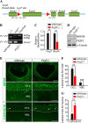
Targeted disruption of Fezf2 leads to a reduction in BRN3B+ RGCs and an increase in apoptosis. A, the Fezf2 second exon harbors two loxp sites flanking the second exon. P1, P2, and P3 are the three genotyping primers located in exon 1–3 of the Fezf2 locus. B, gel electrophoresis of the PCR products from genotyping. The presence of the first loxp site and the absence of exon 2 were determined by PCR with the primer sets of P1-P2 and P1-P3, respectively. Expression of Cre was also confirmed in the knockout mice by PCR (data not shown). C, E17.5 retinas were isolated from wild-type and Fezf2-null mice, and the relative expressions of Brn3b and Fezf2 were measured by q-PCR. The knockout mice had diminished expression of Fezf2 and Brn3b. D, expression of BRN3B and FEZF2 was measured in isolated retinas from control and Fezf2-null mice by Western blotting with α-tubulin as an internal control. E, immunostaining of BRN3B and activated CASPASE III at E17.5 in control and Fezf2 knockout retinas. Knocking out Fezf2 reduced BRN3B+ cells and increased apoptotic cells. Scale bars = 200 and 50 μm for low and high magnitude, respectively (BRN3B) and 50 μm (CASPASE III). F, the numbers of BRN3B+ in the GCL and NBL per field (×40). G, total of CASPASE III+ cells per section in control and Fezf2-null retinas. **, p < 0.005; ***, p < 0.0005.
FIGURE 4.
FIGURE 4.
Reduced BRN3B+ cells and axons in Fezf2−/− mice on P7. A, whole mount retinas from wild-type and Fezf2−/− mice on P7 were examined for the expression of BRN3B. Insets show enlarged images at ×40. Scale bars = 5000 μm and 30 μm (insets). B, histogram showing that the number of BRN3B+ cells per field (×40) was significantly reduced in knockout mice. C, SMI32 staining in the whole mount of retinas from wild-type and Fezf2−/− mice on P7. The knockout retina had weakened axonal processes compared with the wild type. The arrow in the Fezf2−/− retinal section points to an aberrant axon bifurcation. Scale bar = 30 μm. D, eyes with attached optic nerves were dissected out. The optic nerves in Fezf2−/− mice were thinner than in the wild type. The image is a representative of four samples (eyes). *, p < 0.05.
FIGURE 5.
FIGURE 5.
Inhibition of BRN3B expression by Fezf2-siRNA. A, Fezf2-scRNA-1 or Fezf2-siRNA-1 was delivered to one eye cup of E13.5 embryos (green). Retinal sections were examined for the expression of MATH5, ISLET1, and BRN3B (red) at E17.5. Similar to those expressing Fezf2-scRNA-1, Fezf2-siRNA-1-transfected cells partially colocalized with MATH5 and ISLET1 (arrowheads). In contrast, retinal cells expressing Fezf2-siRNA were mostly BRN3B-negative. B, the percentages of colocalization in the Fezf2-siRNA-1- and Fezf2-scRNA-1-transfected cells. C, single cell suspensions prepared at E17.5 from transfected retinas were sorted by FACS. The relative expression of Brn3b and Fezf2 was measured in GFP+ cells by q-PCR. D, Fezf2-scRNA-2 or Fezf2-siRNA-2 was delivered to the eye cups of E13.5 embryos (green). Retinal sections were examined for the expression of BRN3A (red) on E17.5. Arrowheads indicate colocalizations of BRN3A and transfected GFP. E, no significant difference in the percentage of colocalization between scramble- and siRNA-transfected cells. Scale bars = 200 μm and 20 μm (insets). *, p < 0.05; **, p < 0.005.
FIGURE 6.
FIGURE 6.
Overexpression of Fezf2 alters the phenotype of retinal cells. A, retinas transfected with pCAGEN and Fezf2-pCAGEN were examined on E17.5 with IF. Colocalization of MATH5, ISLET1, and BRN3B (red) with GFP was determined by IF. B, the percentages of colocalization in the pCAGEN and Fezf2-pCAGEN groups. Overexpression of Fezf2 abolished the expression of MATH5, ISLET1, and BRN3B. Scale bars = 200 μm and 20 μm (insets). **, p < 0.005; ***, p < 0.0005.
FIGURE 7.
FIGURE 7.
FEZF2 binds to the 5′ regulatory sequence in Brn3b. A, schematic of the genomic structure of Brn3b. A tentative promoter including the 5′ UTR of Brn3b contains predicted FEZF2 binding motifs (yellow bars). B, relative luciferase activities measured in the lysates of N2a cells. N2a cells stably expressing the pGL4.18-Brn3b promoter were assayed for luciferase activity after transient expression of the indicated constructs for 2 days. Fezf2-pCAGEN increased, whereas Fezf2-siRNA decreased, luciferase activity. C and D, ChIP with an anti-V5 antibody precipitated FEZF2-bound DNA from N2a cells expressing V5-tagged FEZF2 but not those expressing V5-GFP. The DNA was analyzed by gel electrophoresis (C) of the PCR products for the presence of the tentative promoter and measured by q-PCR (D) for enrichment of the binding sequence. The input of ChIP and the pulldown without V5 antibody were taken as positive and negative controls of the PCR, respectively. The control sequence for the q-PCR in D is a region in the 3′ UTR of the Brn3b locus. *, p < 0.05; **, p < 0.005.

References

    1. Yang Z., Ding K., Pan L., Deng M., and Gan L. (2003) Math5 determines the competence state of retinal ganglion cell progenitors. Dev. Biol. 264, 240–254 - PubMed
    1. Wang S. W., Kim B. S., Ding K., Wang H., Sun D., Johnson R. L., Klein W. H., and Gan L. (2001) Requirement for math5 in the development of retinal ganglion cells. Genes Dev. 15, 24–29 - PMC - PubMed
    1. Hutcheson D. A., and Vetter M. L. (2001) The bHLH factors Xath5 and XNeuroD can upregulate the expression of XBrn3d, a POU-homeodomain transcription factor. Dev. Biol. 232, 327–338 - PubMed
    1. Liu W., Mo Z., and Xiang M. (2001) The Ath5 proneural genes function upstream of Brn3 POU domain transcription factor genes to promote retinal ganglion cell development. Proc. Natl. Acad. Sci. U.S.A. 98, 1649–1654 - PMC - PubMed
    1. Gan L., Wang S. W., Huang Z., and Klein W. H. (1999) POU domain factor Brn-3b is essential for retinal ganglion cell differentiation and survival but not for initial cell fate specification. Dev. Biol. 210, 469–480 - PubMed

Publication types

MeSH terms

LinkOut - more resources