. 2016 Apr 8;291(15):8173-88.
doi: 10.1074/jbc.M115.641902. Epub 2016 Feb 9.

Co-immunoprecipitation with Tau Isoform-specific Antibodies Reveals Distinct Protein Interactions and Highlights a Putative Role for 2N Tau in Disease

Affiliations

Co-immunoprecipitation with Tau Isoform-specific Antibodies Reveals Distinct Protein Interactions and Highlights a Putative Role for 2N Tau in Disease

Chang Liu et al. J Biol Chem. .

Abstract

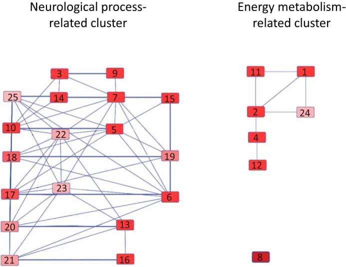
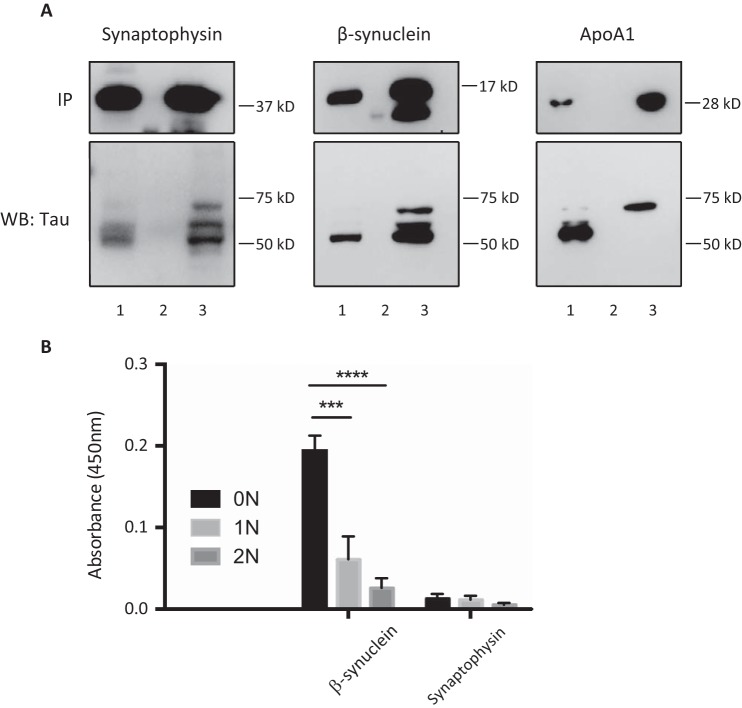
Alternative splicing generates multiple isoforms of the microtubule-associated protein Tau, but little is known about their specific function. In the adult mouse brain, three Tau isoforms are expressed that contain either 0, 1, or 2 N-terminal inserts (0N, 1N, and 2N). We generated Tau isoform-specific antibodies and performed co-immunoprecipitations followed by tandem mass tag multiplexed quantitative mass spectrometry. We identified novel Tau-interacting proteins of which one-half comprised membrane-bound proteins, localized to the plasma membrane, mitochondria, and other organelles. Tau was also found to interact with proteins involved in presynaptic signal transduction. MetaCore analysis revealed one major Tau interaction cluster that contained 33 Tau pulldown proteins. To explore the pathways in which these proteins are involved, we conducted an ingenuity pathway analysis that revealed two significant overlapping pathways, "cell-to-cell signaling and interaction" and "neurological disease." The functional enrichment tool DAVID showed that in particular the 2N Tau-interacting proteins were specifically associated with neurological disease. Finally, for a subset of Tau interactions (apolipoprotein A1 (apoA1), apoE, mitochondrial creatine kinase U-type, β-synuclein, synaptogyrin-3, synaptophysin, syntaxin 1B, synaptotagmin, and synapsin 1), we performed reverse co-immunoprecipitations, confirming the preferential interaction of specific isoforms. For example, apoA1 displayed a 5-fold preference for the interaction with 2N, whereas β-synuclein showed preference for 0N. Remarkably, a reverse immunoprecipitation with apoA1 detected only the 2N isoform. This highlights distinct protein interactions of the different Tau isoforms, suggesting that they execute different functions in brain tissue.

Keywords: Alzheimer disease; Tau protein (Tau); Tauopathy; apolipoprotein; protein/protein interaction.


Figures

FIGURE 1.
FIGURE 1.
Western blot validation of Tau immunoprecipitation reactions. The 1st row indicates the tissue source, and the 2nd row indicates the antibody used for the IP (Tau isoform-specific antibodies 0N, 1N, and 2N and pan-Tau antibodies Dako Tau and M). The input, IP, and flow-through (FT) were loaded for each reaction. An IP from wild-type mouse brain using normal serum (IP:normal serum) and from a Tau KO brain using the Dao antibody (IP:Dako with Tau KO mice) were included as negative controls.
FIGURE 2.
FIGURE 2.
Schematic diagram of the quantitative proteomics workflow by LC-MS/MS. The procedure combines incubation of the indicated antibodies with protein G (ProtG) beads, followed by adding the indicated lysates, digestion into peptides, an estimation of the protein concentration, isobaric tandem mass tag (TMT) labeling of a sample pooled from the eight reactions, separation by liquid chromatography (LC), determining relative TMT tagging, adjustments of the sample volumes based on relative TMT representation, adjustment accordingly of volumes of the eight samples to generate a mixture of equal TMT representation, followed by a measurement by mass spectrometry (MS) and quantification of relative protein intensities by tags. Eight co-IP reactions were performed in parallel, with samples 1 and 2 being labeled with two different tags (Table 1) for normalization.
FIGURE 3.
FIGURE 3.
Validation of identified Tau-interacting proteins by co-immunoprecipitation. A, Tau-interacting proteins were immunoprecipitated under native conditions from brain lysate obtained from a 2-month-old wild-type mouse using antibodies for apoE, synaptophysin, syntaxin 1B, apoA1, β-synuclein, synaptogyrin-3, MtCK, synaptotagmin, and synapsin 1 and then blotted with an anti-Tau antibody. The input is shown in lane 1; the wild-type lysate that has been immunoprecipitated with normal IgG is shown as a negative control in lane 2; and the IP with the specified antibody in lane 3. B, Fyn that is known to interact with tau was not co-immunoprecipitated because of the buffer used for the reaction. Extraction of mouse brain tissue in IP buffer yielded a supernatant (Sup, input for IP) and a pellet that was further extracted with 70% formic acid to obtain the pellet fraction. 20 μg of supernatant fraction (the equivalent of that used in the above IP reactions) contains very little Fyn compared with the pellet.
FIGURE 4.
FIGURE 4.
Classification of Tau-interacting proteins. Charts of the subcellular localization of the identified Tau-interacting proteins (A), the subset of membrane-bound proteins identified as the largest category using the annotation cell compartment (B), and functional classes of the identified proteins (C) are shown. Only major categories are shown; categories with fewer proteins are cumulatively displayed as others.
FIGURE 5.
FIGURE 5.
Ingenuity pathway analysis identifies two highly enriched networks for the Tau-interacting proteins. These networks are the cell-to-cell signaling and interaction (A) and the neurological disease network (B). Tau and its interacting proteins are marked with red dots.
FIGURE 6.
FIGURE 6.
Pathway cluster analysis. The top 25 canonical pathways enriched for Tau-interacting proteins are as follows: 1) glycolysis I; 2) gluconeogenesis I; 3) mitochondrial dysfunction; 4) TCA cycle II (eukaryotic); 5) remodeling of epithelial adherens junctions; 6) clathrin-mediated endocytosis signaling; 7) Huntington disease signaling; 8) creatine-phosphate biosynthesis; 9) oxidative phosphorylation; 10) 14-3-3-mediated signaling; 11) sucrose degradation (mammalian); 12) isoleucine degradation I; 13) glutamate receptor signaling; 14) Parkinson disease signaling; 15) GABA receptor signaling; 16) glutamine biosynthesis I; 17) gap junction signaling; 18) germ cell-Sertoli cell junction signaling; 19) virus entry via endocytic pathway; 20) calcium signaling; 21) amyotrophic lateral sclerosis signaling, 22) breast cancer regulation by stathmin1; 23) fMLP signaling in neutrophils; 24) NADH repair; and 25) axonal guidance signaling. The pathways were ranked by enrichment score (−log(P)) from high to low. The threshold of the enrichment score was 1.3.
FIGURE 7.
FIGURE 7.
Functional distribution and enrichment of Tau-interacting proteins classified according to “biomarkers of diseases” using Metacore analysis.
FIGURE 8.
FIGURE 8.
A, reverse-IP validation of selected interactions. To validate the binding preference of Tau-interacting proteins for specific Tau isoforms as identified by MS, dephosphorylated wild-type extracts (neither heated nor dialyzed) were immunoprecipitated with antibodies for synaptophysin, β-synuclein, and apoA1, respectively, followed by probing with the Dako Tau antibody. Quantification of the Western blot (WB) reveals a ratio of 0N:1N:2N = 1:0.97:0.93 for synaptophysin and 1:0.897:0.5 for β-synuclein. In the case of apoA1, only the 2N isoform was detected. There are three lanes per reaction: lane 1, input; lane 2, flow-through; and lane 3, IP. B, ELISA determination of direct interactions. To determine whether direct binding of Tau-interacting proteins to specific Tau isoforms occurs, ELISA was conducted using plates coated with recombinant 0N, 1N, and 2N Tau and then incubated with recombinant apoA1, β-synuclein, and synaptophysin. Binding was detected using either an anti-apoA1 antibody or an anti-V5 antibody for β-synuclein and synaptophysin. Measurements were conducted in triplicate (n = 2) and were analyzed after subtraction of the background absorbance. β-Synuclein binds preferentially to 0N Tau compared with 1N (***, p < 0.001) and 0N (****, p < 0.0001) Tau with a ratio of 0N:1N:2N = 1:063:026. Synaptophysin showed very weak binding to all isoforms of Tau, whereas apoA1 did not show any interaction with Tau.

References

    1. Dehmelt L., and Halpain S. (2005) The MAP2/Tau family of microtubule-associated proteins. Genome Biol. 6, 204. - PMC - PubMed
    1. Halpain S., and Dehmelt L. (2006) The MAP1 family of microtubule-associated proteins. Genome Biol. 7, 224. - PMC - PubMed
    1. Matus A. (1990) Microtubule-associated proteins and the determination of neuronal form. J. Physiol. 84, 134–137 - PubMed
    1. Tashiro K., Hasegawa M., Ihara Y., and Iwatsubo T. (1997) Somatodendritic localization of phosphorylated Tau in neonatal and adult rat cerebral cortex. Neuroreport 8, 2797–2801 - PubMed
    1. Yoshiyama Y., Lee V. M., and Trojanowski J. Q. (2013) Therapeutic strategies for tau-mediated neurodegeneration. J. Neurol. Neurosurg. Psychiatry 84, 784–795 - PMC - PubMed

Publication types