. 2016 Feb 9:13:35.
doi: 10.1186/s12974-016-0489-7.

Angiotensin-converting enzyme 2 activator diminazene aceturate prevents lipopolysaccharide-induced inflammation by inhibiting MAPK and NF-κB pathways in human retinal pigment epithelium

Affiliations

Angiotensin-converting enzyme 2 activator diminazene aceturate prevents lipopolysaccharide-induced inflammation by inhibiting MAPK and NF-κB pathways in human retinal pigment epithelium

Lifei Tao et al. J Neuroinflammation. .

Abstract

Background: Retinal inflammation is a devastating pathological process in ocular diseases. Functional impairment of retinal pigment epithelium (RPE) is associated with inflammatory retinal diseases. Enhancing the protective axis namely ACE2/Ang-(1-7)/Mas by activation of ACE2 presents anti-inflammatory properties. We investigated whether diminazene aceturate (DIZE), an angiotensin-converting enzyme 2 (ACE2) activator, prevented lipopolysaccharide (LPS)-induced inflammatory response by activating the protective axis and whether the effect was mediated by inhibiting the mitogen-activated protein kinase (MAPK) and the nuclear factor-κB (NF-κB) pathways.

Methods: Cell counting kit-8 (CCK-8) assay and real-time PCR were used to determine the optimum concentration and incubation time of DIZE. ARPE-19 cells and primary cultured human retinal pigment epithelia (hRPE) were incubated with or without 10 μg/mL DIZE for 6 h before stimulated with 5 μg/mL LPS for 24 h. The mRNA expression of inflammatory cytokines, AT1R, and AT2R was analyzed. The protein level of inflammatory cytokines, Ang II, and Ang-(1-7) was detected. Phosphorylation of p38 MAPK, extracellular signal-regulated kinase (ERK)1/2, c-Jun N-terminal kinase (JNK) and phosphorylated transcription inhibition factor-κB-α (p-IκB-α) were measured. Inhibitors of MAPKs and NF-κB were added to verify the involvement of these pathways. A small interfering RNA (siRNA) targeted to ACE2 and a selective Ang-(1-7) antagonist A779 was used to confirm the role of ACE2 and the involvement of ACE2/Ang-(1-7)/Mas axis.

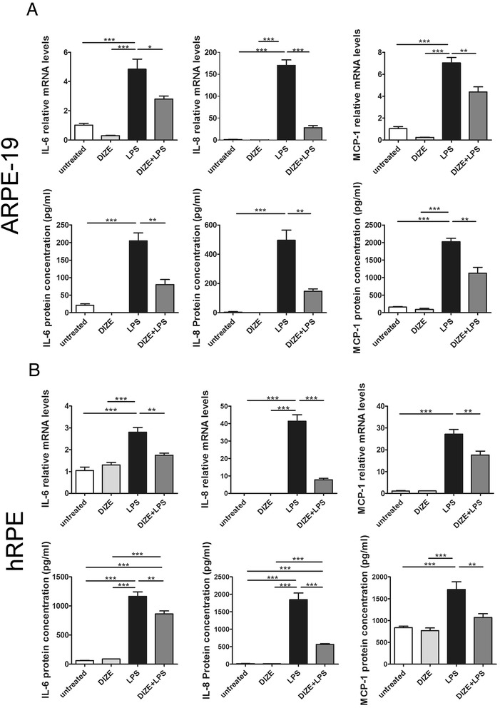
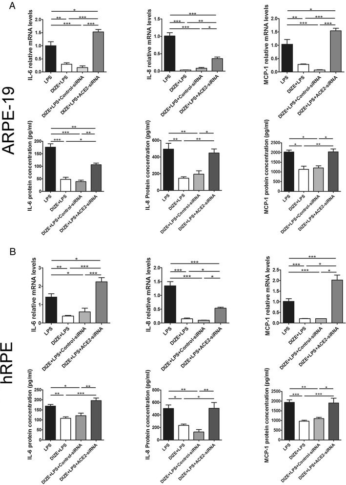
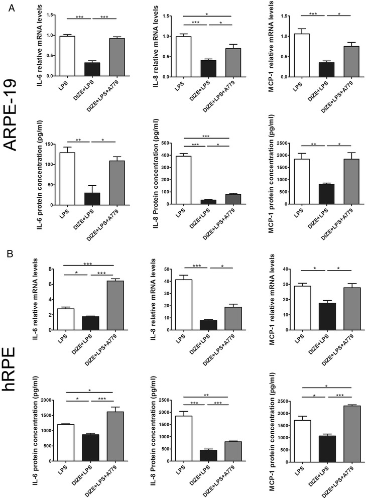
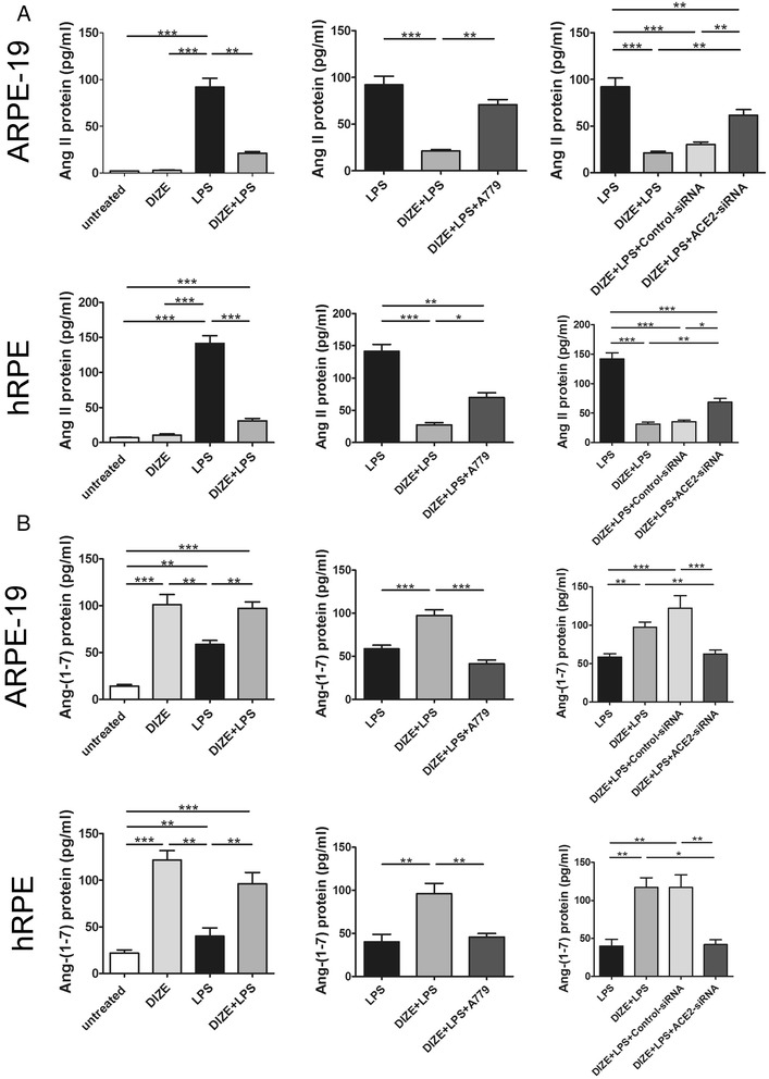
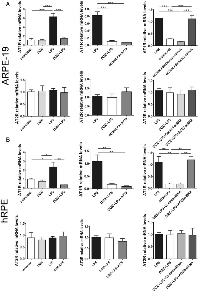
Results: DIZE remarkably increased the expression of ACE2 and inhibited the expression of IL-6, IL-8, and MCP-1 at both mRNA and protein levels in both RPE cell lines stimulated with LPS. Inhibitors of p38, ERK1/2, JNK, and NF-κB significantly decreased LPS-induced overproduction of IL-6, IL-8, and MCP-1. DIZE reduced the expression of Ang II and AT1R, whereas increased Ang-(1-7). Furthermore, DIZE downregulated the phosphorylation of p38MAPK, ERK1/2, JNK, and the activation of NF-κB upon stimulation with LPS. Downregulating ACE2 and pre-treatment with A779 abrogated the effects of DIZE on production of cytokines, the expression of Ang II, Ang-(1-7), AT1R, phosphorylation of MAPKs and activation of NF-κB.

Conclusions: DIZE inhibits LPS-induced inflammatory response by activating ACE2/Ang-(1-7)/Mas axis in human RPE cells. The protective effect is mediated by inhibiting the p38MAPK, ERK1/2, JNK, and NF-κB pathways.


Figures

Fig. 1
Fig. 1
The optimum concentration and incubation time of DIZE in cultured ARPE-19 cell line. a The cell viability was measured by CCK-8 assay. ARPE-19 cells were treated with 0.01, 0.1, 1, 10, and 100 μg/mL of DIZE for 3, 6, and 12 h and then stimulated with LPS for 24 h. (***p < 0.001, ### p < 0.001, +++ p < 0.001, n = 6, “asterisk” “number sign” “plus sign” indicated pre-incubated with 100 μg/mL of DIZE for 3, 6, and 12 h versus time-matched untreated cells, respectively). b ARPE-19 cells were pre-treated with 10 μg/mL of DIZE for 3, 6, and 12 h, following stimulated with LPS for 24 h. The mRNA expression of IL-6, IL-8, and MCP-1 in the DIZE-treated groups were compared with the LPS group by real-time PCR. All data are expressed as mean ± SEM. (***p < 0.001, **p < 0.01, *p < 0.05, n = 4 ~ 6)
Fig. 2
Fig. 2
Identification of cutured hRPE and ARPE-19 cells. The RPE65 is a RPE-specific protein. It was used to identify whether the primary cells isolated from the donor eyes, and the cell line were both derived from RPE. Merged images showed that the RPE65 was localized in the cytoplasm of the cells with DAPI-positive nucleus in both the primary cultured hRPE and the ARPE-19. The RPE65-positive cells were counted in five non-overlapping fields for each type of cell under a fluorescence microscope. The ratio of RPE65-positive cell was calculated as the percentage of RPE65-positive cells in the DAPI-positive cells. The ratio was 96.1 % ± 1.4 % in ARPE-19 cells, and 91.3 % ± 4.7 % in the hRPE cells. The scale bar 100 μm, magnification ×200
Fig. 3
Fig. 3
Expression of ACE2 in DIZE pre-treated hRPE and ARPE-19 cells followed by LPS stimulation. The mRNA and protein expressions of ACE2 detected by real-time PCR and Western blotting showed that the ACE2 levels increased in the DIZE group and the DIZE+LPS group when compared with the untreated group and the LPS-treated group in both ARPE-19 cells (a) and hRPE (b). There was no significant difference between the untreated group and the LPS group. The band intensity of ACE2 was normalized to β-actin. The mRNA level of ACE2 was detected by real-time PCR in both ARPE-19 cells (c) and hRPE (d), which were transfected with ACE2-siRNA or the negative control scrambled siRNA. All data are expressed as mean ± SEM. (***p < 0.001, **p < 0.01, *p < 0.05, n = 4)
Fig. 4
Fig. 4
The mRNA and protein expressions of pro-inflammatory cytokines in DIZE pre-treated hRPE and ARPE-19 cells followed by LPS stimulation. Real-time PCR and ELISA were conducted to determine the production of IL-6, IL-8, and MCP-1 in ARPE-19 cells (a) as well as hRPE (b). Exposure to LPS for 24 h caused a significant overproduction of IL-6, IL-8, and MCP-1 in the ARPE-19 cells and hRPE. Whereas overproduction of the cytokines was markedly inhibited by DIZE. IL-6 and IL-8 protein levels were undetectable in the DIZE group. All data are expressed as mean ± SEM. (***p < 0.001, **p < 0.01, *p < 0.05, n = 4)
Fig. 5
Fig. 5
Effect of ACE2-siRNA on the production of inflammatory cytokines in ARPE-19 cells and hRPE. IL-6, IL-8, and MCP-1 were detected by real-time PCR and ELISA assay in both ARPE-19 cells (a) and hRPE (b). DIZE reduced the expression of IL-6, IL-8, and MCP-1 in LPS-treated cells at both mRNA and protein levels, while ACE2-siRNA reversed this effect. All data are expressed as mean ± SEM (***p < 0.001, **p < 0.01, *p < 0.05, n = 4)
Fig. 6
Fig. 6
Effect of A779 on the production of inflammatory cytokines in ARPE-19 cells and hRPE. IL-6, IL-8, and MCP-1 were detected by real-time PCR and ELISA assay in both ARPE-19 cells (a) and hRPE (b). In LPS-treated cells, DIZE reduced the expression of IL-6, IL-8, and MCP-1, while A779 reversed this effect. All data are expressed as mean ± SEM (***p < 0.001, **p < 0.01, *p < 0.05, n = 4)
Fig. 7
Fig. 7
Effect of DIZE, A779, and ACE2-siRNA on the expression of Ang II and Ang-(1-7) in ARPE-19 cells and hRPE. The protein expression of Ang II and Ang-(1-7) was detected by ELISA assay in both ARPE-19 cells and hRPE. DIZE reduced the expression of Ang II in response to LPS, while A779 and ACE2-siRNA reversed this effect (a). DIZE increased the expression of Ang-(1-7) compared with LPS-treated cells, while A779 and ACE2-siRNA reversed this effect (b). All data are expressed as mean ± SEM (***p < 0.001, **p < 0.01, *p < 0.05, n = 4 ~ 6)
Fig. 8
Fig. 8
Effect of DIZE, A779, and ACE2-siRNA on the expression of AT1R and AT2R in ARPE-19 cells and hRPE. The mRNA level of AT1R and AT2R was detected by real-time PCR in both ARPE-19 cells and hRPE. DIZE reduced the expression of AT1R in response to LPS, while ACE2-siRNA reversed this effect. A779 did not affect the expression of AT1R. There was no significant difference of AT2R expression among each group in ARPE-19 cells (a) and hRPE (b). All data are expressed as mean ± SEM (***p < 0.001, **p < 0.01, *p < 0.05, n = 4)
Fig. 9
Fig. 9
Effect of the MAPK and NF-κB inhibitors on the mRNA and protein expression of inflammatory cytokines in LPS-stimulated ARPE-19 cells. The expression of IL-6, IL-8, and MCP-1 was detected by real-time PCR and ELISA assay in the MAPK pathway inhibitor-treated cells (a) and NF-κB pathway inhibitor-treated cells (b). The p38 inhibitor (SB203580, SB), JNK inhibitor (SP600125, SP), and ERK1/2 inhibitor (PD98059, PD) significantly decreased the production of IL-6, IL-8, and MCP-1 in the ARPE-19 cells stimulated with LPS at both mRNA and protein levels (a). The NF-κB inhibitor (BAY 11-7082, BAY) remarkably reduced the production of IL-6, IL-8, and MCP-1 in LPS-treated group at both mRNA and protein levels (b). All data are expressed as mean ± SEM (***p < 0.001, **p < 0.01, *p < 0.05, n = 4)
Fig. 10
Fig. 10
Effect of DIZE on the phosphorylation levels of MAPKs and the activation of NF-κB in ARPE-19 cells stimulated with LPS. a Western blotting was used to determine the protein levels of p38 MAPK, JNK, ERK1/2 and their phosphorylated forms (p-p38, p-JNK, and p-ERK1/2). b Activation of NF-κB pathway was indicated by p-IκB-α, an indicator for the activation of NF-κB. The expressions of p-p38, p-JNK, and p-ERK1/2 were normalized to p38, JNK, and ERK1/2, respectively. The band intensity of p-IκB-α was normalized to β-actin. DIZE treatment resulted in remarkably reduction of phosphorylation of p38, JNK, ERK1/2, and IκB-α compared with the LPS-stimulated group. All data are expressed as mean ± SEM (***p < 0.001, **p < 0.01, *p < 0.05, n = 3 ~ 4)
Fig. 11
Fig. 11
Effect of A779 on the phosphorylation of MAPKs and the activation of NF-κB in ARPE-19. Western blotting was applied to determine the protein levels of p-p38, p-JNK, p-ERK1/2, p38, JNK, ERK1/2 (a) and p-IκB-α, an indicator for the activation of NF-κB (b). The band intensity of p-p38, p-JNK, and p-ERK1/2 was normalized to p38, JNK, and ERK1/2, respectively. The band intensity of p-IκB-α was normalized to β-actin. DIZE reduced the phosphorylation of p38, JNK, and ERK1/2 and IκB-α induced by LPS, while A779 abrogated DIZE’s effect. All data are expressed as mean ± SEM (***p < 0.001, **p < 0.01, *p < 0.05, n = 3 ~ 4)
Fig. 12
Fig. 12
Effect of ACE2-siRNA on the phosphorylation of MAPKs and the activation of NF-κB in ARPE-19. The protein levels of p-p38, p-JNK, p-ERK1/2, p38, JNK, ERK1/2 (a) and p-IκB-α (b) were determined by Western blotting. The band intensity of p-p38, p-JNK, and p-ERK1/2 was normalized to p38, JNK, and ERK1/2, respectively. The band intensity of p-IκB-α was normalized to β-actin. DIZE reduced the phosphorylation of p38, JNK, and ERK1/2 and IκB-α induced by LPS, while ACE2-siRNA abolished DIZE’s effect. All data are expressed as mean ± SEM (***p < 0.001, **p < 0.01, *p < 0.05, n = 3 ~ 4)

References

    1. Weir MR, Dzau VJ. The renin-angiotensin-aldosterone system: a specific target for hypertension management. Am J Hypertens. 1999;12:205S–13. doi: 10.1016/S0895-7061(99)00103-X. - DOI - PubMed
    1. van Thiel BS, van der Pluijm I, Te Riet L, Essers J, Danser AH. The renin-angiotensin system and its involvement in vascular disease. European journal of pharmacology. 2015. doi:10.1016/j.ejphar.2015.03.090. - PubMed
    1. de Man FS, Tu L, Handoko ML, Rain S, Ruiter G, Francois C, et al. Dysregulated renin-angiotensin-aldosterone system contributes to pulmonary arterial hypertension. Am J Respir Crit Care Med. 2012;186:780–9. doi: 10.1164/rccm.201203-0411OC. - DOI - PMC - PubMed
    1. Kurihara T, Ozawa Y, Ishida S, Okano H, Tsubota K. Renin-angiotensin system hyperactivation can induce inflammation and retinal neural dysfunction. Int J Inflam. 2012;2012:581695. doi: 10.1155/2012/581695. - DOI - PMC - PubMed
    1. Strain WD, Chaturvedi N. The renin-angiotensin-aldosterone system and the eye in diabetes. J Renin Angiotensin Aldosterone Syst. 2002;3:243–6. doi: 10.3317/jraas.2002.045. - DOI - PubMed

Publication types

MeSH terms