. 2016 Apr;33(4):309-23.
doi: 10.1007/s10585-016-9780-3. Epub 2016 Feb 9.

β-catenin-independent WNT signaling and Ki67 in contrast to the estrogen receptor status are prognostic and associated with poor prognosis in breast cancer liver metastases

Affiliations

β-catenin-independent WNT signaling and Ki67 in contrast to the estrogen receptor status are prognostic and associated with poor prognosis in breast cancer liver metastases

Annalen Bleckmann et al. Clin Exp Metastasis. 2016 Apr.

Abstract

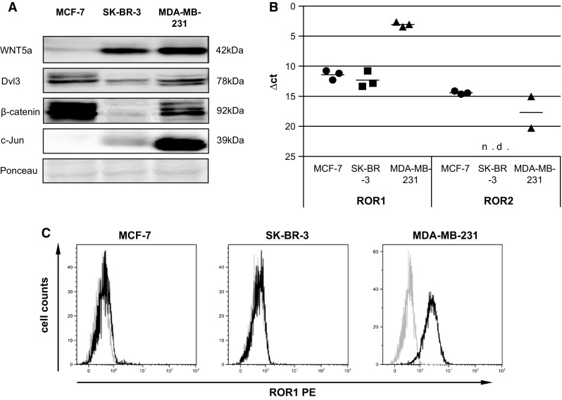
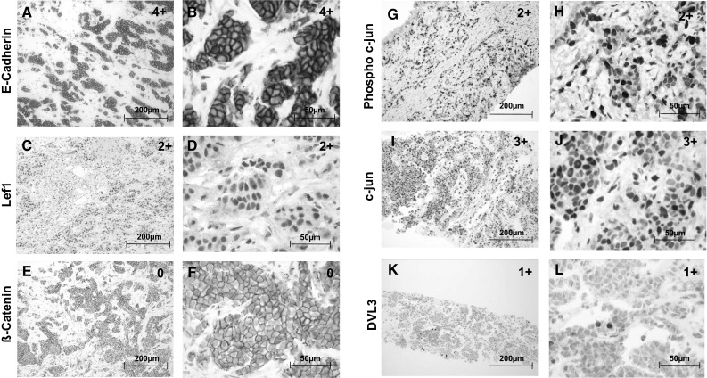
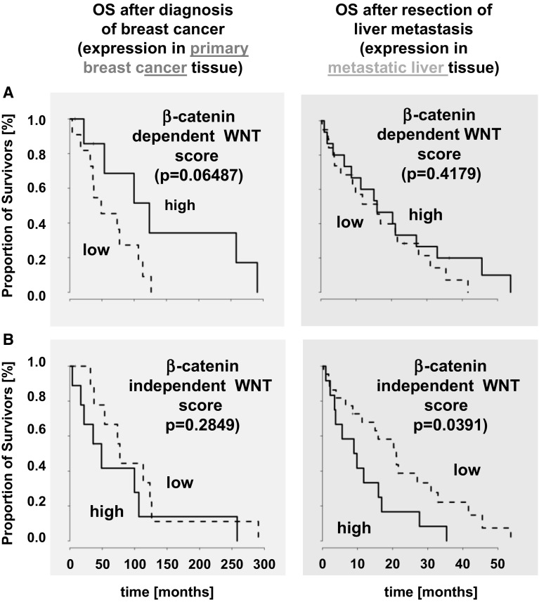
Liver metastasis development in breast cancer patients is common and confers a poor prognosis. So far, the prognostic significance of surgical resection and clinical relevance of biomarker analysis in metastatic tissue have barely been investigated. We previously demonstrated an impact of WNT signaling in breast cancer brain metastasis. This study aimed to investigate the value of established prognostic markers and WNT signaling components in liver metastases. Overall N = 34 breast cancer liver metastases (with matched primaries in 19/34 cases) were included in this retrospective study. Primaries and metastatic samples were analyzed for their expression of the estrogen (ER) and progesterone receptor, HER-2, Ki67, and various WNT signaling-components by immunohistochemistry. Furthermore, β-catenin-dependent and -independent WNT scores were generated and analyzed for their prognostic value. Additionally, the influence of the alternative WNT receptor ROR on signaling and invasiveness was analyzed in vitro. ER positivity (HR 0.09, 95 % CI 0.01-0.56) and high Ki67 (HR 3.68, 95 % CI 1.12-12.06) in the primaries had prognostic impact. However, only Ki67 remained prognostic in the metastatic tissue (HR 2.46, 95 % CI 1.11-5.44). Additionally, the β-catenin-independent WNT score correlated with reduced overall survival only in the metastasized situation (HR 2.19, 95 % CI 1.02-4.69, p = 0.0391). This is in line with the in vitro results of the alternative WNT receptors ROR1 and ROR2, which foster invasion. In breast cancer, the value of prognostic markers established in primary tumors cannot directly be translated to metastases. Our results revealed β-catenin-independent WNT signaling to be associated with poor prognosis in patients with breast cancer liver metastasis.

Keywords: Breast cancer; Metastasis; Prognostic score; WNT signaling.


Figures

Fig. 1
Fig. 1
a Immunoblot for WNT5a, Dvl3, β-catenin, and c-Jun of MCF-7, SK-BR-3 and MDA-MB-231 cell lysates. B qRT-PCR of MCF-7, SK-BR-3 and MDA-MB-231 for ROR1 and ROR2. Each dot represents one independent biological sample (means, n = 3, n.d. = not detectable). Note that in MDA-MB-231 ROR2 was only detectable in 2/3 samples. C Flow cytometry (gray isotype control, black stained cells for MCF-7, SK-BR-3 and MDA-MB-231) measuring the expression of ROR1
Fig. 2
Fig. 2
a Immunoblots showing the expression of ROR1 and ROR2 in MCF-7 and SK-BR-3 cells transfected with either an empty vector (ctl) or a ROR2 overexpression plasmid (pROR2). B In vitro microinvasion assays of ROR2-overexpressing MCF-7 and SK-BR-3 cells compared to wildtype cells (wt) or cells transfected with an empty vector (ctl) (mean ± SD, n = 3, *p < 0.001). C Immunoblot for c-Jun and HDAC of MCF-7 and SK-BR-3 empty vector (ctl) or ROR2-overexpressing cells for cytoplasmic (C) and nuclear (N) cell lysates. D Immunofluorescence staining of c-Jun and F-actin (phalloidin) in MCF-7 empty vector (ctl) and ROR2 cells as well as β-catenin and Dapi in MCF-7 wt and ROR2 cells. E Immunoblot of ROR1 in MDA-MB-231 cells transfected with nonsense control (ctl) or ROR1 (shROR1) shRNA. F In vitro microinvasion assay of MDA-MB-231 wildtype (wt), non-sense control (ctl) and ROR1 knockdown (shROR1) cells (mean ± SD, n = 3, *p < 0.001)
Fig. 3
Fig. 3
Kaplan-Meier curves illustrate that ER expression in the primary tumor but not in the liver metastases is correlated with a good prognosis (a). Positivity for the proliferation index Ki67 in the primary tumor tissue as well as in the liver metastases (b) is associated with reduced overall survival
Fig. 4
Fig. 4
Immunohistochemical stainings for all proteins analysed were performed on primary tumors and metastases respectively. Representative pictures are shown for stainings of primary tumor samples (af) and on metastatic tissue (gl). IHC-staining of membrane E-cadherin showing a positivity in primary tumor cells of >75 % in 20× (a) and 40× magnification (b); nuclear Lef1 positivity is shown at 20× (c) and 40× magnification of primary tumor tissue being positive in 30 % of tumor cells (d); β-catenin staining is shown in panel e (20×) and f without any nuclear activity. Representative examples of nuclear Phospho-c-Jun staining is shown in g (20×) and h (40×) with a positivity rate of >26 % for intrahepatic breast cancer metastases cells; nuclear c-Jun staining is represented in panels i (20×) and j (40×) showing positive stained nuclei in >76 % of metastatic tissue; nuclear Dvl3 staining is also represented in liver metastases beeing positive in <25 % of metastases cells at a magnification of 20× (k) and 40× (l). For all markers, the specific expression pattern was analyzed for primaries and metastases and classified into five groups: 0 = 0 %, 1+ = 1–25 %, 2+ = 26–50 %, 3+ = 51–75 % and 4+ = 76–100 % of the tumor cells with positive (≥1) staining. Bar: 200 μm (a, c, e, g, i and k) and 50 μm (b, d, f, h, j and l)
Fig. 5
Fig. 5
No prognostic impact could be shown for the β-catenin-dependent WNT score (a). In contrast, the β-catenin-independent WNT score has prognostic impact on survival in the metastatic setting (b). Survival rates are depicted with Kaplan–Meier-curves in months

References

    1. De Angelis R, et al. Cancer survival in Europe 1999–2007 by country and age: results of EUROCARE–5-a population-based study. Lancet Oncol. 2014;15(1):23–34. doi: 10.1016/S1470-2045(13)70546-1. - DOI - PubMed
    1. Cardoso F et al. (2014) ESO-ESMO 2nd international consensus guidelines for advanced breast cancer (ABC2) dagger. Annals of oncology: Official Journal of the European Society for Medical Oncology/ESMO vol 25(10), pp 1871–88 - PMC - PubMed
    1. Ruiterkamp J, Ernst MF. The role of surgery in metastatic breast cancer. Eur J Cancer. 2011;47(Suppl 3):S6–S22. doi: 10.1016/S0959-8049(11)70142-3. - DOI - PubMed
    1. Choti MA. Liver-directed treatments for metastatic colorectal cancer. Curr Treat Options Oncol. 2014;15(3):456–464. doi: 10.1007/s11864-014-0297-1. - DOI - PubMed
    1. Elsberger B, Roxburgh CS, Horgan PG. Is there a role for surgical resections of hepatic breast cancer metastases? Hepatogastroenterology. 2014;61(129):181–186. - PubMed

Publication types

MeSH terms