

A case of giant nodular posterior scleritis mimicking choroidal malignancy
- PMID: 26862098
- PMCID: PMC4784081
- DOI: 10.4103/0301-4738.176038
A case of giant nodular posterior scleritis mimicking choroidal malignancy
Abstract
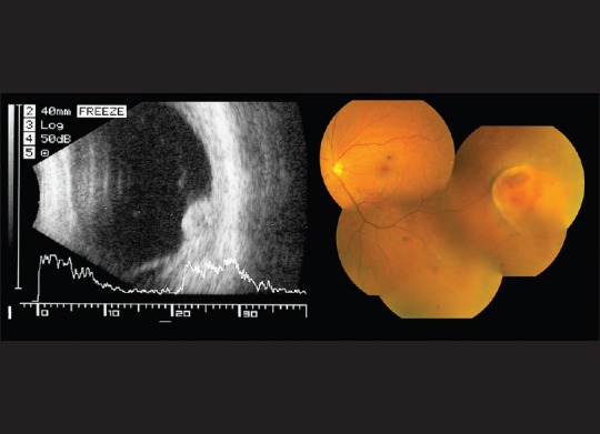
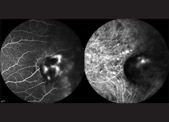
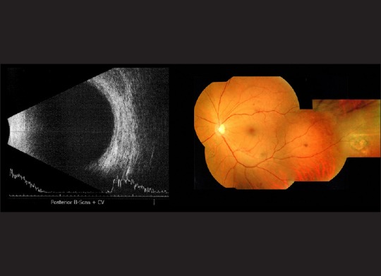
To report a case of giant nodular posterior scleritis mimicking a choroidal tumor. A 42-year-old lady with systemic hypertension presented with a 1-week history of unilateral visual loss, pain and redness in her left eye. Examination showed sectoral anterior episcleritis in her left eye as well as a dome-shaped choroidal mass at the inferior-temporal periphery, associated with retinal hemorrhages and subretinal fluid. Systemic evaluation and imaging of the choroidal mass were performed and could not rule out amelanotic choroidal melanoma. At the same time, she was prescribed a 2-week course of oral nonsteroidal anti-inflammatory drug (NSAID) for her sectoral anterior episcleritis. The choroidal mass was found to have resolved completely right before her scheduled fine needle biopsy. Diagnosis of nodular posterior scleritis and a trial of oral NSAID can be considered in patients presenting with a choroidal mass before any invasive procedure.
Figures
References
-
- McCluskey PJ, Watson PG, Lightman S, Haybittle J, Restori M, Branley M. Posterior scleritis: Clinical features, systemic associations, and outcome in a large series of patients. Ophthalmology. 1999;106:2380–6. - PubMed
-
- Biswas J, Mittal S, Ganesh SK, Shetty NS, Gopal L. Posterior scleritis: Clinical profile and imaging characteristics. Indian J Ophthalmol. 1998;46:195–202. - PubMed
-
- Shields JA, Augsburger JJ, Brown GC, Stephens RF. The differential diagnosis of posterior uveal melanoma. Ophthalmology. 1980;87:518–22. - PubMed
-
- Accuracy of diagnosis of choroidal melanomas in the Collaborative Ocular Melanoma Study. COMS report no 1. Arch Ophthalmol. 1990;108:1268–73. - PubMed
Publication types
MeSH terms
Substances
LinkOut - more resources
Full Text Sources
Other Literature Sources
