Review
. 2016 Feb;44(1):212-27.
doi: 10.1042/BST20150209.

Specificity and disease in the ubiquitin system

Affiliations
Review

Specificity and disease in the ubiquitin system

Viduth K Chaugule et al. Biochem Soc Trans. 2016 Feb.

Abstract

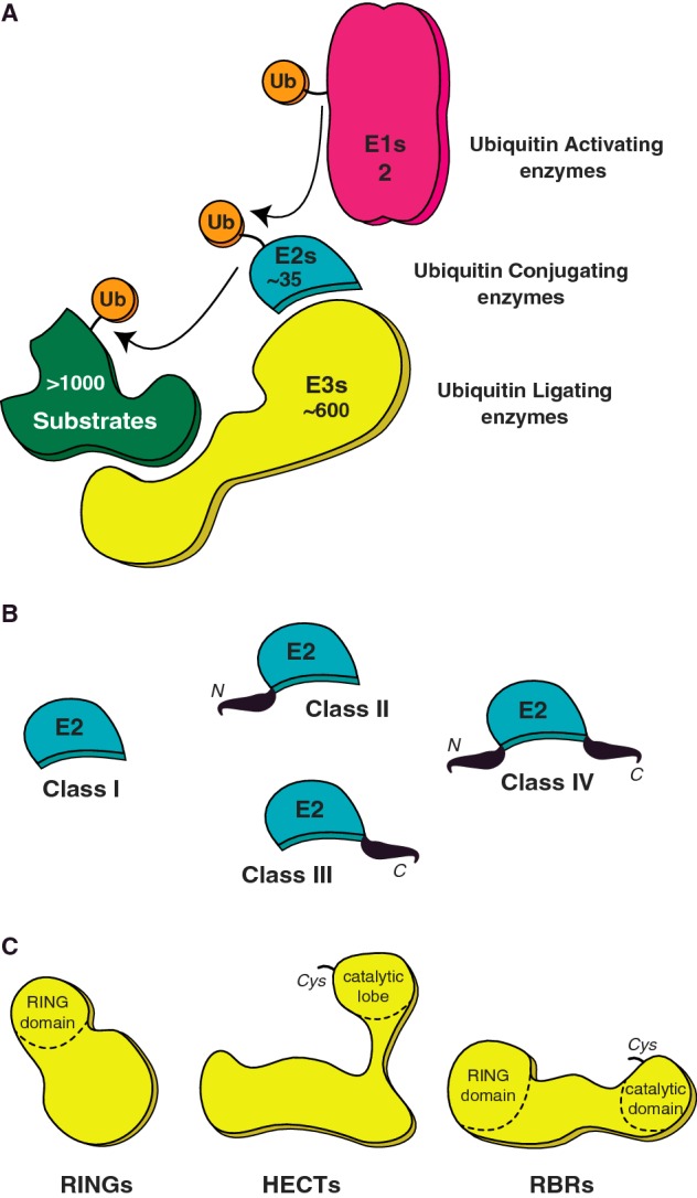
Post-translational modification (PTM) of proteins by ubiquitination is an essential cellular regulatory process. Such regulation drives the cell cycle and cell division, signalling and secretory pathways, DNA replication and repair processes and protein quality control and degradation pathways. A huge range of ubiquitin signals can be generated depending on the specificity and catalytic activity of the enzymes required for attachment of ubiquitin to a given target. As a consequence of its importance to eukaryotic life, dysfunction in the ubiquitin system leads to many disease states, including cancers and neurodegeneration. This review takes a retrospective look at our progress in understanding the molecular mechanisms that govern the specificity of ubiquitin conjugation.

Keywords: E3 ligase; Fanconi anemia; Parkin; RBR; RING; ubiquitin.


Figures

Figure 1
Figure 1. UBC pathway
(A) A schematic of the ubiquitin (Ub) pathway and the members involved at each step; activation (E1), conjugation (E2) and ligation (E3). Ubiquitin activation is catalysed by the E1 in an energy-consuming step. The ubiquitin thioester conjugate is then passed onto the catalytic cysteine site on the E2 and finally ligated on to a target lysine of a substrate, an event mediated by E3 ligases. Also indicated is the numerical hierarchy of the ubiquitin pathway in humans. (B) Ubiquitin E2s are classified into four classes based on the N- or C- terminal extensions of the core UBC domain. (C) Three classes of E3 ubiquitin ligases; RINGs, HECTs and RBRs are classified based on their enzymatic mechanisms, with HECT and RBR ligase possessing a catalytic cysteine for Ub–E3 intermediate formation.
Figure 2
Figure 2. Specificity of the E3 ligase FANCL
(A) Schematic of the FANCL and Ube2T-mediated specific substrate monoubiquitination. (B) Ribbon diagram depicting the FANCL structure (PDB 3K1L). The ELF, DRWD and RING domains are coloured light brown, green/lime and blue respectively. Zinc atoms are represented as grey spheres. (C) Surface representation of the protein interaction surfaces on FANCL. The binding patch for ubiquitin (orange) and substrate (red) reside on the ELF and DRWD domains respectively (left). The E2 binding surface (light blue) is on the RING domain (right).
Figure 3
Figure 3. The broad-spectrum E3 ligase Parkin
(A) Schematic of the variety of substrate ubiquitination events mediated by Parkin through several E2 enzymes. (B) A cartoon depicting the domain/motif arrangement of full-length Parkin (top) and the multiple inter-domain interactions that stabilize the tertiary structure (bottom). The RBR module comprises RING1, BRcat and Rcat domains with the catalytic cysteine (yellow star) present in the Rcat domain. Additional regulatory domains/motifs are the Ubl domain, a zinc-chelating RING0 domain and the small helical REP. (C) Surface representation of Parkin (grey, PDB 5C23) shows the distal location of phospho-serine (pink) on the Ubl (lime) domain and basic patch (dark blue) created on the surface of the RING0/RING1 interface (left). Binding of phospho-ubiquitin (orange, PDB 4WZP) to the basic patch on Parkin (right) leads to the complete displacement of the phospho-Ubl domain exposing the ubiquitin-binding patch on Parkin's RING1 domain (brown). This exposed patch on Parkin can support interactions with multiple E2–Ub intermediates and hence catalyse diverse ubiquitin signals.

References

    1. Pickart C.M. Mechanisms underlying ubiquitination. Annu. Rev. Biochem. 2001;70:503–533. doi: 10.1146/annurev.biochem.70.1.503. - DOI - PubMed
    1. Hochstrasser M. Origin and function of ubiquitin-like proteins. Nature. 2009;458:422–429. doi: 10.1038/nature07958. - DOI - PMC - PubMed
    1. Zou H., McGarry T.J., Bernal T., Kirschner M.W. Identification of a vertebrate sister-chromatid separation inhibitor involved in transformation and tumorigenesis. Science. 1999;285:418–422. doi: 10.1126/science.285.5426.418. - DOI - PubMed
    1. Hoege C., Pfander B., Moldovan G.L., Pyrowolakis G., Jentsch S. RAD6-dependent DNA repair is linked to modification of PCNA by ubiquitin and SUMO. Nature. 2002;419:135–141. doi: 10.1038/nature00991. - DOI - PubMed
    1. Stelter P., Ulrich H.D. Control of spontaneous and damage-induced mutagenesis by SUMO and ubiquitin conjugation. Nature. 2003;425:188–191. doi: 10.1038/nature01965. - DOI - PubMed

Publication types