

Biliary and pancreatic stenting: Devices and insertion techniques in therapeutic endoscopic retrograde cholangiopancreatography and endoscopic ultrasonography
- PMID: 26862364
- PMCID: PMC4734973
- DOI: 10.4253/wjge.v8.i3.143
Biliary and pancreatic stenting: Devices and insertion techniques in therapeutic endoscopic retrograde cholangiopancreatography and endoscopic ultrasonography
Abstract
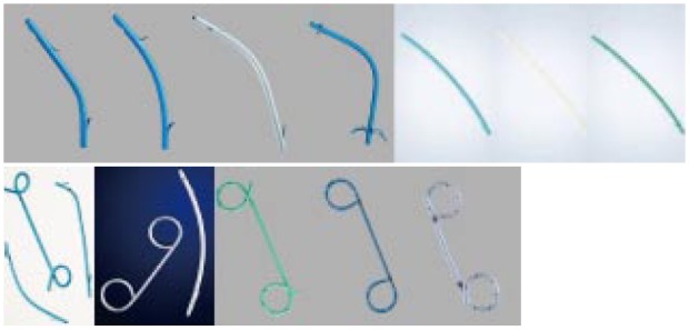
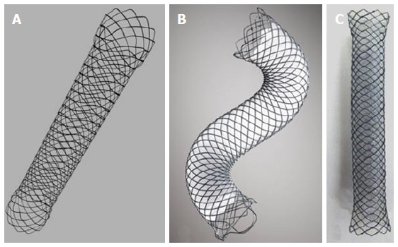
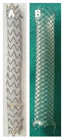
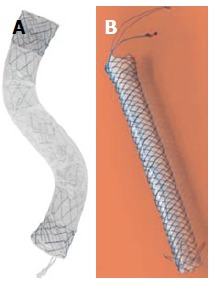
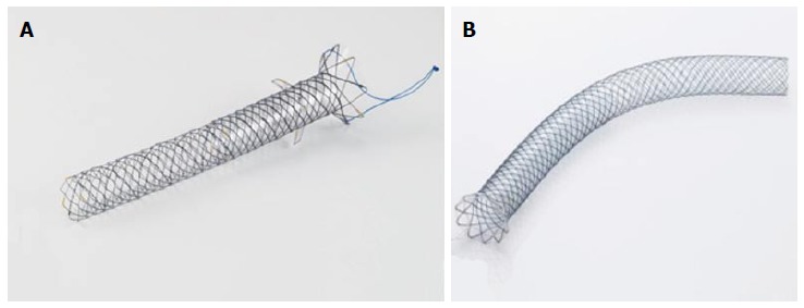
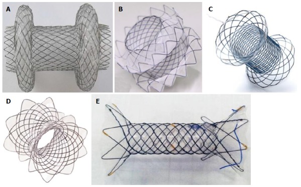
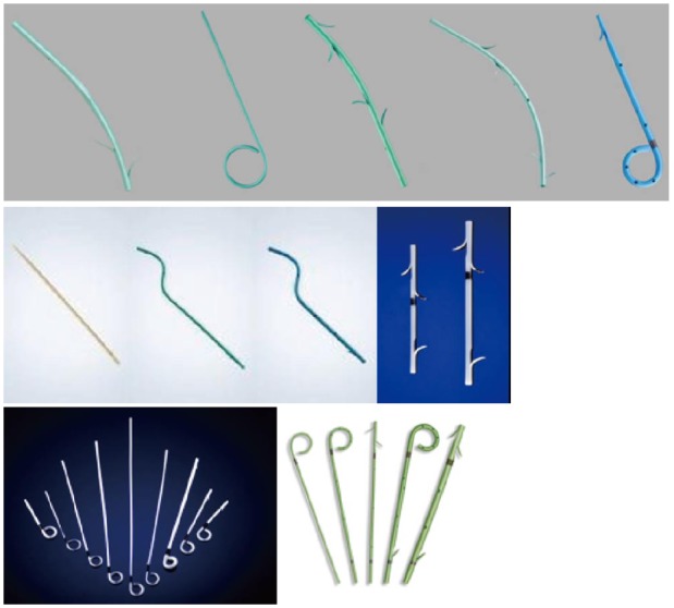
Stents are tubular devices made of plastic or metal. Endoscopic stenting is the most common treatment for obstruction of the common bile duct or of the main pancreatic duct, but also employed for the treatment of bilio-pancreatic leakages, for preventing post- endoscopic retrograde cholangiopancreatography pancreatitis and to drain the gallbladder and pancreatic fluid collections. Recent progresses in techniques of stent insertion and metal stent design are represented by new, fully-covered lumen apposing metal stents. These stents are specifically designed for transmural drainage, with a saddle-shape design and bilateral flanges, to provide lumen-to-lumen anchoring, reducing the risk of migration and leakage. This review is an update of the technique of stent insertion and metal stent deployment, of the most recent data available on stent types and characteristics and the new applications for biliopancreatic stents.
Keywords: Biliary stent; Endoscopic retrograde cholangiopancreatography; Endoscopic ultrasonography; Pancreatic stent; Self-expandable metal stent.
Figures
References
-
- Soehendra N, Reynders-Frederix V. Palliative bile duct drainage - a new endoscopic method of introducing a transpapillary drain. Endoscopy. 1980;12:8–11. - PubMed
-
- Cotton PB. Duodenoscopic placement of biliary prostheses to relieve malignant obstructive jaundice. Br J Surg. 1982;69:501–503. - PubMed
-
- Huibregtse K, Haverkamp HJ, Tytgat GN. Transpapillary positioning of a large 3.2 mm biliary endoprosthesis. Endoscopy. 1981;13:217–219. - PubMed
-
- Neuhaus H, Hagenmüller F, Classen M. Self-expanding biliary stents: preliminary clinical experience. Endoscopy. 1989;21:225–228. - PubMed
-
- Huibregtse K, Cheng J, Coene PP, Fockens P, Tytgat GN. Endoscopic placement of expandable metal stents for biliary strictures--a preliminary report on experience with 33 patients. Endoscopy. 1989;21:280–282. - PubMed
Publication types
LinkOut - more resources
Full Text Sources
Other Literature Sources
