Review
. 2016 Feb 8:5:6.
doi: 10.1186/s13630-016-0026-4. eCollection 2015.

Paramecium tetraurelia basal body structure

Affiliations
Review

Paramecium tetraurelia basal body structure

Anne-Marie Tassin et al. Cilia. .

Abstract

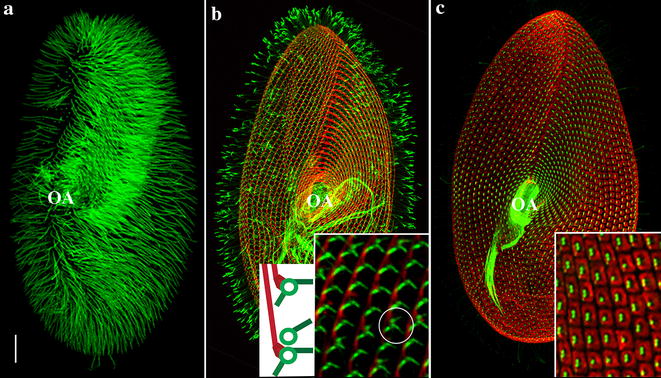
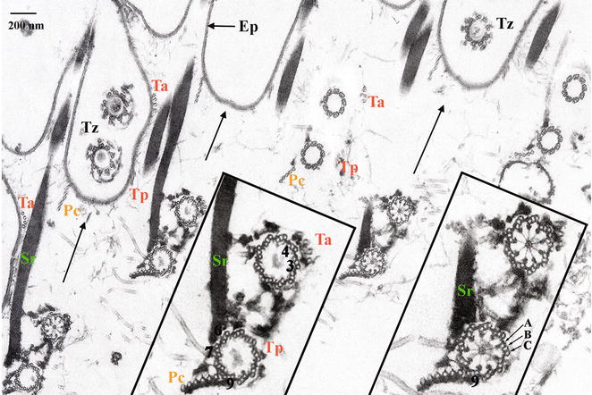
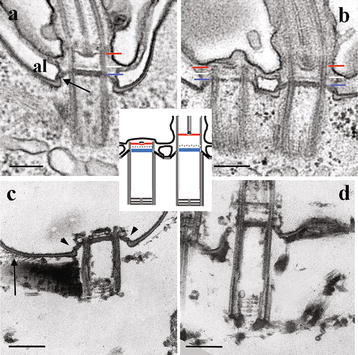
Paramecium is a free-living unicellular organism, easy to cultivate, featuring ca. 4000 motile cilia emanating from longitudinal rows of basal bodies anchored in the plasma membrane. The basal body circumferential polarity is marked by the asymmetrical organization of its associated appendages. The complex basal body plus its associated rootlets forms the kinetid. Kinetids are precisely oriented within a row in correlation with the cell polarity. Basal bodies also display a proximo-distal polarity with microtubule triplets at their proximal ends, surrounding a permanent cartwheel, and microtubule doublets at the transition zone located between the basal body and the cilium. Basal bodies remain anchored at the cell surface during the whole cell cycle. On the opposite to metazoan, there is no centriolar stage and new basal bodies develop anteriorly and at right angle from the base of the docked ones. Ciliogenesis follows a specific temporal pattern during the cell cycle and both unciliated and ciliated docked basal bodies can be observed in the same cell. The transition zone is particularly well organized with three distinct plates and a maturation of its structure is observed during the growth of the cilium. Transcriptomic and proteomic analyses have been performed in different organisms including Paramecium to understand the ciliogenesis process. The data have incremented a multi-organism database, dedicated to proteins involved in the biogenesis, composition and function of centrosomes, basal bodies or cilia. Thanks to its thousands of basal bodies and the well-known choreography of their duplication during the cell cycle, Paramecium has allowed pioneer studies focusing on the structural and functional processes underlying basal body duplication. Proteins involved in basal body anchoring are sequentially recruited to assemble the transition zone thus indicating that the anchoring process parallels the structural differentiation of the transition zone. This feature offers an opportunity to dissect spatio-temporally the mechanisms involved in the basal body anchoring process and transition zone formation.

Keywords: Basal body assembly; Basal body docking; Ciliogenesis; Paramecium; Transition zone.


Figures

Fig. 1
Fig. 1
Pattern of cilia and basal bodies in Paramecium. Images are projections of confocal images taken at the level of the ventral side of the cell. For details in immunofluorescence procedures, see [1]. Bars 20 μm. Insets:  ×5. a Ciliary pattern. The cell is labelled with an antibody directed against monoglycylated tubulin. The anterior left half-quarter appears brighter because it is more densely ciliated than other parts of the cell. Beating of these cilia guides the water current towards the cell centre where the oral apparatus (OA) is located. At the posterior pole of the cell are few longer non-motile cilia. b Pattern of the basal body-associated rootlets. Green: microtubular rootlets, decorated with an anti-acetylated tubulin; red: striated rootlets, decorated with an antibody specific for striatins [2]. Striated rootlets of successive basal bodies cluster to form a continuous bundle along the right of the basal body row. Cartoon: one (top) or two (bottom) transverse microtubular ribbons are detected in association with single or paired basal bodies, respectively. Circle: paired basal bodies with two transverse ribbons. OA oral apparatus. c Pattern of cortical units. Red: epiplasm units, decorated with an antibody specific for epiplasmins [3]; green: basal bodies labelled with an anti-polyglutamylated tubulin [4]. OA oral apparatus
Fig. 2
Fig. 2
Organization of the cell surface at the ultrastructural level; tangential section of a detergent-extracted Paramecium cell fixed in the presence of tannic acid (modified from 12). Bar 200 μm. Insets: ×2. The arrows point towards the anterior of the basal body rows. Basal bodies are transversally cut at the level of the cartwheel (right inset) or at the level of the transition zone (Tz) (left inset). At their base, paired basal bodies are connected together by a complex set of links (insets). The post-ciliary rootlet (Pc) originates close to the triplet 9 (according to the Grain’s triplet numbering in Ciliata [25]), the transverse anterior (Ta) and posterior (Tp) rootlets close to the triplets 3 and 4, and the striated rootlet (Sr) is connected to the triplets 6 and 7. These three rootlets, associated with each basal body pairs, extend from the basal body bases towards the cell surface where they connect the epiplasm (Ep). The anterior basal body is connected in its proximal part to the striated rootlet (insets). At the proximal level, the Pc rootlet is connected to the ciliary rootlet by a set of links (insets). At the Tz level, links are detected in association with each microtubule doublets. Tubules A, B and C composing the basal body wall are indicated on the right inset
Fig. 3
Fig. 3
Longitudinal sections through Paramecium basal bodies after glutaraldehyde/osmium classical fixation (a, b) or with an additional tannic acid treatment performed after cell permeabilization (c, d). Bars 200 nm. a Connection between the Tz and the cell surface. Inside the basal body, the Tz is organized in three plates: the terminal plate (blue line), the intermediate plate (white) and the axosomal plate (red line). Outside the basal body, the terminal plate extends to link the epiplasm (arrow). Inside the basal body, dense granules are observed. Al alveolar sac , a vacuolar system located beneath the outer cell membrane found in all representatives of the Chromalveolata. b Comparison of Tz of non-ciliated and ciliated basal bodies: Tz of ciliated basal bodies is more extended than that of non-ciliated basal bodies, but the three plates and the connection with the epiplasm are detected in both of them. c, d (modified from [12]): short (c) and long (d) basal bodies. The cartwheel is longer in the long basal body. The three plates, as well as the connection with the epiplasm (arrowheads) can be observed on the short non-ciliated basal body; connections between the striated rootlet and the epiplasm appear as delicate links (arrow). A schematic representation of anchored ciliated and non-ciliated basal bodies has been inserted in this figure showing the transition zone with its three plates: the terminal plate (blue), the intermediate plate (discontinuous line) and axosomal plate (red)

References

    1. Aubusson-Fleury A, Cohen J, Lemullois M. Ciliary heterogeneity within a single cell: the Paramecium model. Methods Cell Biol. 2015;127:457–85. - PubMed
    1. Sperling L, Keryer G, Ruiz F, Beisson J. Cortical morphogenesis in Paramecium: a transcellular wave of protein phosphorylation involved in ciliary rootlet disassembly. Dev Biol. 1991;148:205–18. - PubMed
    1. Nahon P, Coffe G, Le Guyader H, Darmanaden-Delorme J, Jeanmaire-Wolf R, Clérot J-C, Adoutte A. Identification of the epiplasmins, a new set of cortical proteins of the membrane cytoskeleton in Paramecium. J Cell Sci. 1993;104:975–90.
    1. Janke C, Bulinski JC. Post-translational regulation of the microtubule cytoskeleton: mechanisms and functions. Nat Rev Mol Cell Biol. 2011;12:773–86. - PubMed
    1. Hufnagel LA. Cortical ultrastructure of Paramecium aurelia. studies on isolated pellicles. J Cell Biol. 1969;40:779–801. doi: 10.1083/jcb.40.3.779. - DOI - PMC - PubMed

LinkOut - more resources