"True" posterior communicating aneurysms: Three cases, three strategies
- PMID: 26862441
- PMCID: PMC4722528
- DOI: 10.4103/2152-7806.173307
"True" posterior communicating aneurysms: Three cases, three strategies
Abstract
Background: The authors provide a review of true aneurysms of the posterior communicating artery (PCoA). Three cases admitted in our hospital are presented and discussed as follows.
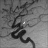
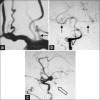
Case descriptions: First patient is a 51-year-old female presenting with a Fisher II, Hunt-Hess III (headache and confusion) subarachnoid hemorrhage (SAH) from a ruptured true aneurysm of the right PCoA. She underwent a successful ipsilateral pterional craniotomy for aneurysm clipping and was discharged on postoperative day 4 without neurological deficit. Second patient is a 53-year-old female with a Fisher I, Hunt-Hess III (headache, mild hemiparesis) SAH and multiple aneurisms, one from left ophthalmic carotid artery and one (true) from right PCoA. These lesions were approached and successfully treated by a single pterional craniotomy on the left side. The patient was discharged 4 days after surgery, with complete recovery of muscle strength during follow-up. Third patient is a 69-year-old male with a Fisher III, Hunt-Hess III (headache and confusion) SAH, from a true PCoA on the right. He had a left subclavian artery occlusion with flow theft from the right vertebral artery to the left vertebral artery. The patient underwent endovascular treatment with angioplasty and stent placement on the left subclavian artery that resulted in aneurysm occlusion.
Conclusion: In conclusion, despite their seldom occurrence, true PCoA aneurysms can be successfully treated with different strategies.
Keywords: Etiology; physiopathology; treatment; true posterior communicating artery aneurysms.
Figures


References
-
- Ferns SP, Sprengers ME, van Rooij WJ, van den Berg R, Velthuis BK, de Kort GA, et al. De novo aneurysm formation and growth of untreated aneurysms: A 5-year MRA follow-up in a large cohort of patients with coiled aneurysms and review of the literature. Stroke. 2011;42:313–8. - PubMed
-
- Gibo H, Lenkey C, Rhoton AL., Jr Microsurgical anatomy of the supraclinoid portion of the internal carotid artery. J Neurosurg. 1981;55:560–74. - PubMed
-
- He W, Gandhi CD, Quinn J, Karimi R, Prestigiacomo CJ. True aneurysms of the posterior communicating artery: A systematic review and meta-analysis of individual patient data. World Neurosurg. 2011;75:64–72. - PubMed
-
- He W, Hauptman J, Pasupuleti L, Setton A, Farrow MG, Kasper L, et al. True posterior communicating artery aneurysms: Are they more prone to rupture? A biomorphometric analysis. J Neurosurg. 2010;112:611–5. - PubMed
LinkOut - more resources
Full Text Sources
Other Literature Sources

