. 2016 Feb 10;11(2):e0147687.
doi: 10.1371/journal.pone.0147687. eCollection 2016.

Fusion Patterns in the Skulls of Modern Archosaurs Reveal That Sutures Are Ambiguous Maturity Indicators for the Dinosauria

Affiliations

Fusion Patterns in the Skulls of Modern Archosaurs Reveal That Sutures Are Ambiguous Maturity Indicators for the Dinosauria

Alida M Bailleul et al. PLoS One. .

Abstract

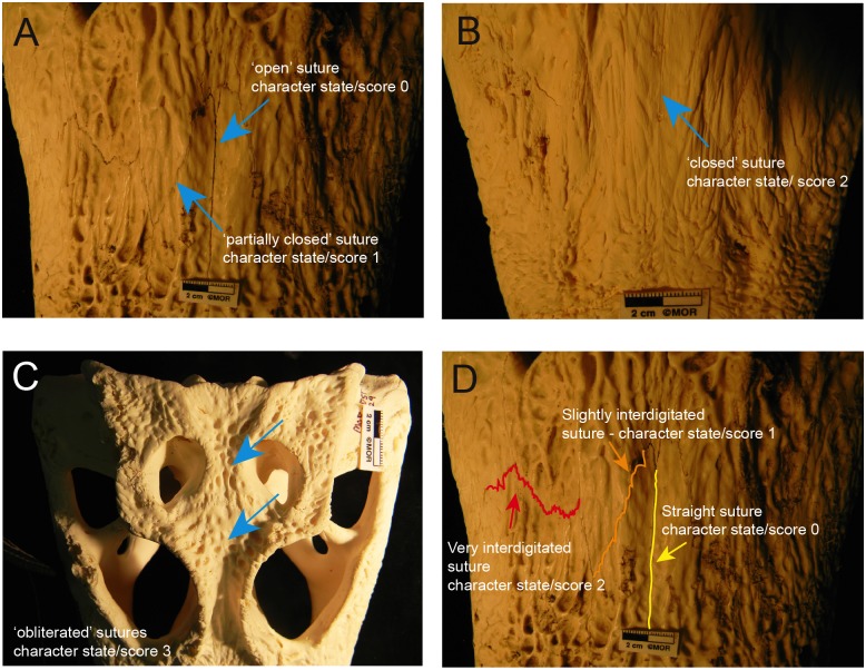
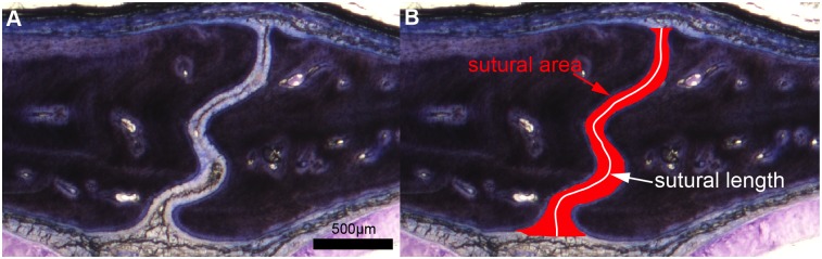
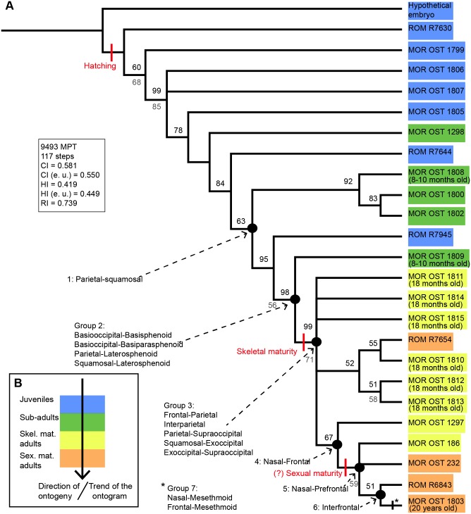
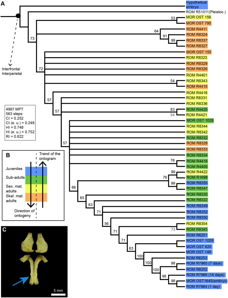
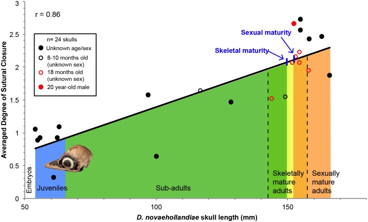
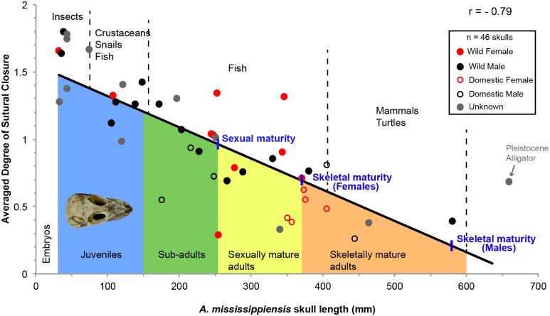
The sutures of the skulls of vertebrates are generally open early in life and slowly close as maturity is attained. The assumption that all vertebrates follow this pattern of progressive sutural closure has been used to assess maturity in the fossil remains of non-avian dinosaurs. Here, we test this assumption in two members of the Extant Phylogenetic Bracket of the Dinosauria, the emu, Dromaius novaehollandiae and the American alligator, Alligator mississippiensis, by investigating the sequence and timing of sutural fusion in their skulls. As expected, almost all the sutures in the emu skull progressively close (i.e., they get narrower) and then obliterate during ontogeny. However, in the American alligator, only two sutures out of 36 obliterate completely and they do so during embryonic development. Surprisingly, as maturity progresses, many sutures of alligators become wider in large individuals compared to younger, smaller individuals. Histological and histomorphometric analyses on two sutures and one synchondrosis in an ontogenetic series of American alligator confirmed our morphological observations. This pattern of sutural widening might reflect feeding biomechanics and dietary changes through ontogeny. Our findings show that progressive sutural closure is not always observed in extant archosaurs, and therefore suggest that cranial sutural fusion is an ambiguous proxy for assessing maturity in non-avian dinosaurs.


Conflict of interest statement

Competing Interests: The authors have declared that no competing interests exist.

Figures

Fig 1
Fig 1. Character states and scores describing the degree of sutural closure and interdigitation in some American alligator skulls.
A, The blue arrows show an open and a partially closed suture on MOR OST 155. B, The blue arrow shows a closed suture (with its suture line still visible) on MOR OST 1029. C, The blue arrows show two obliterated sutures (with their suture lines morphologically invisible) on MOR OST 1029. D, The colored lines show the different degrees of interdigitation: a straight (yellow line), slightly interdigitated (orange line) and a very interdigitated suture (red line) on MOR OST 155. These interdigitation degrees are based on those in Herring [82].
Fig 2
Fig 2. Photomicrographs of a transverse section through an alligator internasal suture showing measurements of sutural area and sutural length.
A, An alligator internasal suture. B, Measurements of sutural area and sutural length. The ratio between sutural area (in red) and sutural length (in white) gives the averaged width of the suture. The sections are stained with Toluidine-blue.
Fig 3
Fig 3. Ontogram of D. novaehollandiae (n = 24).
A, This ontogram is the 50% majority rule consensus tree for 9493 MPT. The onset of hatching, skeletal maturity and (approximate) sexual maturity are mapped on this diagram. A precise sequence of sutural obliteration is given by synontomorphies on the left of the ontogram (i.e., the sutures or group of sutures that obliterate consistently in all the specimens of the analysis).Bootstrap support values are in grey below nodes, and the percent occurrence for nodes are in black above horizontal lines.B, Color code indicating the ontogenetic category of each specimen. Categories were estimated prior to phylogenetic analysis in each specimen and were based on known age or on skull length (Table 1) and already-published growth patterns on this species. This color code is the same in A. Note that the ‘trend’ of the ontogram follows the direction of ontogeny in general. Abbreviations: CI, consistency index; e.u., excluding uninformative characters; HI, homoplasy index; MPT, most parcimonious trees; RI, retention index; Skel. mat., skeletally mature; Sex. mat., sexually mature.
Fig 4
Fig 4. Ontogram of A. mississippiensis (n = 49).
A, This ontogram is the 50% majority rule consensus tree for 4997 MPT. The analysis only produced two synontomorphies concerning sutural obliterations (the interfrontal and interparietal sutures). Bootstrap support values are in grey below nodes, and the percent occurrence for nodes are in black above horizontal lines. B, Color code indicating the ontogenetic category of each specimen in A. These categories were estimated prior to phylogenetic analysis in each specimen, based on known skull length, total length, sex and geographic provenance (Table 2), as well as previously published growth patterns for this species. Note that in general, the ‘trend’ of the ontogram is opposite to the correct direction of ontogeny. C, Paired frontals (bottom) and parietals (top) of an unhatched embryo (MOR OST 1646). The blue arrow indicates an anterior-posterior gradient of fusion. Abbreviations: CI, consistency index; e.u., excluding uninformative characters; HI, homoplasy index; MPT, most parcimonious trees; Pleistoc., Pleistocene; RI, retention index; Skel. mat., skeletally mature; Sex. mat., sexually mature.
Fig 5
Fig 5. Linear relationship between ontogeny (using skull length as a proxy) and the averaged degree of sutural closure in D. novaehollandiae (n = 24).
If the previously mentioned character states of each suture are transformed into set scores and if averages of closure are calculated for each specimen consequently, this type of data visualization can be generated. An average of zero for one specimen would mean that all its sutures are open, while an average of three would mean that all its sutures are obliterated. This relationship has a Pearson’s coefficient of correlation (r) of 0.86. The approximate onsets of skeletal and sexual maturity are mapped on the trend line. Below the trend line are the different ontogenetic categories, indicated with the same color code as that in Fig 3B. Each data point (i.e., each skull) is characterized by its age (see legend in black square). This relationship shows an obvious increase in the degree of sutural closure as ontogeny proceeds, in other words sutures become more closed.
Fig 6
Fig 6. Linear relationship between ontogeny (using skull length as a proxy) and the averaged degree of sutural closure in A. mississippiensis (n = 46).
This relationship has a Pearson’s coefficient of correlation (r) of -0.79. The approximate onsets of sexual maturity and skeletal maturity for females and males are mapped on the trend line. Below the trend line are the different ontogenetic categories, indicated with the same color code as that used in previous figures. Above the trend line are exposed the estimated diet of these animals at the time of death, based on previous studies [71,72]. Each data point (i.e., each skull) is characterized by its sex and domestic or wild status (see legend in black square). This relationship shows an obvious decrease in the degree of sutural closure as ontogeny proceeds, in other words, sutures become wider.
Fig 7
Fig 7. Parasagittal sections through the frontoparietal suture in an ontogenetic series of emus, stained with Toluidine-blue.
A, Open suture in MOR OST 1799, a specimen a few weeks old. B, Open suture in MOR OST 1801, an 8 to 10 month old specimen. C, Obliterated suture in MOR OST 1803, a 20 year-old male. This suture is replaced by trabeculae of lamellar bone. The average sutural widths are marked in red on each photomicrograph.
Fig 8
Fig 8. Transverse sections through the frontoparietal suture, the internasal suture and the basioccipital-exoccipital synchondrosis in an ontogenetic series of three American alligator heads, stained with Toluidine-blue.
A-C, Frontoparietal suture; D-F, internasal suture; G-H, Basioccipital-exoccipital synchondrosis. The youngest specimen MOR OST 1647 is a few days old and is shown in the first column (A, D, G); the second column shows a 4 to 5 year-old specimen MOR OST 1797 (B, E, H); the third column shows a 9 to 12 year-old sexually mature specimen, MOR OST 1798 (C, F, I). Sutures of the youngest specimen show two thin bone struts overlapping each other (A, D, red arrows). The basioccipital-exoccipital synchondrosis (G) in this same specimen is not fully formed (and appears to have a synovial cavity, red arrow). Interdigitations increase drastically in the frontoparietal suture, but only slightly in the internasal suture (being a midline suture) and in the basioccipital-exoccipital synchondrosis. These sections do not show any sutural bony fusion. The average sutural and synchondroseal widths are marked in red on each photomicrograph, and it is clear that these widths increase as ontogeny progresses.

References

    1. Marieb EN (2014) Essentials of Human Anatomy & Physiology: Pearson Education.
    1. Chopra SRK. The cranial suture closure in monkeys; 1957. Wiley Online Library; pp. 67–112.
    1. Cole A, Fedak T, Hall B, Olson W, Vickaryous M (2003) Sutures joining ontogeny and fossils. The Palaeontological Association Newsletter 52: 29–32.
    1. Dwight T (1890) The closure of the cranial sutures as a sign of age. The Boston Medical and Surgical Journal 122: 389–392.
    1. Herring S (2000) Sutures and craniosynostosis: a comparative, functional, and evolutionary perspective Craniosynostosis: diagnosis, evaluation, and management, 2nd ed New York: Oxford University Press; pp. 3–10.

Publication types