. 2016 Jul 15;194(2):185-97.
doi: 10.1164/rccm.201505-0999OC.

A Chronic Obstructive Pulmonary Disease Susceptibility Gene, FAM13A, Regulates Protein Stability of β-Catenin

Affiliations

A Chronic Obstructive Pulmonary Disease Susceptibility Gene, FAM13A, Regulates Protein Stability of β-Catenin

Zhiqiang Jiang et al. Am J Respir Crit Care Med. .

Erratum in

Abstract

Rationale: A genetic locus within the FAM13A gene has been consistently associated with chronic obstructive pulmonary disease (COPD) in genome-wide association studies. However, the mechanisms by which FAM13A contributes to COPD susceptibility are unknown.

Objectives: To determine the biologic function of FAM13A in human COPD and murine COPD models and discover the molecular mechanism by which FAM13A influences COPD susceptibility.

Methods: Fam13a null mice (Fam13a(-/-)) were generated and exposed to cigarette smoke. The lung inflammatory response and airspace size were assessed in Fam13a(-/-) and Fam13a(+/+) littermate control mice. Cellular localization of FAM13A protein and mRNA levels of FAM13A in COPD lungs were assessed using immunofluorescence, Western blotting, and reverse transcriptase-polymerase chain reaction, respectively. Immunoprecipitation followed by mass spectrometry identified cellular proteins that interact with FAM13A to reveal insights on FAM13A's function.

Measurements and main results: In murine and human lungs, FAM13A is expressed in airway and alveolar type II epithelial cells and macrophages. Fam13a null mice (Fam13a(-/-)) were resistant to chronic cigarette smoke-induced emphysema compared with Fam13a(+/+) mice. In vitro, FAM13A interacts with protein phosphatase 2A and recruits protein phosphatase 2A with glycogen synthase kinase 3β and β-catenin, inducing β-catenin degradation. Fam13a(-/-) mice were also resistant to elastase-induced emphysema, and this resistance was reversed by coadministration of a β-catenin inhibitor, suggesting that FAM13A could increase the susceptibility of mice to emphysema development by inhibiting β-catenin signaling. Moreover, human COPD lungs had decreased protein levels of β-catenin and increased protein levels of FAM13A.

Conclusions: We show that FAM13A may influence COPD susceptibility by promoting β-catenin degradation.

Keywords: FAM13A; cell proliferation; emphysema; protein stability; β-catenin.


Figures
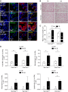
Figure 1.
Figure 1.
Depletion of Fam13a confers resistance to emphysema development in murine models. (A) FAM13A is localized in human airway cells including mucosal cells (MUC5AC), Club cells (CC10), pan-cytokeratin (PanCK, airway epithelial cells), and alveolar cells positive for surfactant protein-C (SP-C, alveolar type II cells). Representative images are from patients with very severe chronic obstructive pulmonary disease (n = 3) (FEV1 <40%). Goat = anti-goat; Ms = anti-mouse; Rb = anti-rabbit. (B) Representative hematoxylin and eosin staining images (200×) in Fam13a null mice (Fam13a−/−) and wild-type littermate control mice (Fam13a+/+) exposed to cigarette smoke (CS) or room air (Air) for 6 months. (C) Mean alveolar chord length measurement in four groups of mice. Data shown are means ± SEM derived from 11–22 mice per group (the number of mice studied in each group is indicated inside each column). (D) Total leukocytes and leukocyte subsets in bronchoalveolar lavage (BAL) samples from Fam13a−/− and Fam13a+/+ mice exposed to CS or air for 1 month. Two-way analysis of variance tests were used to analyze the effects of treatment and genotype in (C) and (D). Unpaired Student’s t test, **P < 0.01.
Figure 2.
Figure 2.
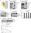
FAM13A interacts with PP2A/β-catenin complex and promotes degradation of β-catenin. (A) Affinity purification of cellular protein complexes associated with Flag-tagged FAM13A (FL) in HEK 293 cells as revealed by silver staining. Vector-transfected cells (V) were used as a control. The * indicates Flag-tagged FAM13A. Immunoprecipitation (IP) of FAM13A (B) and reverse immunoprecipitation (IP) targeting β-catenin (short for β) (C) was performed in HEK 293 cells. Examination of the levels and phosphorylation of β-catenin in human bronchial epithelial (16HBE) cells transfected with either increasing amount of FAM13A (D) or short hairpin RNAs (shRNAs) targeting FAM13A (E). NT = nontarget control hairpins; columns B, C, and D = three independent hairpins targeting FAM13A. (F) Expression of FAM13A and β-catenin in cells measured by reverse transcriptase–polymerase chain reaction. Glyceraldehyde 3-phosphate dehydrogenase (GAPDH) was used as a reference gene. (G) Half-life of β-catenin was measured in 16HBE cells stably infected with nontargeting control shRNA (NT) or FAM13A shRNA B and treated with cycloheximide (CHX, 5 nM) for various time periods. Means ± SD were from two independent repeats normalized to α-tubulin. Linear mixed effects model was used to compare differences in slope of β-catenin reductions in NT-shRNA and FAM13A shRNA–transfected cells (P < 0.01).
Figure 3.
Figure 3.
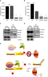
FAM13A down-regulates β-catenin activity and regulates cell proliferation. (A) Assessments of the β-catenin activity by TOPFLASH reporter assay in human bronchial epithelial (16HBE) cells transfected with full-length human FAM13A. Cells treated with β-catenin inhibitor PKF118-310 (7.5 μg/ml) overnight were used as negative controls and cells transfected with constitutively active β-catenin mutant (β-catenin::CS2) as positive controls in this reporter assay. Statistical comparisons in controls were not shown. Means ± SD were from triplicate assays with separately transfected and treated wells per condition. *P <  0.05, **P < 0.01. Unpaired Student’s t test was applied. One representative repeat from three independent biologic repeats was shown here. The other two repeats were shown in Figure E4D. (B) Cell growth curve in 16HBE cells transfected with different constructs. Z-test was used to compare the slopes between the curves using linear regression model. The significance was indicated right to the last time point of each curve: *Comparison between FAM13A shRNA-pCMV and NT shRNA-pCMV, P < 0.05; ##Comparison between FAM13A shRNA-pCMV and FAM13A shRNA-FL, P < 0.05. FL = full-length FAM13A; NT shRNA = nontargeting shRNA; pCMV = empty vector as controls; shRNA = short hairpin RNA.
Figure 4.
Figure 4.
FAM13A is required for the interaction between β-catenin, and PP2A thus promotes degradation of β-catenin. (A and B) Phosphatase activity of PP2A in 16HBE cells stably infected with nontargeting control shRNA (NT) or FAM13A shRNA B (B) was measured using PP2A Immunoprecipitation Phosphatase Assay Kit (Millipore). Cells treated with okadaic acid (OA) for 18 hours were used as negative controls in this assay. Cellular proteins were immunoprecipitated with PP2Ac antibody (A) or β-catenin antibody (B) separately. Note that the same amount of cellular proteins was used for immunoprecipitation (IP) in NT cells as in B cells. (C) Detection of β-catenin and PP2Ab associated with PP2Ac in NT cells and B cells. (D) Detection of PP2Ac and PP2Ab associated with β-catenin in NT cells and B cells. **P < 0.05. Unpaired Student’s t test was applied. (E) Schematic illustration on how FAM13A regulates β-catenin protein stability through interacting with PP2A. 16HBE = human bronchial epithelial cells; GSK-3β = glycogen synthase kinase 3β; ns = not significant; shRNA = short hairpin RNA.
Figure 5.
Figure 5.
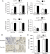
Depletion of Fam13a leads to activation of β-catenin signaling in murine lungs. (A) Quantification of total and phosphorylated β-catenin and GSK-3β by Western blotting in murine lungs from the same groups of mice exposed to air or cigarette smoke (CS) for 6 months as in Figure 6A. Representative Ki67 staining images (200× magnification with 600× magnification in insets) (B) and quantifications (C) in lung sections from Fam13a−/− and Fam13a+/+ mice exposed to air or cigarette smoke for 6 months. Cells positive for Ki67 staining were counted in six randomly acquired fields per mouse. Data are mean ± SEM from four mice per group (number of mice in each group is indicated inside each column). *P < 0.05; **P < 0.01, unpaired Student’s t test analysis in A and C. GSK3β = glycogen synthase kinase 3β.
Figure 6.
Figure 6.
Inhibition on transcriptional activity of β-catenin reverted the resistance to emphysema in Fam13a−/− mice in elastase-induced experimental emphysema. (A) Airspace size assessment in Fam13a+/+ and Fam13a−/− mice treated with phosphate-buffered saline, porcine pancreatic elastase (PPE), or β-catenin inhibitor PKF118-310 (200× magnification). (B) Mean chord length was quantified on randomly selected images in a blinded fashion. Data shown are means ± SEM derived from n = 6–8 mice per group (number in each column represents the number of mice in each group). (C) Ki67 staining measurement in lung tissue from mice treated with PPE. Data shown are mean ± SEM from four mice per group with six randomly acquired images of lung sections stained for Ki67. (D) Total leukocytes counted in bronchoalveolar lavage (BAL) samples from Fam13a−/− and Fam13a+/+ mice treated with PPE with or without PKF118-310. *P < 0.05; **P < 0.01, unpaired Student’s t test in BD.
Figure 7.
Figure 7.
Increased FAM13A levels and reduced β-catenin levels are associated with human emphysema. (A) Increased levels of FAM13A protein and decreased levels of β-catenin protein (B) in human chronic obstructive pulmonary disease lungs compared with healthy ex-smokers. Data shown are mean ± SEM from six subjects for each group. **P < 0.01, unpaired Student’s t tests were used to analyze the data. (C and D) Measurements on FAM13A (C) and β-catenin (D) mRNA levels in human chronic obstructive pulmonary disease lungs by reverse transcriptase polymerase chain reaction. GAPDH = glyceraldehyde 3-phosphate dehydrogenase; S = healthy ex-smokers; SC = very severe COPD.

Comment in

References

    1. Miniño AM. Death in the United States, 2011. NCHS Data Brief. 2013;115:1–8. - PubMed
    1. Miniño AM, Kochanek KD. National Vital Statistics Reports. 2010. Division of Vital Statistics. Deaths: preliminary data for 2008. U.S. Department of Health and Human Services Centers for Disease Control and Prevention, National Center for Health Statistics, National Vital Statistics System; pp. 1–72. - PubMed
    1. Burrows B, Knudson RJ, Cline MG, Lebowitz MD. Quantitative relationships between cigarette smoking and ventilatory function. Am Rev Respir Dis. 1977;115:195–205. - PubMed
    1. Fletcher C, Peto R, Tinker C, Speizer FE. The natural history of chronic bronchitis and emphysema. Oxford: Oxford University Press; 1976. Factors related to the development of airflow obstruction; pp. 70–105.
    1. McCloskey SC, Patel BD, Hinchliffe SJ, Reid ED, Wareham NJ, Lomas DA. Siblings of patients with severe chronic obstructive pulmonary disease have a significant risk of airflow obstruction. Am J Respir Crit Care Med. 2001;164:1419–1424. - PubMed

Publication types