. 2016 Mar 8;7(10):11463-77.
doi: 10.18632/oncotarget.7201.

Reduced RKIP enhances nasopharyngeal carcinoma radioresistance by increasing ERK and AKT activity

Affiliations

Reduced RKIP enhances nasopharyngeal carcinoma radioresistance by increasing ERK and AKT activity

Li Yuan et al. Oncotarget. .

Abstract

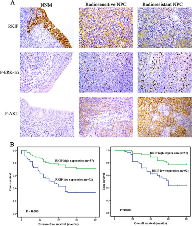
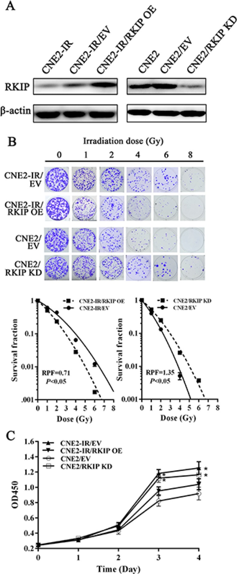
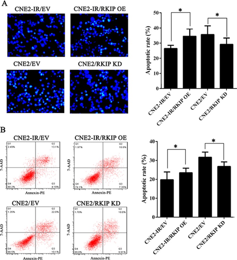
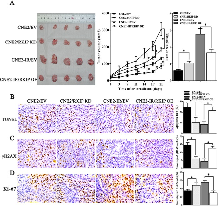
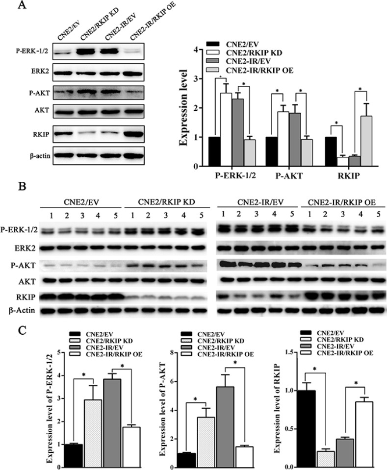
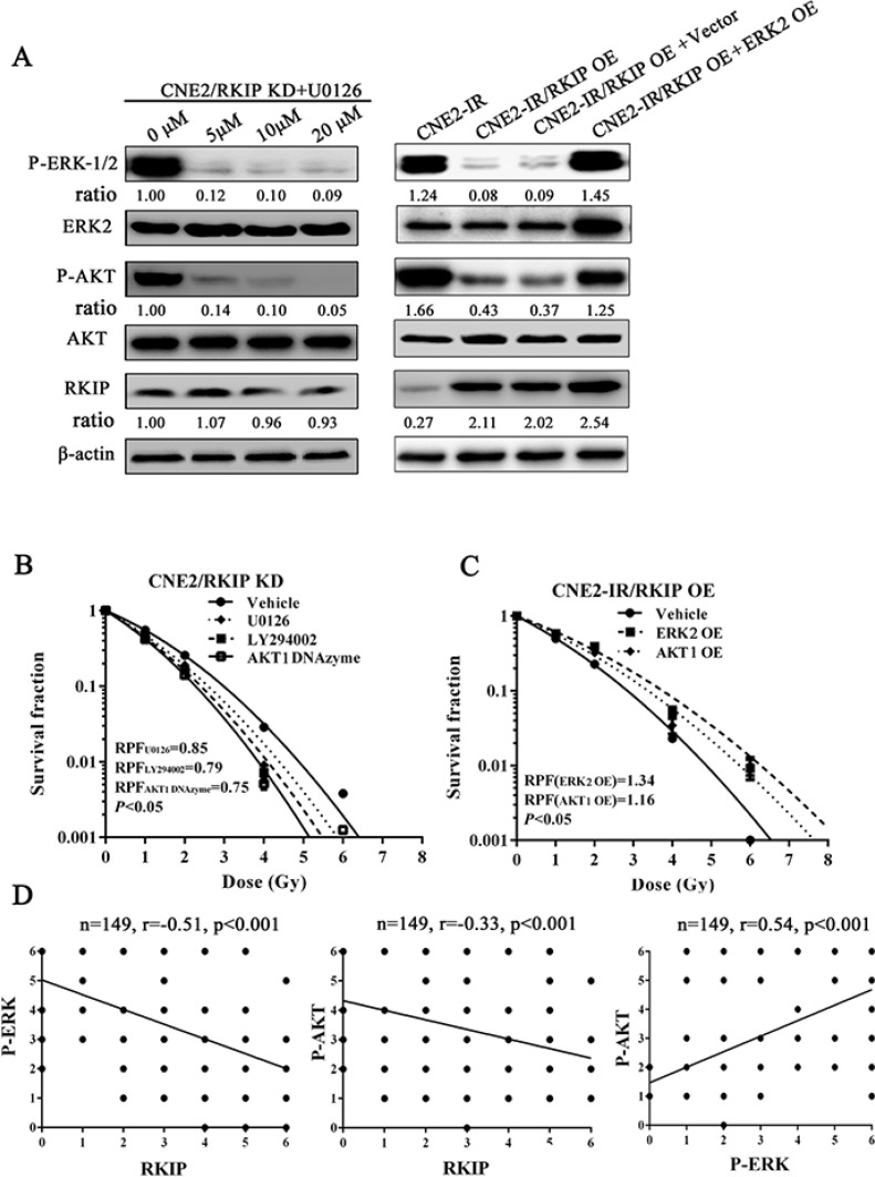
Raf kinase inhibitory protein (RKIP) functions as a chemo-immunotherapeutic sensitizer of cancers, but regulation of RKIP on tumor radiosensitivity remains largely unexplored. In this study, we investigate the role and mechanism of RKIP in nasopharyngeal carcinoma (NPC) radioresistance. The results showed that RKIP was frequently downregulated in the radioresistant NPC tissues compared with radiosensitive NPC tissues, and its reduction correlated with NPC radioresistance and poor patient survival, and was an independent prognostic factor. In vitro radioresponse assay showed that RKIP overexpression decreased while RKIP knockdown increased NPC cell radioresistance. In the NPC xenografts, RKIP overexpression decreased while RKIP knockdown increased tumor radioresistance. Mechanistically, RKIP reduction promoted NPC cell radioresistance by increasing ERK and AKT activity, and AKT may be a downstream transducer of ERK signaling. Moreover, the levels of phospho-ERK-1/2 and phospho-AKT were increased in the radioresistant NPC tissues compared with radiosensitive ones, and negatively associated with RKIP expression, indicating that RKIP-regulated NPC radioresponse is mediated by ERK and AKT signaling in the clinical samples. Our data demonstrate that RKIP is a critical determinant of NPC radioresponse, and its reduction enhances NPC radioresistance through increasing ERK and AKT signaling activity, highlighting the therapeutic potential of RKIP-ERK-AKT signaling axis in NPC radiosensitization.

Keywords: AKT; ERK−1/2; RKIP; nasopharyngeal carcinoma; radioresistance.


Conflict of interest statement

CONFLICTS OF INTEREST

The authors disclose no potential conflicts of interest.

Figures

Figure 1
Figure 1. RKIP reduction is correlated with NPC radioresistance and poor patient survival
(A) a representative result of RKIP, phospho-ERK−1/2 and phospho-AKT immunohistochemical staining in normal nasopharyngeal mucosa (NNM), and radiosensitive and radioresistant NPC tissues. Original magnification, ×200. (B) Kaplan-Meier survival analysis for NPC patients according to the expression levels of RKIP. NPC patients with low RKIP expression have a significantly worse disease-free survival (left) and overall survival (right) than those with high RKIP expression.
Figure 2
Figure 2. RKIP reduction increases NPC cell radioresistance in vitro
(A) a representative result of Western blotting shows the levels of RKIP expression in the RKIP OE CNE2-IR cells, RKIP KD CNE2 cells and their corresponding EV-transfected cells. (B) a clonogenic survival assay shows that radioresponse of RKIP OE CNE2-IR cells, RKIP KD CNE2 cells and their corresponding EV-transfected cells. (top) cells were irradiated with a range of 1–8Gy radiation doses, and colonies that formed after incubation of 12 days were stained with crystal violet and photographed; (bottom) dose survival curves were created by fitting surviving fractions to the linear quadratic equation. (C) CCK-8 assay shows that proliferation of RKIP OE CNE2-IR cells, RKIP KD CNE2 cells and their corresponding EV-transfected cells after 4Gy irradiation. Three experiments were done; Means, SDs, and statistical significance are denoted; *P < 0.01. OE, overexpression; KD, knockdown; EV, empty vector.
Figure 3
Figure 3. RKIP reduction inhibits irradiation-induced NPC cell apoptosis
(A) (left) RKIP OE CNE2-IR cells, RKIP KD CNE2 cells and their corresponding EV-transfected cells were irradiated with 6Gy irradiation, incubated for 72 hours, and stained with Hoechst 33258 and photographed; (right) a histogram shows the apoptotic rate of these cells. (B) (left) RKIP OE CNE2-IR cells, RKIP KD CNE2 cells and their corresponding EV-transfected cells were irradiated with 6Gy irradiation, incubated for 72 hours, subjected to analysis of apoptosis using flow cytometry, and a representative result of cell apoptosis is showed; (right) a histogram shows the apoptotic rate of these cells. Three experiments were done; Means, SDs, and statistical significance are denoted; *P < 0.01. OE, overexpression; KD, knockdown; EV, empty vector.
Figure 4
Figure 4. RKIP reduction increases NPC cell radioresistance in vivo
(A) the growth and weight of tumors generated by RKIP KD CNE2 cells, RKIP OE CNE2-IR cells and their corresponding EV-transfected cells after 8Gy irradiation. (left) 3 weeks after irradiation, the mice were killed, and the tumors were photographed; (middle) the growth curves of the tumors after irradiation (n = 5 each group) at the sacrifice with respect to the first measurements; (right) the average weights of the tumors after irradiation (n = 5 each group) at the sacrifice. (B) (left) a representative image of TUNEL detection of apoptotic cells in the tumors generated by RKIP KD CNE2 cells, RKIP OE CNE2-IR cells and their corresponding EV-transfected cells after irradiation; (right) a histogram shows percentages of apoptotic cells in the tumors (n = 5 each group). (C) (left) a representative image of immunohistochemical staining of γH2AX in the tumors generated by RKIP KD CNE2 cells, RKIP OE CNE2-IR cells and their corresponding EV-transfected cells after irradiation; (right) a histogram shows percentages of γ-H2AX positive cells in the tumors (n = 5 each group). (D) (left) a representative image of immunohistochemical staining of Ki-67 in the tumors generated by RKIP KD CNE2 cells, RKIP OE CNE2-IR cells and their corresponding EV-transfected cells after irradiation; (right) a histogram shows percentages of Ki-67 positive cells in the tumors (n = 5 each group). Means, SDs, and statistical significance are denoted; *P < 0.01. Original magnification, ×400. OE, overexpression; KD, knockdown; EV, empty vector.
Figure 5
Figure 5. RKIP inhibits activity of ERK-1/2 and AKT signaling in the irradiated NPC cells and xenograft tumors
(A) (left) a representative result of Western blotting shows the levels of phospho-ERK−1/2, phospho-AKT and RKIP in the RKIP KD CNE2 cells, RKIP OE CNE2-IR cells and their corresponding EV-transfected cells after 4Gy irradiation; (right) a histogram shows the levels of phospho-ERK−1/2, phospho-AKT and RKIP in these cells. (B) (top) a representative result of Western blotting shows the levels of phospho-ERK−1/2, phospho-AKT and RKIP in the tumors generated by RKIP KD CNE2 cells, RKIP OE CNE2-IR cells and their corresponding EV-transfected cells after 8Gy irradiation; (bottom) a histogram shows the expression levels of phospho-ERK-1/2, phosphor-AKT and RKIP in the xenograft tumors. Means, SDs, and statistical significance are denoted; *P < 0.01. OE, overexpression; KD, knockdown; EV, empty vector.
Figure 6
Figure 6. RKIP-regulated NPC cell radioresponse is mediated through ERK and AKT signaling
(A) (left) a representative result of Western blotting shows the levels of the phospho-ERK-1/2, phospho-AKT and RKIP in the RKIP KD CNE2 treated with a range of 5–20 μmol/L MEK inhibitor U0126; (right) a representative result of Western blotting shows the levels of phospho-ERK-1/2, phospho-AKT and RKIP in the RKIP OE CNE2-IR cells transfected with 4 μg/mL ERK-2 expression vector (OE). (B) a clonogenic survival assay shows that inhibition of ERK and AKT signaling significantly abrogated CNE2 KD cell radioresistance induced by RKIP knockdown. Cells treated with 10 μmol/L U0126, 10 μmol/L LY294002 or 2 μmol/L AKT-1 DNAzyme were irradiated with a range of 1–8Gy radiation doses, and dose survival curves were created by fitting surviving fractions to the linear quadratic equation. (C) a clonogenic survival assay shows that activation of ERK and AKT signaling restored CNE2-IR OE cell radioresistance reduced by RKIP overexpression. Cells transfected with 4 μmol/L of ERK-2 or AKT-1 expression plasmid were irradiated with a range of 1–8Gy radiation doses, and dose survival curves were created by fitting surviving fractions to the linear quadratic equation. (C) Spearman rank correlation analysis shows that RKIP level was negatively associated with phospho-ERK−1/2 level (a) and phospho-AKT level (b), and phospho-ERK−1/2 level was positively associated with phospho-AKT level in 149 cases of NPCs. OE, overexpression; KD, knockdown.

References

    1. Lo KW, To KF, Huang DP. Focus on nasopharyngeal carcinoma. Cancer Cell. 2004;5:423–428. - PubMed
    1. Suarez C, Rodrigo JP, Rinaldo A, Langendijk JA, Shaha AR, Ferlito A. Current treatment options for recurrent nasopharyngeal cancer. Eur Arch Otorhinolaryngol. 2010:2671811–1824. - PMC - PubMed
    1. Lee AW, Poon Y, Foo W, Law SC, Cheung FK, Chan DK, Tung SY, Thaw M, Ho JH. Retrospective analysis of 5037 patients with nasopharyngeal carcinoma treated during 1976–1985: overall survival and patterns of failure. Int J Radiat Oncol Biol Phys. 1992;23:261–270. - PubMed
    1. Yeung K, Seitz T, Li S, Janosch P, McFerran B, Kaiser C, Fee F, Katsanakis KD, Rose DW, Mischak H, Sedivy JM, Kolch W. Suppression of Raf-1 kinase activity and MAP kinase signalling by RKIP. Nature. 1999;401:173–177. - PubMed
    1. Yeung KC, Rose DW, Dhillon AS, Yaros D, Gustafsson M, Chatterjee D, McFerran B, Wyche J, Kolch W, Sedivy JM. Raf kinase inhibitor protein interacts with NF-kappaB-inducing kinase and TAK1 and inhibits NF-kappaB activation. Mol Cell Biol. 2001;21:7207–7217. - PMC - PubMed

MeSH terms

Substances