Review
. 2016 Apr;37(2):135-87.
doi: 10.1210/er.2015-1106. Epub 2016 Feb 10.

Role of Thyroid Hormones in Skeletal Development and Bone Maintenance

Affiliations
Review

Role of Thyroid Hormones in Skeletal Development and Bone Maintenance

J H Duncan Bassett et al. Endocr Rev. 2016 Apr.

Abstract

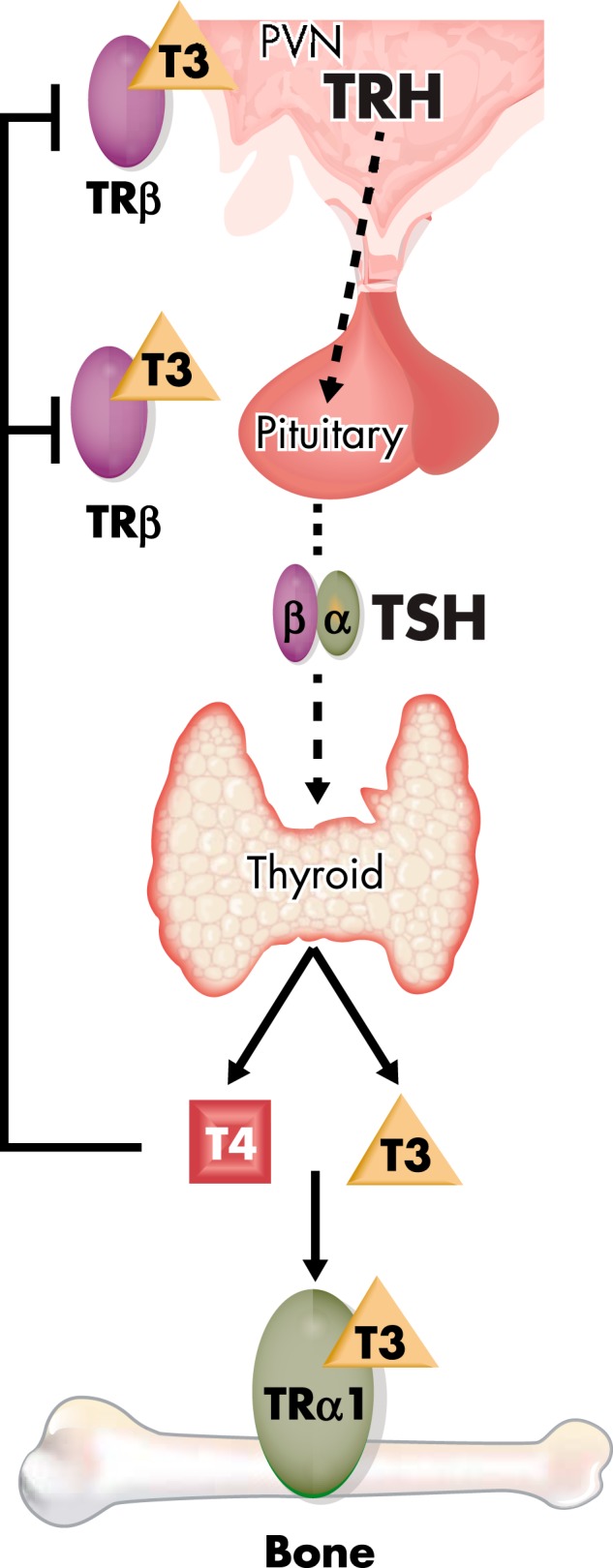
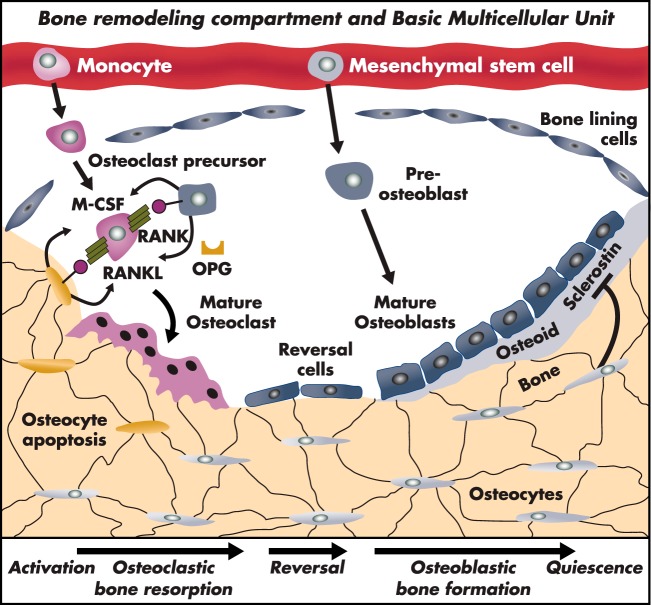
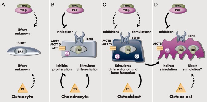
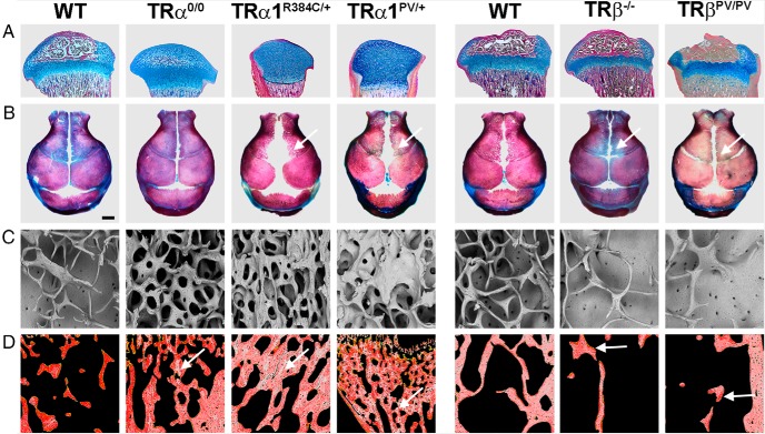
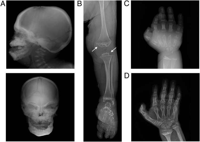
The skeleton is an exquisitely sensitive and archetypal T3-target tissue that demonstrates the critical role for thyroid hormones during development, linear growth, and adult bone turnover and maintenance. Thyrotoxicosis is an established cause of secondary osteoporosis, and abnormal thyroid hormone signaling has recently been identified as a novel risk factor for osteoarthritis. Skeletal phenotypes in genetically modified mice have faithfully reproduced genetic disorders in humans, revealing the complex physiological relationship between centrally regulated thyroid status and the peripheral actions of thyroid hormones. Studies in mutant mice also established the paradigm that T3 exerts anabolic actions during growth and catabolic effects on adult bone. Thus, the skeleton represents an ideal physiological system in which to characterize thyroid hormone transport, metabolism, and action during development and adulthood and in response to injury. Future analysis of T3 action in individual skeletal cell lineages will provide new insights into cell-specific molecular mechanisms and may ultimately identify novel therapeutic targets for chronic degenerative diseases such as osteoporosis and osteoarthritis. This review provides a comprehensive analysis of the current state of the art.


Figures

Figure 1.
Figure 1.
HPT axis. The thyroid gland secretes the prohormone T4 and the active hormone T3, and circulating concentrations are regulated by a classical endocrine negative feedback loop that maintains an inverse physiological relationship between TSH, and T4 and T3. PVN, paraventricular nucleus.
Figure 2.
Figure 2.
TSH action. Binding of TSH to the TSHR results in activation of G protein-coupled downstream signaling including: 1) the adenylyl cyclase (AC), cAMP, protein kinase A (PKA), and the cAMP response element binding (CREB) protein; or 2) phospholipase C (PLC), inositol triphosphate (IP3), and intracellular calcium pathway; or 3) the PLC, diacylglycerol (DAG), protein kinase C (PKC), and signal transducer and activator of transcription 3 (STAT3) pathway. PIP2, phosphatidylinositol 4,5-bisphosphate.
Figure 3.
Figure 3.
Thyroid hormone action in bone cells. A, In hypothyroidism, despite maximum DIO2 (D2) and minimum DIO3 (D3) activities, TRα1 remains unliganded and bound to corepressor, thus inhibiting T3-target gene transcription. B, In the euthyroid state, D2 and D3 activities are regulated to optimize ideal intracellular T3 availability, resulting in displacement of corepressor and physiological transcriptional activity of TRα1. C, In thyrotoxicosis, despite maximum D3 and minimum D2 activities, supraphysiological intracellular T3 concentrations result in increased TRα1 activation and enhanced T3-target gene responses.
Figure 4.
Figure 4.
Intramembranous and endochondral ossification. A, Postnatal day 1 skull vault stained with alizarin red (bone) and alcian blue (cartilage) showing sutures and fontanelles. B, Schematic representation of intramembranous bone formation at a skull suture. C, Proximal tibial section at P21 growth plate stained with alcian blue (cartilage) and Van Gieson (bone matrix, red). D, Schematic representation of the growth plate.
Figure 5.
Figure 5.
Bone remodeling compartment and “basic multicellular unit” of the bone-remodeling cycle. The bone remodeling cycle is initiated and orchestrated by osteocytes. Bone remodeling results from changes in mechanical load, structural microdamage, or exposure to systemic or paracrine factors. Monocyte/macrophage precursors differentiate to mature osteoclasts and resorb bone. Differentiation is induced by M-CSF and RANKL and inhibited by OPG. During reversal, osteoblastic progenitors are recruited to the site of resorption, synthesize osteoid, and mineralize new bone to repair the defect.
Figure 6.
Figure 6.
Actions of T3 and TSH in skeletal cells. A, T3 and TSH actions in osteocytes have not been investigated, and it is unknown whether osteocytes express thyroid hormone transporters, deiodinases, TRs, or the TSHR. B, Chondrocytes express MCT8, MCT10, and LAT1 transporters, DIO3 (D3), TRs (predominantly TRα), and TSHR. T3 inhibits proliferation and stimulates prehypertrophic and hypertrophic chondrocyte differentiation, whereas TSH might inhibit proliferation and matrix synthesis. C, Osteoblasts express MCT8 and LAT1/2 transporters, the DIO2 (D2) and D3, TRs (predominantly TRα), and TSHR. Most studies indicate that T3 stimulates osteoblast differentiation and bone formation. Contradictory data suggest that TSH may stimulate, inhibit, or have no effect on osteoblast differentiation and function. D, Osteoclasts express MCT8, D3, TRs and the TSHR. Currently, it is unclear whether T3 acts directly in osteoclasts or whether indirect effects in the osteoblast lineage mediate its actions. Most studies indicate that TSH inhibits osteoclast differentiation and function.
Figure 7.
Figure 7.
Skeletal phenotype of TRα and TRβ mutant mice. A, Proximal tibias stained with alcian blue (cartilage) and van Gieson (bone, red) showing delayed formation of the secondary ossification center in TRα-deficient mice (TRα0/0) and grossly delayed formation in mice with dominant-negative TRα mutations (TRα1R384C/+ and TRα1PV/+). Mice with mutation or deletion of TRβ have advanced ossification with premature growth plate narrowing. B, Skull vaults stained with alizarin red (bone) and alcian blue (cartilage) showing skull sutures and fontanelles. Arrows indicate delayed intramembranous ossification in mice with dominant-negative TRα mutations (TRα1R384C/+ and TRα1PV/+) and advanced ossification in mice with mutation or deletion of TRβ. C, Trabecular bone microarchitecture in adult TR mutant mice. Backscattered electron scanning electron microscopy images show increased trabecular bone in TRα0/0 mice and severe osteosclerosis in TRα1R384C/+ and TRα1PV/+ mice. By contrast, TRβ mutant mice have reduced trabecular bone volume and osteoporosis. D, Trabecular bone micromineralization in adult TR mutant mice. Pseudo-colored quantitative backscattered electron scanning electron microscopy images showing mineralization densities in which high mineralization density is gray and low density is red. Mice with deletion or mutation of TRα have retention of highly mineralized calcified cartilage (arrows) demonstrating a persistent remodeling defect. By contrast, mice with deletion or mutation of TRβ have reduced bone mineralization (arrow) secondary to increased bone turnover.
Figure 8.
Figure 8.
X-rays of a 36-year-old female with severe untreated congenital hypothyroidism. (X-rays were kindly provided by Dr. Jonathan LoPresti, Keck School of Medicine, University of Southern California). A, Lateral and anteroposterior skull images showing persistently patent sutures and fontanelles and delayed tooth eruption. B, Lower limb x-ray showing severe epiphyseal dysgenesis with grossly delayed formation of secondary ossification centers (arrows). C, Hand x-ray demonstrating a 35-year delay in bone maturation (bone age, 14 months). D, Bone age advanced by 8 years after T4 replacement for a period of only 18 months, demonstrating rapid acceleration of endochondral ossification and “catch-up growth.”

References

    1. Medvei VC. A history of endocrinology. Lancaster, UK: MTP Press; 1982.
    1. Braverman LE, Cooper DS. Werner & Ingbar's the Thyroid: a Fundamental and Clinical Text. 10th ed Philadelphia, PA: Lippincott Williams Wilkins; 2013.
    1. Wass JA, Stewart PM. Oxford Textbook of Endocrinology and Diabetes. 2nd ed New York: Oxford University Press; 2011.
    1. Delling G, Kummerfeldt K. Friedrich Daniel von Recklinghausen. A reminiscence on the occasion of the centenary of his publication Osteitis fibrosa or deformans, osteomalacia and osteoplastic carcinosis in their interrelationships [in German]. Dtsch Med Wochenschr. 1991;116:1976–1979. - PubMed
    1. Von Recklinghausen FD. Die Fibrose oder deformierende Ostitis, die Osteomalacie und die osteoplastische Carcinose in ihren gegenseitigen Beziehungen. In: Festschrift Rudolf Virchow zum 13. Berlin: Georg Reimer Verlag; 1891.

Publication types

Substances