. 2016 Feb 10;11(2):e0148730.
doi: 10.1371/journal.pone.0148730. eCollection 2016.

Colon Cancer Tumorigenesis Initiated by the H1047R Mutant PI3K

Affiliations

Colon Cancer Tumorigenesis Initiated by the H1047R Mutant PI3K

Alexander E Yueh et al. PLoS One. .

Abstract

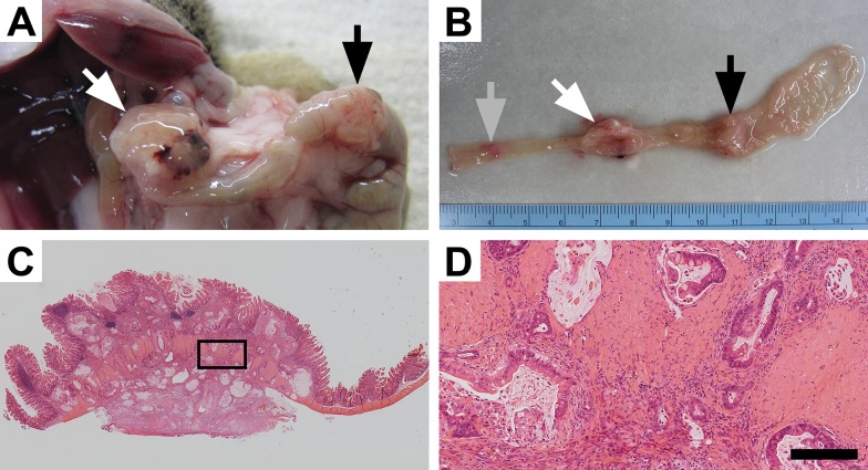
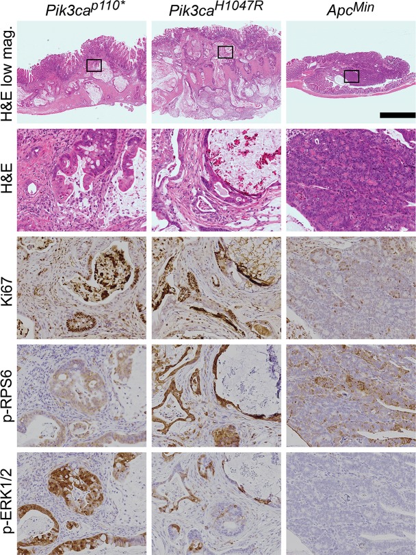
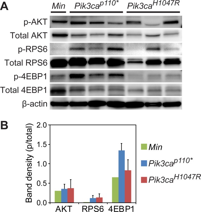
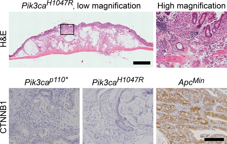
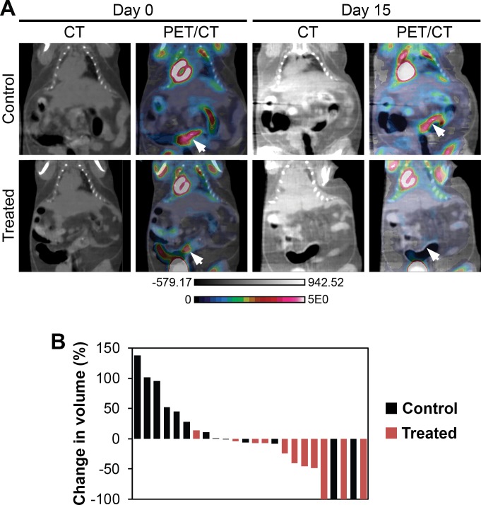
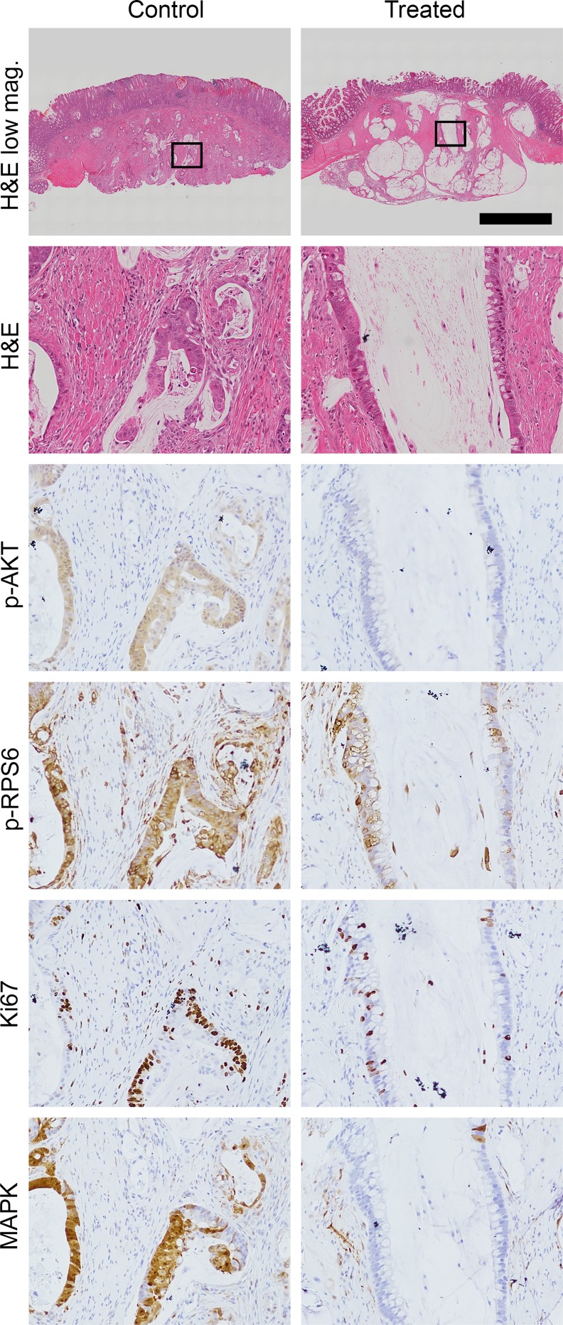
The phosphoinositide 3-kinase (PI3K) signaling pathway is critical for multiple important cellular functions, and is one of the most commonly altered pathways in human cancers. We previously developed a mouse model in which colon cancers were initiated by a dominant active PI3K p110-p85 fusion protein. In that model, well-differentiated mucinous adenocarcinomas developed within the colon and initiated through a non-canonical mechanism that is not dependent on WNT signaling. To assess the potential relevance of PI3K mutations in human cancers, we sought to determine if one of the common mutations in the human disease could also initiate similar colon cancers. Mice were generated expressing the Pik3caH1047R mutation, the analog of one of three human hotspot mutations in this gene. Mice expressing a constitutively active PI3K, as a result of this mutation, develop invasive adenocarcinomas strikingly similar to invasive adenocarcinomas found in human colon cancers. These tumors form without a polypoid intermediary and also lack nuclear CTNNB1 (β-catenin), indicating a non-canonical mechanism of tumor initiation mediated by the PI3K pathway. These cancers are sensitive to dual PI3K/mTOR inhibition indicating dependence on the PI3K pathway. The tumor tissue remaining after treatment demonstrated reduction in cellular proliferation and inhibition of PI3K signaling.


Conflict of interest statement

Competing Interests: The authors have declared that no competing interests exist.

Figures

Fig 1
Fig 1. Mutant PI3K can lead to colon cancer development.
Approximately 60% of Fc+ Pik3caH1047R mice develop tumors within the colon which can result in these mice becoming moribund. At necropsy large colon tumors are found extending through the colonic wall (A). Upon resection, multiple lesions can be identified within the colon and can be over 1 cm in size (A and B). Following histological sectioning, H&E staining demonstrates that these lesions are invasive mucinous adenocarcinomas of the colon without a predominant intra-luminal component (C). Higher magnification demonstrates an abundant desmoplastic reaction with surrounding mucin lakes lined with epithelial cancer cells (D). D size bar = 200 μm.
Fig 2
Fig 2. Fc+ Pik3caH1047R colon cancers are similar to those in Fc+ Pik3cap110* mice.
In both of these Pik3ca mutant models, deeply invasive cancers are seen with the vast majority of the tumors having penetrated below the muscularis mucosa. This is in contrast to ApcMin colon tumors, which typically are adenomatous tumors with no or just superficial invasion. Abundant mucin is present within both the Fc+ Pik3caH1047R and Fc+ Pik3cap110* colon cancers. These Pik3ca mutant cancers also demonstrate increased proliferation, as measured by nuclear Ki67, in comparison to ApcMin tumors. In addition, phosphorylated RPS6 and ERK1/2 are increased above that seen in the ApcMin colon lesions. Scale bar for low magnification images = 1mm. Enlargements are 10x magnifications of the areas outlined in the low magnification images. Min, ApcMin.
Fig 3
Fig 3. The PI3K pathway is activated in ApcMin/+, Fc+ Pik3cap110*, and Fc+ Pik3caH1047R mice.
Immunoblotting demonstrates robust phosphorylation of AKT in ApcMin, Fc+ Pik3cap110* and Fc+ Pik3caH1047R colon tumors (A). Increased phosphorylation of RPS6 and 4EBP1 beyond that seen in the ApcMin lesions is observed in the Fc+ Pik3caH1047R and Fc+ Pik3cap110* colon tumors. The more aggressive phenotype seen in the Fc+ Pik3cap110* mice, with a greater number and decreased latency, is associated with an increased phosphorylation of 4EBP1 compared to Fc+ Pik3caH1047R tumors (B).
Fig 4
Fig 4. Fc+ Pik3caH1047R colon cancers develop through a non-canonical pathway.
Histological examination demonstrates that these cancers are flat without a polypoid component. Low grade dysplasia (tubular adenoma) has not been identified in these or the Fc+ Pik3cap110* mice. At higher magnification, malignant glands above the muscularis mucosa can be identified which appear to be originating from the crypt bases without identification of surface low grade dysplasia. CTNNB1 staining demonstrates that nuclear CTNNB1 is absent in both of the Pik3ca mutant models, but is present in the ApcMin controls. Scale bar for low magnification image = 1mm. High magnification H&E image is a 6x magnification of the area indicated in the image to the left. Scale bar for CTNNB1 images = 100μm.
Fig 5
Fig 5. Dual PI3K/mTOR inhibition induces treatment responses in Fc+ Pik3caH1047R colon cancers.
Fc+ Pik3caH1047R mice were treated with NVP-BEZ235 (35mg/kg/day) or control once daily by oral gavage for 14 days. These mice underwent dual hybrid 18F-FDG PET/CT imaging at baseline and then 24 hours following the last dose of study drug (A, arrows denote tumors pre- and post-treatment). A significant reduction in tumor volume, as measured on the CT images, was detected in the NVP-BEZ235 group (p = 0.008; B). In addition, there was a trend for a slight decrease in the median SUV for those cancers treated with the PI3K/mTOR inhibitor. In one control mouse, two tumors were observed on baseline imaging, but not detected on follow-up PET/CT imaging.
Fig 6
Fig 6. Histological examination demonstrates a significant treatment effect following dual PI3K/mTOR inhibition of Fc+ Pik3caH1047R cancers.
In comparison to controls, a dramatic treatment effect was identified on H&E staining: loss of epithelial cells surrounding the areas of cystic change/degeneration and areas of increased fibrosis. A decrease in Ki67 staining and phosphorylation of AKT, RPS6 and ERK1/2 are also seen. Scale bar for low magnification images = 1mm. High magnification panels are 10x magnifications of the areas indicated by the rectangles.

References

    1. Goss KH, Groden J. Biology of the adenomatous polyposis coli tumor suppressor. J Clin Oncol. 2000;18:1967–79. - PubMed
    1. Kinzler KW, Vogelstein B. Cancer-susceptibility genes. Gatekeepers and caretakers. Nature. 1997;386:761, 3. - PubMed
    1. Segditsas S, Tomlinson I. Colorectal cancer and genetic alterations in the Wnt pathway. Oncogene. 2006;25:7531–7. - PubMed
    1. Fearon ER, Vogelstein B. A genetic model for colorectal tumorigenesis. Cell. 1990;61:759–67. - PubMed
    1. Markowitz SD, Bertagnolli MM. Molecular origins of cancer: Molecular basis of colorectal cancer. N Engl J Med. 2009;361:2449–60. 10.1056/NEJMra0804588 - DOI - PMC - PubMed

Publication types

MeSH terms

LinkOut - more resources