Review
. 2016:78:437-61.
doi: 10.1146/annurev-physiol-021115-105412.

Vascular Growth Factors and Glomerular Disease

Affiliations
Review

Vascular Growth Factors and Glomerular Disease

Christina S Bartlett et al. Annu Rev Physiol. 2016.

Abstract

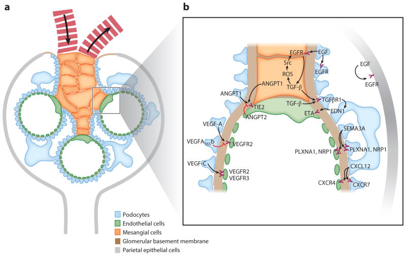
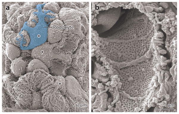
The glomerulus is a highly specialized microvascular bed that filters blood to form primary urinary filtrate. It contains four cell types: fenestrated endothelial cells, specialized vascular support cells termed podocytes, perivascular mesangial cells, and parietal epithelial cells. Glomerular cell-cell communication is critical for the development and maintenance of the glomerular filtration barrier. VEGF, ANGPT, EGF, SEMA3A, TGF-β, and CXCL12 signal in paracrine fashions between the podocytes, endothelium, and mesangium associated with the glomerular capillary bed to maintain filtration barrier function. In this review, we summarize the current understanding of these signaling pathways in the development and maintenance of the glomerulus and the progression of disease.

Keywords: VEGF; angiopoietin; endothelial cell; podocyte.


Figures

Figure 1
Figure 1
Schematic of glomerular structure and signaling. (a) A mature glomerulus in cross section. Fewer capillary loops than normal are shown for clarity, and the image is not to scale. The four major cell types of the glomerulus are podocytes, mesangial cells, endothelial cells, and parietal epithelial cells. The glomerulus has a network of capillary loops with mesangial cells forming a nexus at the base of the capillary network. The glomerular basement membrane lies between the podocytes and the endothelial cells and divides the glomerulus into an inner compartment containing capillaries and mesangial cells and an outer one containing podocytes and Bowman’s space, into which the filtrate passes. The arrows in the capillaries indicate the flow of blood into and out of the glomerulus. (b) Summary of signaling pathways between the different cellular compartments of the glomerulus discussed in this review. Abbreviations: ANGPT1, angiopoietin 1; ANGPT2, angiopoietin 2; CXCL12, C-X-C chemokine ligand 12; CXCR, C-X-C chemokine receptor; EDN1, endothelin-1; EGF, epidermal growth factor; EGFR, epidermal growth factor receptor; ETA, endothelin-1 receptor A; NRP1, neuropilin-1; PLXNA1, plexin-A1; ROS, reactive oxygen species; SEMA3A, semaphorin 3A; Src, Src tyrosine kinase; TGF-β, transforming growth factor-β; TGFβR1, transforming growth factor-βreceptor 1; VEGF, vascular endothelial growth factor; VEGFR, vascular endothelial growth factor receptor.
Figure 2
Figure 2
Scanning electron micrographs of mouse glomeruli. (a) View from the urinary space showing several capillary loops and podocyte cell bodies (marked by asterisks) with their foot processes wrapping around capillaries. (b) View from the capillary lumen showing a fenestrated glomerular capillary.
Figure 3
Figure 3
Schematic of glomerular development. Glomerular development is generally described in five steps: (1) vesicle, (2) comma-shaped body, (3) S-shaped body, (4) glomerular capillary loop stage, and (5) mature glomerulus. During the capillary loop stage, presumptive podocytes express VEGF-A, which induces the migration of VEGFR2-positive endothelial cell precursors in the renal mesenchyme. Endothelial cells migrate into the vascular cleft and proliferate and differentiate in intimate association with VEGF-A-producing podocytes. Mesangial cells express PDGFRβ and are attracted into the developing glomerular tuft by PDGF-β-expressing glomerular endothelial cells. Connections to the tubule system were omitted for clarity. Abbreviations: PDGF-β, platelet-derived growth factor-β; PDGFRβ, platelet-derived growth factor-βreceptor; VEGF, vascular endothelial growth factor; VEGFR, vascular endothelial growth factor receptor.

References

    1. Haraldsson B, Nystrom J, Deen WM. Properties of the glomerular barrier and mechanisms of proteinuria. Physiol Rev. 2008;88:451–87. - PubMed
    1. Herrera GA. Plasticity of mesangial cells: a basis for understanding pathological alterations. Ultrastruct Pathol. 2006;30:471–79. - PubMed
    1. Ohyama K, Seyer JM, Raghow R, Kang AH. Extracellular matrix phenotype of rat mesangial cells in culture. Biosynthesis of collagen types I, III, IV, and V and a low molecular weight collagenous component and their regulation by dexamethasone. J Lab Clin Med. 1990;116:219–27. - PubMed
    1. Johnson RJ, Floege J, Yoshimura A, Iida H, Couser WG, Alpers CE. The activated mesangial cell: a glomerular “myofibroblast”? J Am Soc Nephrol. 1992;2:S190–97. - PubMed
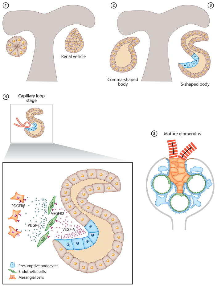
    1. Abrahamson DR. Glomerulogenesis in the developing kidney. Semin Nephrol. 1991;11:375–89. - PubMed

Publication types

MeSH terms

Substances