. 2016 Feb 11:6:20213.
doi: 10.1038/srep20213.

Tofla virus: A newly identified Nairovirus of the Crimean-Congo hemorrhagic fever group isolated from ticks in Japan

Affiliations

Tofla virus: A newly identified Nairovirus of the Crimean-Congo hemorrhagic fever group isolated from ticks in Japan

Satoshi Shimada et al. Sci Rep. .

Abstract

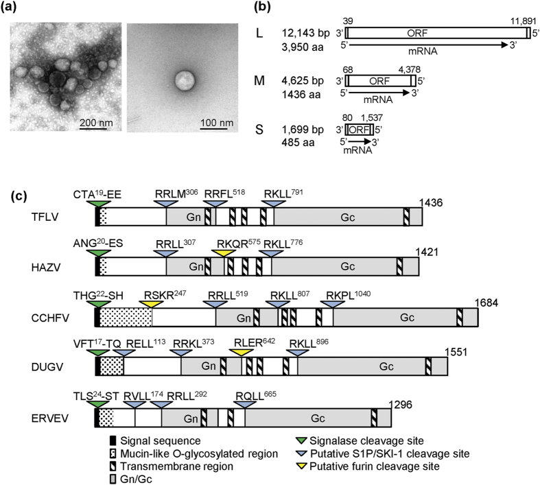
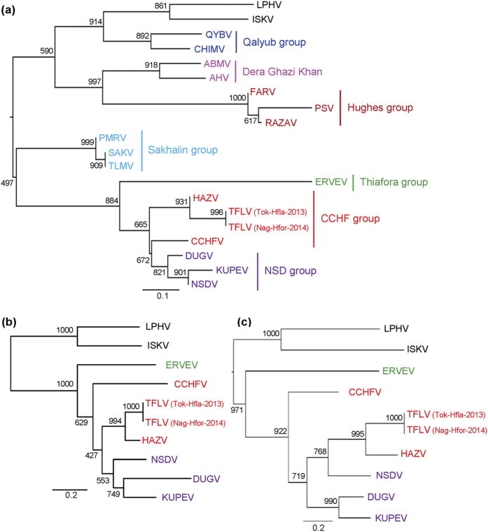
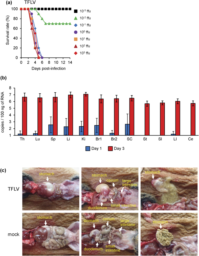
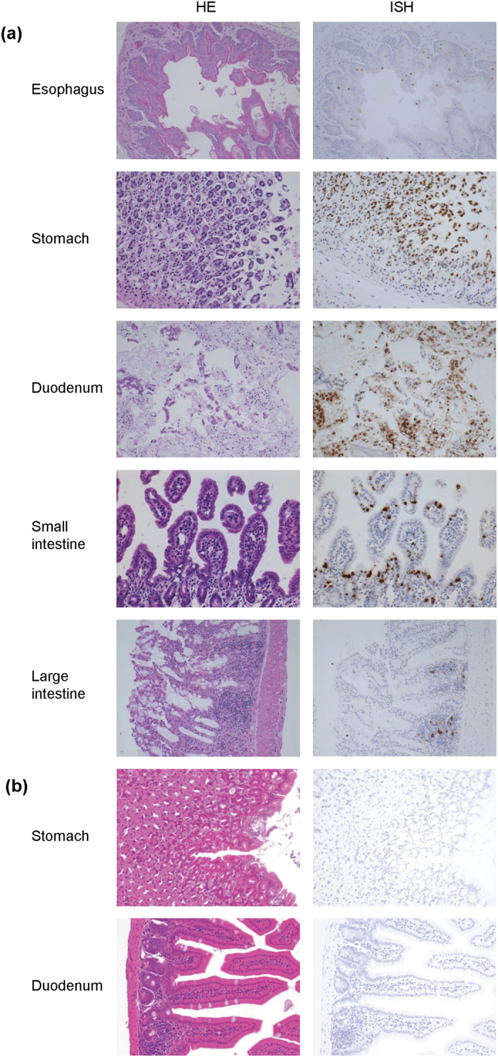
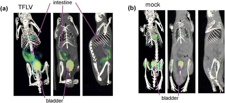
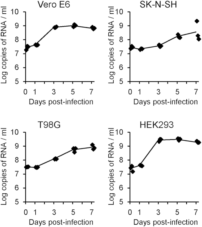
Ixodid ticks transmit several important viral pathogens. We isolated a new virus (Tofla virus: TFLV) from Heamaphysalis flava and Heamaphysalis formsensis in Japan. The full-genome sequences revealed that TFLV belonged to the genus Nairovirus, family Bunyaviridae. Phylogenetic analyses and neutralization tests suggested that TFLV is closely related to the Hazara virus and that it is classified into the Crimean-Congo hemorrhagic fever group. TFLV caused lethal infection in IFNAR KO mice. The TFLV-infected mice exhibited a gastrointestinal disorder, and positron emission tomography-computed tomography images showed a significant uptake of (18)F-fluorodeoxyglucose in the intestinal tract. TFLV was able to infect and propagate in cultured cells of African green monkey-derived Vero E6 cells and human-derived SK-N-SH, T98-G and HEK-293 cells. Although TFLV infections in humans and animals are currently unknown, our findings may provide clues to understand the potential infectivity and to develop of pre-emptive countermeasures against this new tick-borne Nairovirus.


Figures

Figure 1
Figure 1
(a) Electron micrograph of TFLV. (b) Schematic diagram of three segments (L, M and S) of TFLV. (c) Schematic diagram of cleavage sites in the glycoproteins of the genus Nairovirus. HAZV, Hazara virus; CCHFV, Crimean–Congo hemorrhagic fever virus; DUGV, Dugbe virus; ERVEV, Erve virus.
Figure 2
Figure 2. Phylogenetic trees of the genus Nairovirus.
The phylogenetic trees were constructed based on the conserved amino acid sequences (a) Phylogenetic tree based on the L segment (amino acids 2218–2535 in the L segment of TFLV) using the substitution model LG + I + G. (b) Phylogenetic tree based on the M segment (amino acids 791–948 in TFLV) using the substitution model LG + G. (c) Phylogenetic tree based on the sequences of the S segment (2–179 of TFLV) using the substitution model LG + G. Nairoviruses are designated by the following abbreviations: ABHV, Abu Hammad virus; AMV, Abu Mina virus; CCHFV, Crimean–Congo hemorrhagic fever virus; CHIMV, Chim virus; DUGV, Dugbe virus; ERVEV, Erve virus; HAZV, Hazara virus; ISKV, Issyk-Kul virus; KUPEV, Kupe virus; LPHV, Leopards Hill virus; NSDV, Nairobi sheep disease virus; PMRV, Paramushir virus; PSV, Punte Salinas virus; QYBV, Qalyub virus; RAZAV, Raza virus; SAKV, Sakhalin virus; and TLMV, Tillamok virus.
Figure 3
Figure 3
(a) Survival curves of A129 mice (n = 10) subcutaneously inoculated with 10−3 and 103 ffu of TFLV (Tok-Hfla-2013). The mice were observed for 14 days, and no mice died after 14 days. (b) Viral loads in the tissues of A129 mice infected with 102 ffu of TFLV (Tok-Hfla-2013). The copy numbers in the thymus (Th), lung (Lu), spleen (Sp), liver (Li), kidney (Ki), brain cortex (Br1) and non-cortex (Br2), spinal cord (SC), stomach (St), small intestine (SI), large intestine (LI) and cecum (Ce) of mice 1- and 3-days post-infection were determined using real-time RT-PCR (n = 5). (c) Gross pathology and viral loads of A129 mice inoculated with TFLV (Tok-Hfla-2013). Internal organs of TFLV-and mock-infected A129 3 days pi.
Figure 4
Figure 4. Histological and ISH features of the gastrointestinal tract.
TFLV (Tok-Hfla-2013)-infected (a) and mock-infected (b) A129 mice were dissected at 3 days pi. Left panels: hematoxylin and eosin. Right panels: ISH using AT-tailed sense and antisense cocktail probes (positive signals are labeled in brown). The gastric and duodenal mucosae highly involved by the viral infection show marked erosive change. Non-eroded small intestinal mucosa reveal positive signals in both the crypts and villi. Some keratinocytes in the esophageal mucosa are also involved. In the large bowel mucosa, the germinal center of the lymphoid follicles represents the site of infection. The gastroduodenal mucosa in mock-infected control animals demonstrates normal histology without viral infection.
Figure 5
Figure 5. Representative whole body PET/CT images.
TFLV (Tok-Hfla-2013)-infected (a) and mock-infected (b) A129 mice were observed at 3 days pi (n = 4). The PET/CT images were acquired 30–60 min after intravenous injection of 18F-FDG (10 MBq in 400 μl of saline per mouse).
Figure 6
Figure 6. TFLV (Tok-Hfla-2013) propagation in the Vero E6, SK-N-SH, T98-G and HEK-293 cells.
CFs were harvested at 0, 1, 3, 5 and 7 days pi. Growth curves in the CFs are indicated by the viral RNA copy numbers.

References

    1. Lani R. et al. Tick-borne viruses: a review from the perspective of therapeutic approaches. Ticks and tick-borne diseases 5, 457–465, 10.1016/j.ttbdis.2014.04.001 (2014). - DOI - PubMed
    1. Butenko A. M., Gromashevsky V. L., L’Vov, D. K. & Popov V. F. First isolations of Barur virus (Rhabdoviridae) from ticks (Acari: Ixodidae) in Africa. J Med Entomol 18, 232–234 (1981). - PubMed
    1. Davies C. R., Jones L. D. & Nuttall P. A. Experimental studies on the transmission cycle of Thogoto virus, a candidate orthomyxovirus, in Rhipicephalus appendiculatus. Am J Trop Med Hyg 35, 1256–1262 (1986). - PubMed
    1. Bente D. A. et al. Crimean-Congo hemorrhagic fever: history, epidemiology, pathogenesis, clinical syndrome and genetic diversity. Antiviral Res 100, 159–189, 10.1016/j.antiviral.2013.07.006 (2013). - DOI - PubMed
    1. Yu X. J. et al. Fever with thrombocytopenia associated with a novel bunyavirus in China. N Engl J Med 364, 1523–1532, 10.1056/NEJMoa1010095 (2011). - DOI - PMC - PubMed

Publication types

MeSH terms

Substances