. 2016 Feb 11;530(7589):233-6.
doi: 10.1038/nature16936.

Structure- and function-based design of Plasmodium-selective proteasome inhibitors

Affiliations

Structure- and function-based design of Plasmodium-selective proteasome inhibitors

Hao Li et al. Nature. .

Abstract

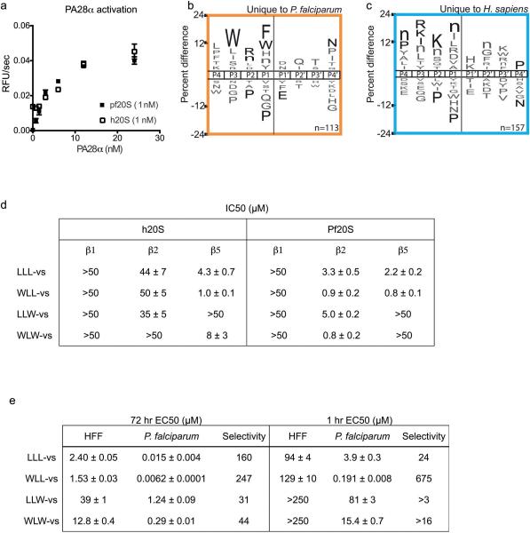
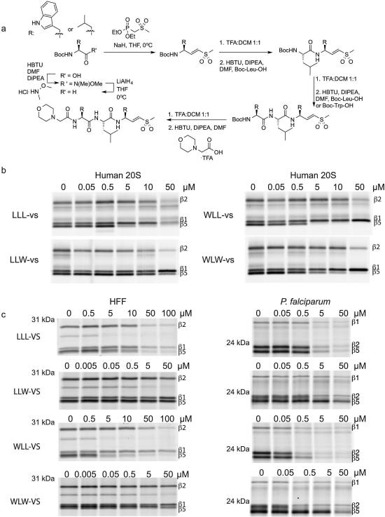
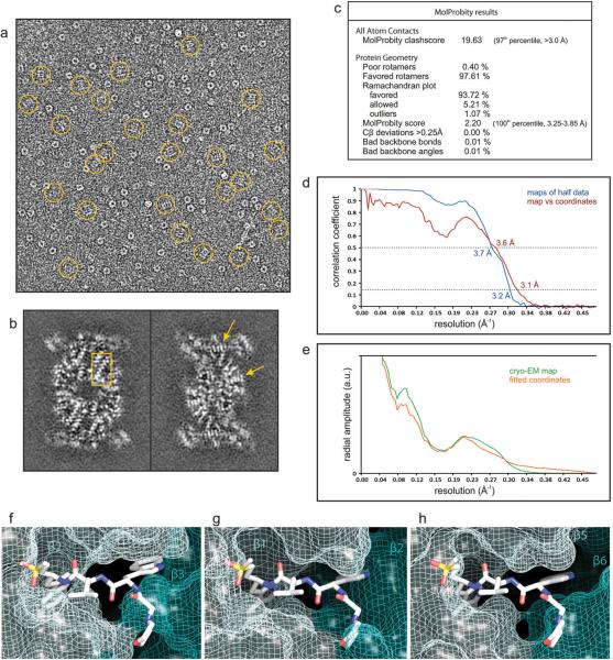
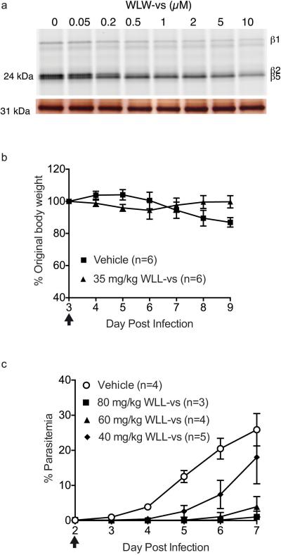
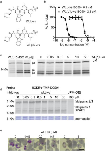
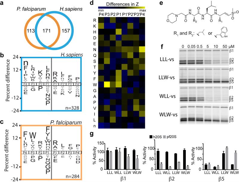
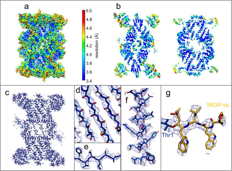
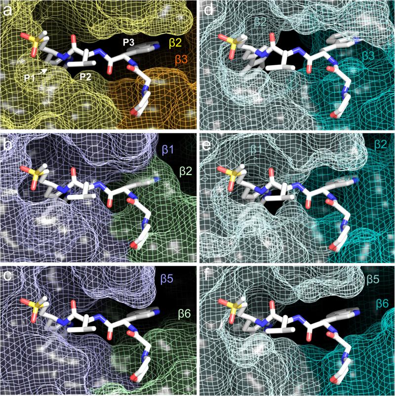
The proteasome is a multi-component protease complex responsible for regulating key processes such as the cell cycle and antigen presentation. Compounds that target the proteasome are potentially valuable tools for the treatment of pathogens that depend on proteasome function for survival and replication. In particular, proteasome inhibitors have been shown to be toxic for the malaria parasite Plasmodium falciparum at all stages of its life cycle. Most compounds that have been tested against the parasite also inhibit the mammalian proteasome, resulting in toxicity that precludes their use as therapeutic agents. Therefore, better definition of the substrate specificity and structural properties of the Plasmodium proteasome could enable the development of compounds with sufficient selectivity to allow their use as anti-malarial agents. To accomplish this goal, here we use a substrate profiling method to uncover differences in the specificities of the human and P. falciparum proteasome. We design inhibitors based on amino-acid preferences specific to the parasite proteasome, and find that they preferentially inhibit the β2-subunit. We determine the structure of the P. falciparum 20S proteasome bound to the inhibitor using cryo-electron microscopy and single-particle analysis, to a resolution of 3.6 Å. These data reveal the unusually open P. falciparum β2 active site and provide valuable information about active-site architecture that can be used to further refine inhibitor design. Furthermore, consistent with the recent finding that the proteasome is important for stress pathways associated with resistance of artemisinin family anti-malarials, we observe growth inhibition synergism with low doses of this β2-selective inhibitor in artemisinin-sensitive and -resistant parasites. Finally, we demonstrate that a parasite-selective inhibitor could be used to attenuate parasite growth in vivo without appreciable toxicity to the host. Thus, the Plasmodium proteasome is a chemically tractable target that could be exploited by next-generation anti-malarial agents.


Figures

Extended Data Figure 1
(a-c) Substrate cleavage profile of activated human and P. falciparum 20S proteasome. a, Activation of the human and P. falciparum 20S proteasome by human PA28α. Activity was determined by cleavage of the fluorogenic substrate Suc-LLVY-amc. Error bars represent s.d. and n=3 purified proteasome in technical replicates. b and c, iceLogos of cleavage sequences that are uniquely processed by either the P. falciparum (b) or human proteasome (c). Amino acids that are most and least favored at each position are shown above and below the axis, respectively. Lowercase ‘n’ corresponds to norleucine and amino acids in black text are statistically significant (p < 0.05, unpaired two-tailed Student's t-test). (d-e) Inhibition potencies of the vinyl sulfone inhibitors. d, Table of IC50 values for each inhibitor in 1 hr P. falciparum and human 20S proteasome. IC50 values are determined from 3 independent experiments of inhibitor pretreatment followed by activity labeling of the 20S proteasome (n=3 purified proteasome). Gels in Fig. 1f and Extended Data Figure 2b were quantified to calculate the IC50 values (for gel source data and replicates, see Supplementary Fig. 1 a-b). Data is mean ± s.d. e, Table of EC50 values for each of the inhibitors in 1 hr and 72 hr treatment of P.falciparum at ring stage or non-confluent human foreskin fibroblasts (HFF). Data is mean ± s.d. n=6 parasite cultures from 2 independent experiment of triplicates for P. falciparum treatments. n=9 cell cultures from 3 independent experiments of triplicates for HFF treatment, except for 1hr WLW-vs, 1hr LLW-vs and 72 hr LLL-vs, where n=6 cell cultures from 2 independent experiment of triplicates.
Extended Data Figure 2
Proteasome inhibitors preferentially inhibit β2 of the P. falciparum proteasome. a, Vinyl sulfone inhibitors are synthesized from the Boc-protected amino acid by first generating the Weinreb amide, followed by the Horner-Wadsworth-Emmons reaction and standard peptide coupling. b, Purified human 20S proteasome was pre-treated for 1 hr at 37°C with each inhibitor followed by addition of activity based probe MV151 to assess for human proteasome activities (for gel source data, see Supplementary Fig. 1b). c, Human foreskin fibroblasts (HFF) or P. falciparum culture was treated for 1 hr at 37°C with each inhibitor, followed by compound washout and post-lysis activity based probe labeling. Gel shown for WLL-vs in P.falciparum is derived from Fig 4c at the indicated concentrations to allow for direct comparison with other compounds (for gel source data, see Supplementary Fig. 1e).
Extended Data Figure 3
(a-e) Evaluation of the single particle analysis of the P. falciparum 20S proteasome core bound to the inhibitor WLW-vs. a, cryo-EM image of the sample analyzed, with molecular images of side views of the complex (normal to its long axis) indicated by rings. The image greyscale was inverted in order to show the protein densities in white. b, Individual sections of the 3D map, as determined by the 3D reconstruction algorithm (without further sharpening, masking or Fourier filtering), are represented as grey scale. These sections are 1 Å thick and reveal the quality of the reconstruction, as the protein densities are clearly resolved against a very smooth background, with regions showing the pattern of α-helices (box) and the clear separation of sheet forming β strands (arrows) indicated. c, Evaluation of the model of the P. falciparum 20S proteasome core using MolProbity. d, Resolution estimate of the cryo-EM map by Fourier shell correlation. The curves correspond to the correlation obtained against the protein model (red) and the correlation between maps determined from two halves of the data (blue). The resolution was estimated from the curve against the model where the 0.5 correlation coefficient criterion yields an estimate of 3.6 Å. The correlation coefficient can be seen to fall to a local minimum at ~6 Å and then recover at higher resolutions for both FSC curves. This behavior is consistent with the rotationally averaged amplitude spectra of both the cryo-EM map and the coordinates (e). This region of the amplitude spectra contains reduced structural information, typical of protein scattering, indicating that these effects in the FSC curves arise from a genuine local reduction in the signal:noise ratio. (f-h) Accessibility of the human 20S proteasome active sites to the inhibitor WLW-vs, using the protein model of the human proteasome core complex bound to a LLL-vs inhibitor. Protein coordinates of the human proteasome 20S core (PDB accession code 5a0q) β2 (f), β1 (g) and β5 (h) active sites were aligned to the coordinates of the P. falciparum proteasome β2 subunit bound to the WLW-vs inhibitor. The model of the human 20S proteasome active sites is represented as van der Waals surfaces with the superimposed WLW-vs inhibitor shown as sticks.
Extended Data Figure 4
Intact cell treatment and in vivo treatment of vinyl sulfone inhibitors. a, WLW-vs was incubated in early trophozoite P. falciparum culture for 3 hrs, washed out and the parasite lysate was incubated with probe BMV037. Top gel shows the fluorescent scan and bottom gel shows the silver stain. For gel source data, see Supplementary Fig. 1f. b, Body weight of WLL-vs and vehicle treated Balb/c mice after compound treatment via tail vein injection (Figure 4 e) and is expressed as a percentage of the original body weight on Day 3 before compound treatment. Body weight of vehicle treated mice decreased after day 6 of infection as part of the response to the natural resolution of the P. chabaudi infection. Treatment day is indicated by arrow. n= 6 mice for each group and error bars represent s.d. e, Balb/c female mice infected with 1 million P. chabaudi parasites from passage host on Day 0 were treated with a single bolus dose of vehicle (45% polyethylene glycol (M.W. 400), 35% propylene glycol, 10% ethanol, 10% DMSO and 10% w/v 2-hydroxyproyl-β-cyclodextrin; n=4 mice) or WLL-vs at 40 mg/kg (n= 5 mice), 60 mg/kg( n=4 mice) and 80 mg/kg (n=3 mice) formulated in the vehicle. Treatment was performed on Day 2 post infection as indicated by the arrow and administered by intra-peritoneal injection. Parasitemia was monitored daily by Giemsa stain of thin blood smears. Error bars represent s.d.
Extended Data Figure 5
Assessing off-target activities of WLL-vs a. Structures of WLL-vs and its diastereomer WL(d)L-vs. b, Dose response curves of WLL-vs and WL(d)L-vs after 72 hr treatment in P. falciparum. Error bars represent s.d. (n=6 parasite cultures for WLL-vs from triplicates of 2 independent experiments and n=8 parasite cultures for WL(d)L-vs over 3 independent experiments) . c, Purified P. falciparum 20S proteasome was treated for 1 hr at 37°C with 10 μM of WLL-vs and WL(d)L-vs (left) or a range of concentrations of WL(d)L-vs. Residual activity was assessed by probe BMV037 (for gel source data, see Supplementary Fig. 1g). d, A mixed-stage culture of P. falciparum was treated for 1 hr with WLL-vs at 37°C, followed by BODIPY-TMRDCG04 for a further 1 hr. Samples were directly loaded onto the SDS-PAGE for analysis. 100 μM JPM-OEt was included as positive control. The fluorescent scan is shown on top and the coomassie stain is shown at the bottom. For gel source data, see Supplementary Fig. 1h). e, Geimsa stain of 1 hr treated P.falciparum ring 24 hr after inhibitor was added. Scale bar: 600 μm.
Figure 1
Figure 1
Substrate profile of the activated human and P. falciparum 20S proteasome guides inhibitor design. a, Total number of cleavage sites detected after 4 hr incubation of the activated human and P. falciparum proteasome with the peptide library. The iceLogos generated from the cleavages are shown in (b) for human and (c) for P. falciparum proteasome. Amino acids that are most and least favored at each position are shown above and below the axis, respectively. Lowercase ‘n’ corresponds to norleucine and amino acids in black text are statistically significant (p < 0.05, unpaired two-tailed Student's t-test). d, The Z-score for amino acid at each position (P4-P4′) was calculated for both human and parasite proteasome based on the cleavages in a, and the difference between the Z-scores is shown as a heatmap. e, Inhibitors are designed by substituting Trp at either P1 and/or P3 position in the morpholino-capped tri-leucine vinyl sulfone. f, Inhibition of purified P. falciparum 20S as assessed by activity based probe labeling. The same experiment was repeated for the human 20S proteasome (Extended Data Figure 2b). g, Activity of each subunit in human or P. falciparum proteasome after 10 μM inhibitor treatment was determined by image quantification of the intensity of probe labeling and normalized to mock treated control. Error bars represent standard deviation (s.d.) and n=3 purified proteasome from 3 independent experiments (for gel source data, see Supplementary Fig. 1a and b).
Figure 2
Figure 2
Structure of the P. falciparum 20S proteasome core bound to the inhibitor WLW-vs, determined by cryo-EM and single particle analysis. a, The cryo-EM map is surface rendered and color coded according to local resolution as determined by ResMap. b, Sections of the map as shown in a, revealing the higher resolution at the internal regions of the complex, compared with solvent exposed surfaces. c, Section of the cryo-EM map, shown as mesh representation, showing the overall fitting of the protein model. d-g, Detailed views of the map showing clear separation of sheet forming β strands (d), good recovery of side chains, both in β strands (e) and α helices (f), and the densities for the WLW-vs inhibitor at the proteasome β2 active site (g).
Figure 3
Figure 3
Structural comparison of the P. falciparum and human proteasome 20S core active sites. a, Coordinates of the P. falciparum proteasome β2 active site bound to WLW-vs. The inhibitor P1, P2 and P3 positions are indicated. b-c, Coordinates of the P. falciparum proteasome β1 (b) and β5 (c) active sites with superimposed coordinates of the WLW-vs inhibitor, as shown in (a). d-f, Coordinates of the apo human proteasome 20S core (PDB accession code 4R3O) β2 (d), β1 (e) and β5 (f) active sites, with superimposed coordinates of the WLW-vs inhibitor, as shown in (a). In all panels the protein is represented as van der Waals surfaces and the inhibitor as sticks.
Figure 4
Figure 4
Exploiting differences in the β2 subunits of the two proteasome species for selective parasite killing. a-b, β2 subunit selective inhibitor WLW-vs can synergize with DHA treatment in ART-resistant P. falciparum. a, Viability 72 hr after a 1 hr treatment of ART sensitive (PL2) and resistant (PL7) parasites at early ring stage (2 hr post-invasion, n=2 parasite cultures) with WLW-vs (abbreviated as WLW). b, Left panel shows the dose-response to DHA in the absence and presence of sublethal doses of WLW-vs. Right panel shows the isobologram of the two inhibitors at the 50% lethal dose (LD50). PL7 parasites were treated for 3 hr at early ring stage (4 hr post-invasion). a-b, data shown are for individual data points that is representative of two technical replicates. c-e, WLL-vs co-inhibits β2 along with the other two catalytic subunits, resulting in potent parasite killing. c, Human foreskin fibroblasts (HFF) or P. falciparum schizonts were treated with WLL-vs for 1 hr, and activity based probe labeling was performed post-lysis (for gel source data, see Supplementary Fig. 1c and d). Fluorescent gel scan (top) and silver stain (bottom) are shown. d, Non-confluent, replicating HFFs (n=9 cell culture, from 3 independent experiments of triplicates) or P. falciparum at ring stage (n=6 parasite cultures, from 2 independent experiments of triplicates) was pulse-treated for 1 hr with WLL-vs and viability was determined after 71 hr. Activities of the P. falciparum proteasome subunits after inhibitor treatment were determined as described in methods. (n= 3 parasite lysates from 3 independent experiments). Error bars represent s.d. e, Balb/c female mice were infected with P. chabaudi and treated with a single I.V. dose of vehicle (n=6 mice) or 35 mg/kg WLL-vs (n=6 mice) on day 3 post-infection (arrow). Parasitemia and weight of the mice were monitored daily until the infection was resolved naturally by the host (day 7 onwards). Error bars represent s.d.

Comment in

References

    1. Voges D, Zwickl P, Baumeister W. The 26S proteasome: a molecular machine designed for controlled proteolysis. Annu. Rev. Biochem. 1999;68:1015–1068. - PubMed
    1. Gantt SM, et al. Proteasome inhibitors block development of Plasmodium spp. Antimicrob. Agents Chemother. 1998;42:2731–2738. - PMC - PubMed
    1. Czesny B, Goshu S, Cook JL, Williamson KC. The Proteasome Inhibitor Epoxomicin Has Potent Plasmodium falciparum Gametocytocidal Activity. Antimicrob. Agents Chemother. 2009;53:4080–4085. - PMC - PubMed
    1. Kreidenweiss A, Kremsner PG, Mordmüller B. Comprehensive study of proteasome inhibitors against Plasmodium falciparum laboratory strains and field isolates from Gabon. Malar. J. 2008;7:187. - PMC - PubMed
    1. Prudhomme J, et al. Marine Actinomycetes: A New Source of Compounds against the Human Malaria Parasite. PLoS ONE. 2008;3:e2335. - PMC - PubMed

Extended Data References

    1. Verdoes M, et al. A Fluorescent Broad-Spectrum Proteasome Inhibitor for Labeling Proteasomes In Vitro and In Vivo. Chem. Biol. 2006;13:1217–1226. - PubMed
    1. Chen VB, et al. MolProbity: all-atom structure validation for macromolecular crystallography. Acta Crystallographica Section D. 2010;66:12–21. - PMC - PubMed
    1. Rosenthal PB, Henderson R. Optimal determination of particle orientation, absolute hand, and contrast loss in single-particle electron cryomicroscopy. J Mol Biol. 2003;333:721–745. - PubMed
    1. da Fonseca PCA, Morris EP. Cryo-EM reveals the conformation of a substrate analogue in the human 20S proteasome core. Nat Commun. 2015;6:7573–7576. - PMC - PubMed

Publication types

MeSH terms

Associated data