Review
. 2016 Mar;270(1):8-19.
doi: 10.1111/imr.12396.

Regulation of germinal center B-cell differentiation

Affiliations
Review

Regulation of germinal center B-cell differentiation

Yang Zhang et al. Immunol Rev. 2016 Mar.

Erratum in

  • Corrigendum.
    [No authors listed] [No authors listed] Immunol Rev. 2016 Jul;272(1):202. doi: 10.1111/imr.12442. Immunol Rev. 2016. PMID: 27319352 Free PMC article. No abstract available.

Abstract

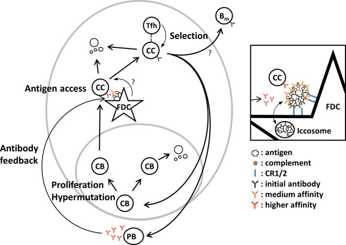
Germinal centers (GC) are the main sites where antigen-activated B-cell clones expand and undergo immunoglobulin gene hypermutation and selection. Iterations of this process will lead to affinity maturation, replicating Darwinian evolution on the cellular level. GC B-cell selection can lead to four different outcomes: further expansion and evolution, apoptosis (non-selection), or output from the GC with differentiation into memory B cells or plasma cells. T-helper cells in GC have been shown to have a central role in regulating B-cell selection by sensing the density of major histocompatibility complex (MHC):peptide antigen complexes. Antigen is provided on follicular dendritic cells in the form of immune complex. Antibody on these immune complexes regulates antigen accessibility by shielding antigen from B-cell receptor access. Replacement of antibody on immune complexes by antibody generated from GC-derived plasma cell output will gradually reduce the availability of antigen. This antibody feedback can lead to a situation where a slow rise in selection stringency caused by a changing environment leads to directional evolution toward higher affinity antibody.

Keywords: B-cell selection; Tfh cells; affinity maturation; cytokines; germinal center; immune complex.


Figures

Figure 1
Figure 1
Entry and disappearance of IgM a after immunization with anti‐ NP IgM a NP CGG immune complex. Following injected IgMa over the first 5 days after immunization, carrier primed IgMb‐expressing C57BL/6 mice were immunized with low affinity anti‐NP IgMaNPCGG immune complex. (A) B‐cell follicle and surrounding area before injection showing T zone (TZ), IgD on B cell follicles (F) is stained in brown, surrounded by marginal zone (MZ). (B) IgMa‐containing immune complex (blue) is easily detectable in marginal sinus and follicle 2 h after injection, and (C) localizes on FDCs in the follicle center within 24 h. (D) Three days post‐immunization GCs have formed in the follicle center containing lacy IgMa‐containing immune complex on the FDC network. (E) Five days after immunization the originally injected low affinity anti‐NP IgMa is undetectable. (F) An adjacent section of the same germinal center shows strong staining for endogenously produced IgMb (blue, right) on the FDC networks and IgMb producing extrafollicular plasma cells. Scale bar = 50 μm. Further experimental detail described in 90.
Figure 2
Figure 2
Affinity dependence of antibody retention in immune complexes on follicular dendritic cells. Five days post immunization using a protocol similar to Fig. 1, but with immune complexes containing anti‐NP IgM of Low, IntLow and IntHigh affinity. For experimental details, see 90. (A) Low (left), IntLow (middle) or IntHigh (right) affinity anti‐NP IgMa (blue). F: follicle; GC: germinal center. Scale bar: 25 μm. (B) High magnification detail of light zone FDC from the images above, showing punctate iccosome‐like staining remaining 5 days after injection of IntLow immune complex (middle) and iccosome plus dendritic‐like staining after injection of IntHigh immune complex (right). (C) Four parameter confocal microscopy 5 days after IntHigh affinity IgMaIC immunization. Germinal center is outlined with dashed line. Square denotes area magnified in (D)–(G). TZ: T zone; F: follicle; GC: germinal center. (D) Injected IgMa (green) shows mainly punctate iccosome‐like staining. (E) Endogenous IgMb (red) is seen in a dendritic pattern on FDC and on B cells. (F) Double exposure of IgMa and IgMb shows punctate pattern (open arrowheads) mainly in green (IgMa only) and some dendritic staining of immune complexes on FDC (closed arrowheads) in yellow (IgMa+IgMb) and red (IgMb). (G) FDC‐M1 staining (blue) shows dendritic FDC staining and tingible body macrophages. Scale bar: 20 μm.
Figure 3
Figure 3
Differentiation and selection of germinal center B cells. Model for affinity‐based differentiation of B cells in germinal center, modified from 15, 79, including antibody feedback 90. B cells proliferate and hypermutate in the germinal center dark zone as centroblasts (CB). CB with non‐functional receptors may undergo apoptosis. CB migrate to the germinal center light zone differentiating into centrocytes (CC). These are dependent on interaction with antigen held on follicular dendritic cells (FDC). Additional signals from FDC are possible. In order to avoid apoptosis, CC must be able to take up antigen and present this to Tfh cells. Dependent on the amount of antigen presented they will receive differentiation signals from Tfh cells. Differentiation will guide them back into the dark zone CB pool, leading to further expansion and mutation, or differentiation into plasma cells leaving via the germinal center dark zone. Memory B cells may be a further result of this interaction. Germinal center‐derived plasma cells produce antibody of higher affinity that may replace antibody in immune complexes held on FDC, leading to a more stringent barrier for antigen access. Inset: FDC hold immune complex via splice variants of CR2 and Fc receptors (blue). Antigen is initially deposited on FDC complexed with low affinity antibody (black) derived from the early extrafollicular plasma cell response. Over time this is replaced by higher affinity variants (orange and red) that are produced by germinal center output cells. Some of the immune complex cycles through iccosomes, possibly representing long‐term reservoirs of antigen and antibody. Note that published experiments on antibody feedback were performed with IgM 90, not with bivalent antibody as schematically depicted here.

Similar articles

Cited by

References

    1. Tarlinton DM. Evolution in miniature: selection, survival and distribution of antigen reactive cells in the germinal centre. Immunol Cell Biol 2008;86:133–138. - PubMed
    1. Sabouri Z, et al. Redemption of autoantibodies on anergic B cells by variable‐region glycosylation and mutation away from self‐reactivity. Proc Natl Acad Sci USA 2014;111:E2567–E2575. - PMC - PubMed
    1. Regner M. Cross‐reactivity in T‐cell antigen recognition. Immunol Cell Biol 2001;79:91–100. - PubMed
    1. Dal Porto JM, Haberman AM, Shlomchik MJ, Kelsoe G. Antigen drives very low affinity B cells to become plasmacytes and enter germinal centers. J Immunol 1998;161:5373–5381. - PubMed
    1. Shih TA, Meffre E, Roederer M, Nussenzweig MC. Role of BCR affinity in T cell dependent antibody responses in vivo. Nat Immunol 2002;3:570–575. - PubMed

Publication types