

doi: 10.1186/s11671-016-1300-5.
Epub 2016 Feb 11.
Synthesis of Capped A(II)B(VI) Nanoparticles for Fluorescent Biomarkers
Affiliations
- PMID: 26864278
- PMCID: PMC4749517
- DOI: 10.1186/s11671-016-1300-5
Item in Clipboard
Synthesis of Capped A(II)B(VI) Nanoparticles for Fluorescent Biomarkers
Nanoscale Res Lett.
2016 Dec.
Abstract
The conditions for growing CdS nanoparticles suitable for the visualization of biological tissues were theoretically studied and experimentally checked. The optimal ranges for pH values and precursors' concentrations were determined. The applicability of the mercaptoethanol-capped nanoparticles for in vitro luminescence visualization of several cellular forms in histological specimens of human placenta has been proven.
Keywords: Biomarker; CdS; Fluorescence; Mercaptoethanol; Nanoparticles.
Figures
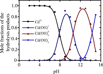
Dependences of the mole fractions of the products of CdCl2 salt hydrolysis—Cd2+, Cd(OH)+, Cd(OH)0
2, and Cd(OH)−
2—vs. pH of the solution

Dependences of the mole fractions of the products of Na2S salt hydrolysis (S2−, HS−, and H2S) vs. pH of the solution

Dependences of the concentrations of chloride complexes of Cd vs. concentration of Cl− ions

Dependencies of pHmin (1) and pHS
min (2) on the concentration of Cd2+ ions

PL and microscopic images: typical photoluminescence spectrum of the colloidal solution of mercaptoethanol-coated CdS NPs (a) and microscopic images (b–f) of histological sections of placenta tissue (fixed and preserved) visualized using mercapto-coated NPs: b unaltered human placental terminal villi, c unaltered human intermediate villus with dichotomy to terminal villi, d intervillous fibrinoid, e human intermediate stem villus with artery and vena and rough collagen, and f human intermediate villus with damaged surface with fibrinoid. The identified forms are 1—trophoblast, 2—endotheliocyte, and 3—erythrocytes
References
-
- Linga D, Hacketta MJ, Hyeon T. Surface ligands in synthesis, modification, assembly and biomedical applications of nanoparticles. Nanotoday. 2014;9(4):457–477. doi: 10.1016/j.nantod.2014.06.005. - DOI
LinkOut - more resources
Full Text Sources
Other Literature Sources
