Review
. 2016 Apr 19;11(8):738-56.
doi: 10.1002/cmdc.201500495. Epub 2016 Feb 11.

Current Experimental Methods for Characterizing Protein-Protein Interactions

Affiliations
Review

Current Experimental Methods for Characterizing Protein-Protein Interactions

Mi Zhou et al. ChemMedChem. .

Abstract

Protein molecules often interact with other partner protein molecules in order to execute their vital functions in living organisms. Characterization of protein-protein interactions thus plays a central role in understanding the molecular mechanism of relevant protein molecules, elucidating the cellular processes and pathways relevant to health or disease for drug discovery, and charting large-scale interaction networks in systems biology research. A whole spectrum of methods, based on biophysical, biochemical, or genetic principles, have been developed to detect the time, space, and functional relevance of protein-protein interactions at various degrees of affinity and specificity. This article presents an overview of these experimental methods, outlining the principles, strengths and limitations, and recent developments of each type of method.

Keywords: biochemical methods; biophysical methods; genetic methods; protein-protein interactions.


Figures

Figure 1
Figure 1
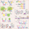
Illustration of some biophysical methods for PPI detection. A) Fluorescence polarization (FP). When a fluorescently labelled small molecule is excited by polarized light, it emits lights with a certain degree of polarization that is inversely proportional to the rotation rate of the fluorescent molecule. A small molecule rotates fast, so the emitted lights are depolarized. Its rotation is greatly decreased when it binds to a larger molecule, so the emitted light remains polarized. B) Surface plasmon resonance (SPR). In an SPR biosensor, an incident light beam hits a prism covered with a thin gold film at a certain angle, leading to the phenomenon of SPR. Perturbations at the gold surface on the sensor chip, such as the interaction between the bait immobilized on the surface and the prey flown over the surface, result in detectable SPR angle shifts. C) Isothermal titration calorimetry (ITC). An ITC instrument is composed of a reference cell and a sample cell, both of which are surrounded by an adiabatic jacket. During measurement, a protein molecule loaded in the syringe is titrated into the sample cell containing its binding partner, causing heat changes in the sample cell. This heat change can be quantified by the electric power required to maintain the isothermal condition between two cells. D) Microscale thermophoresis (MST). The sample solution containing fluorescent molecules inside a capillary is heated by a focused IR‐laser, which is coupled into the path of fluorescence excitation and emission. A series of processes, including initial state, T‐jump, thermophoresis, steady state, inverse T‐jump, and back‐diffusion of fluorescent molecules can be detected through the temperature gradient.
Figure 2
Figure 2
Illustration of some biochemical methods for PPI detection. A) Fluorescence resonance energy transfer (FRET) between a CFP‐fused bait and a YFP‐fused prey, where CFP and YFP act as the donor and the acceptor fluorophore, respectively. If the bait and the prey do not interact, excitation of CFP results in light emission (475 nm) by CFP only. When bait–prey interaction occurs, CFP and YFP are brought into proximity, leading to energy transfer from CFP to nearby YFP. Then, YFP emission can be detected at 528 nm. B) Bioluminescence resonance energy transfer (BRET) between a Rluc‐fused bait and a EYFP‐fused prey, where Rluc and EYFP act as the donor luciferase and the acceptor fluorophore, respectively. If bait and prey do not interact, only the blue‐emitting spectrum (480 nm) of RLuc can be detected. When bait–prey interaction occurs, bioluminescence energy generated by Rluc in the presence of its substrate (coelenterazine) is transferred to EYFP, which in turn generates light emission at 530 nm. C) AlphaScreen. Upon excitation at 680 nm, a photosensitizing agent on the donor bead converts ambient oxygen to singlet oxygen. When bait–prey interaction brings the donor and the acceptor bead into close proximity, energy is transferred from singlet oxygen to thioxene derivatives on the acceptor bead, resulting in light emission at 520–620 nm. D) Protein‐fragment complementation assay (PCA). Complementary C‐terminal and N‐terminal fragments of GFP are fused to the bait and the prey, respectively. If the bait and the prey do not interact, the fragments remain unstructured and lack functional activity. When the bait–prey interaction occurs, two fragments are brought together to form a full‐length GFP with the native structure and function. E) Pull‐down assay. In a GST pull‐down assay, the bait is expressed as a GST‐fused protein, and it is incubated with cell or tissue lysate containing the prey. The GST–bait–prey complex is captured by glutathione‐coupled beads and eluted for analysis by SDS‐PAGE. F) Tandem affinity purification (TAP). In this type of assay, the TAP tag (including CBP, a TEV cleavage site, and ProtA) is fused to the bait, and then the whole construction is introduced into the host cell. The TAP–bait–prey complex is isolated from cell lysate via affinity column containing IgG beads. After washing, TEV protease is added to release the CBP–bait–prey complex, which is then immobilized on calmodulin‐coated beads. After washing, the native complex is eluted by chelating calcium using EGTA. G) Co‐immunoprecipitation (Co‐IP). The bait–prey complex in cell or tissue lysate is captured by beads functionalized with a bait‐specific antibody, and then co‐precipitated, washed, eluted, and analyzed by SDS‐PAGE. H) ELISA. In the case of direct ELISA, the bait is immobilized on a microwell plate, and then cell lysate containing the prey is applied to the wells. After incubation and washing away unbound proteins, the bait–prey complex is detected by an HRP‐conjugated antibody against the prey. Finally, a substance of the HRP enzyme is added, which in turn produces detectable fluorescence‐ or absorbance‐based signal via the enzymatic reaction. I) Proximity ligation assay (PLA). The bait and prey are recognized by their respective PLA probes consisting of antibodies and single‐stranded DNA oligonucleotides, where the free oligonucleotide ends are brought into proximity. An oligonucleotide connecter is added to hybridize to the complementary oligonucleotides on the PLA probes. A ligase seals the nick between two PLA probes and forms a new DNA strand that can be amplified by PCR.
Figure 3
Figure 3
Illustration of some genetic methods for PPI detection. A) Phage display. A library of phage, each displaying a different protein including the prey, is exposed to a plate immobilized with the bait. After washing, phages displaying the prey bound to the bait will remain while nonbinding phages are removed. The remaining phages are then eluted, amplified by bacterial infection, and enriched in repeated selection cycles, that is, the “panning” process. B) The yeast two‐hybrid (Y2H) system. The binding domain (BD) and activation domain (AD) are two separate fragments of the transcription factor GAL4. They are fused to the bait and prey, respectively, where BD is responsible for binding to the upstream activating sequence (UAS) and AD is responsible for activation of transcription. The bait–prey interaction brings BD and AD into proximity to form an intact and functional transcriptional activator, which in turn recruits the basal transcriptional machinery and induces transcription of a downstream reporter gene lacZ. C) Protein microarray. A library of proteins is spotted onto a solid surface. This array is then incubated with a labelled bait. The interaction between the bait and the prey can be detected by a readout system employing a label‐based technique such as fluorescence.

Similar articles

Cited by

References

    1. Ezkurdia I., Juan D., Rodriguez J. M., Frankish A., Diekhans M., Harrow J., Vazquez J., Valencia A., Tress M. L., Hum. Mol. Genet. 2014, 23, 5866–5878. - PMC - PubMed
    1. Berggård T., Linse S., James P., Proteomics 2007, 7, 2833–2842. - PubMed
    1. Braun P., Gingras A. C., Proteomics 2012, 12, 1478–1498. - PubMed
    1. Pelletier J., Sidhu S., Curr. Opin. Biotechnol. 2001, 12, 340–347. - PubMed
    1. Vidal M., Cusick M. E., Barabasi A. L., Cell 2011, 144, 986–998. - PMC - PubMed

Publication types