

MicroRNA-21 induces 5-fluorouracil resistance in human pancreatic cancer cells by regulating PTEN and PDCD4
- PMID: 26864640
- PMCID: PMC4831288
- DOI: 10.1002/cam4.626
MicroRNA-21 induces 5-fluorouracil resistance in human pancreatic cancer cells by regulating PTEN and PDCD4
Abstract
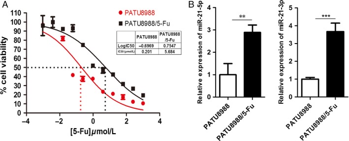
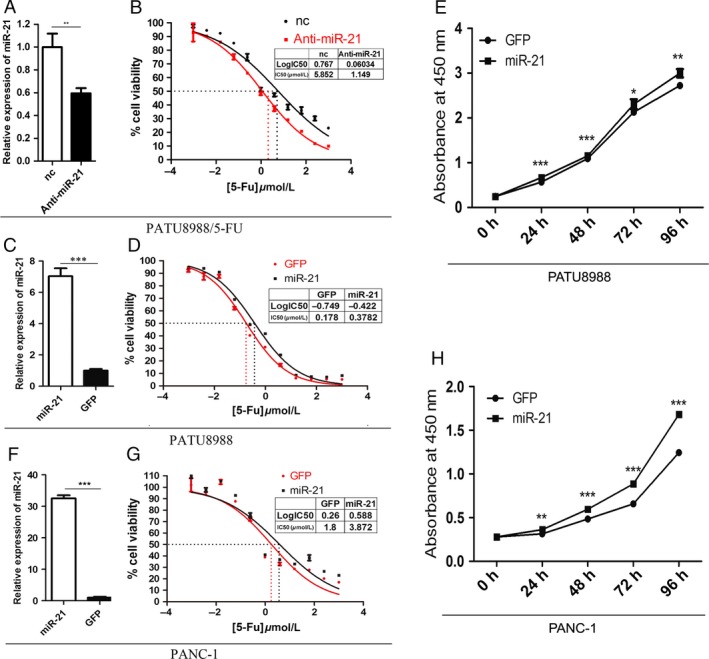
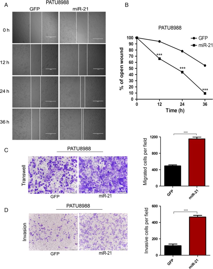
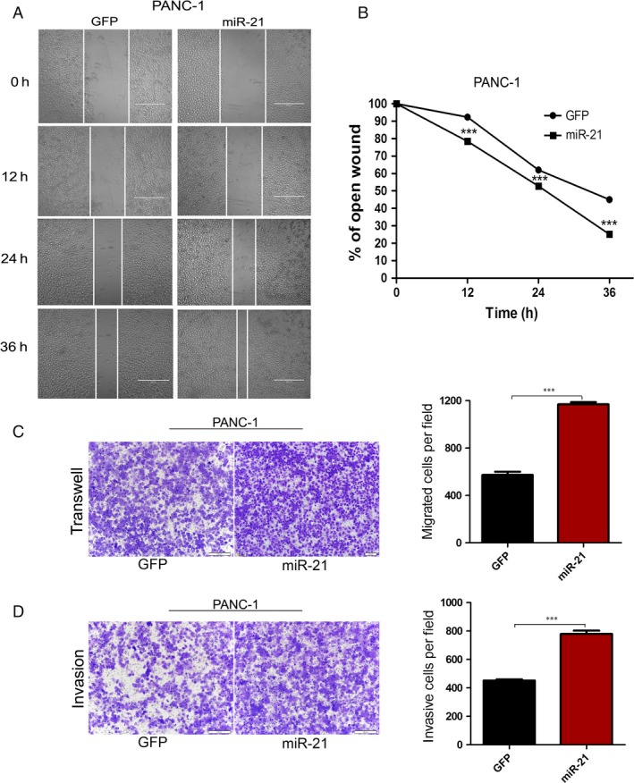
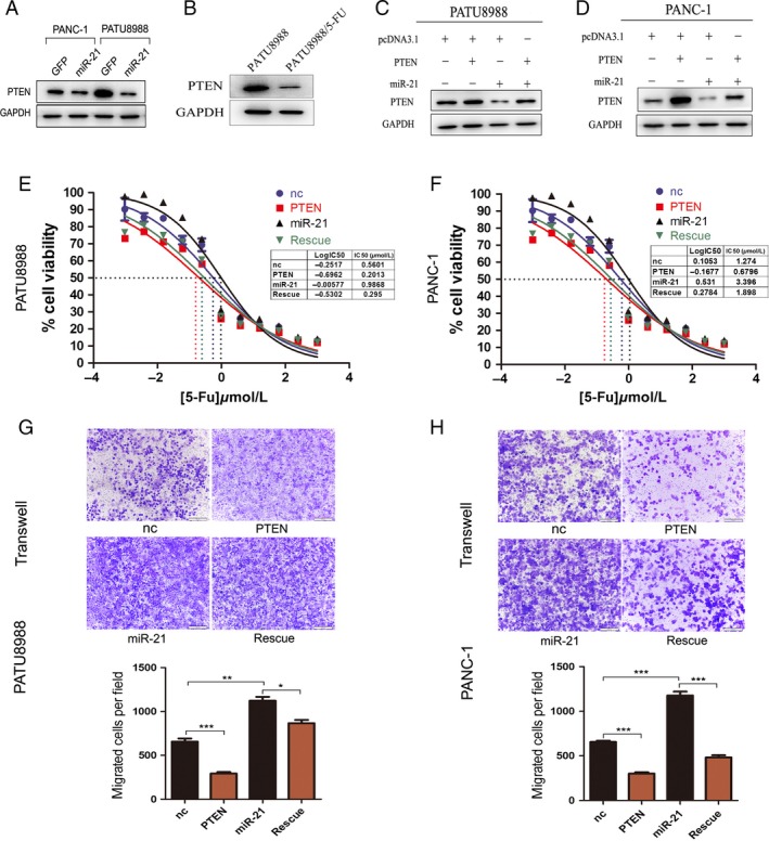
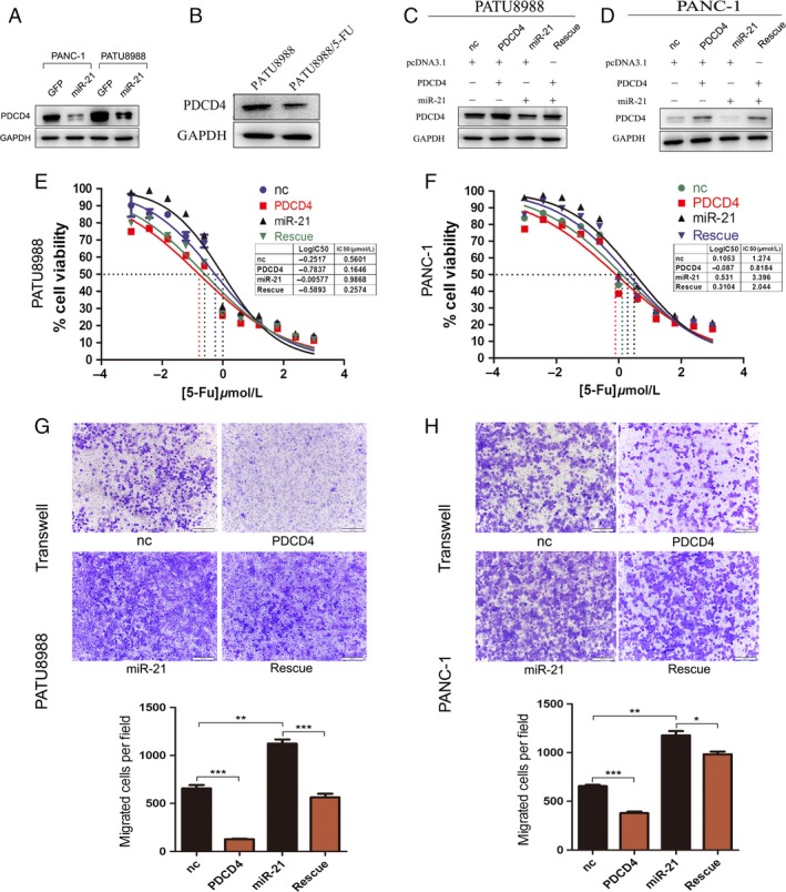
Pancreatic cancer patients are often resistant to chemotherapy treatment, which results in poor prognosis. The objective of this study was to delineate the mechanism by which miR-21 induces drug resistance to 5-fluorouracil (5-FU) in human pancreatic cancer cells (PATU8988 and PANC-1). We report that PATU8988 cells resistant to 5-FU express high levels of miR-21 in comparison to sensitive primary PATU8988 cells. Suppression of miR-21 expression in 5-Fu-resistant PATU8988 cells can alleviate its 5-FU resistance. Meanwhile, lentiviral vector-mediated overexpression of miR-21 not only conferred resistance to 5-FU but also promoted proliferation, migration, and invasion of PATU8988 and PANC-1 cells. The proresistance effects of miR-21 were attributed to the attenuated expression of tumor suppressor genes, including PTEN and PDCD4. Overexpression of PTEN and PDCD4 antagonized miR-21-induced resistance to 5-FU and migration activity. Our work demonstrates that miR-21 can confer drug resistance to 5-FU in pancreatic cancer cells by regulating the expression of tumor suppressor genes, as the target genes of miR-21, PTEN and PDCD4 can rescue 5-FU sensitivity and the phenotypic characteristics disrupted by miR-21.
Keywords: 5-Fluorouracil; PDCD4; PTEN; miR-21; pancreatic cancer.
© 2016 The Authors. Cancer Medicine published by John Wiley & Sons Ltd.
Figures
Similar articles
-
MicroRNA-320a promotes 5-FU resistance in human pancreatic cancer cells.Sci Rep. 2016 Jun 9;6:27641. doi: 10.1038/srep27641. Sci Rep. 2016. PMID: 27279541 Free PMC article.
-
MicroRNA-21 stimulates gastric cancer growth and invasion by inhibiting the tumor suppressor effects of programmed cell death protein 4 and phosphatase and tensin homolog.J BUON. 2014 Jan-Mar;19(1):228-36. J BUON. 2014. PMID: 24659669
-
MicroRNA-21 regulates the ERK/NF-κB signaling pathway to affect the proliferation, migration, and apoptosis of human melanoma A375 cells by targeting SPRY1, PDCD4, and PTEN.Mol Carcinog. 2017 Mar;56(3):886-894. doi: 10.1002/mc.22542. Epub 2016 Sep 22. Mol Carcinog. 2017. PMID: 27533779
-
MiR-132 promotes the proliferation, invasion and migration of human pancreatic carcinoma by inhibition of the tumor suppressor gene PTEN.Prog Biophys Mol Biol. 2019 Nov;148:65-72. doi: 10.1016/j.pbiomolbio.2017.09.019. Epub 2017 Sep 20. Prog Biophys Mol Biol. 2019. PMID: 28941804 Review.
-
5-Fluorouracil: mechanisms of resistance and reversal strategies.Molecules. 2008 Aug 5;13(8):1551-69. doi: 10.3390/molecules13081551. Molecules. 2008. PMID: 18794772 Free PMC article. Review.
Cited by
-
Using microRNAs Networks to Understand Pancreatic Cancer-A Literature Review.Biomedicines. 2024 Aug 1;12(8):1713. doi: 10.3390/biomedicines12081713. Biomedicines. 2024. PMID: 39200178 Free PMC article. Review.
-
Repressing PDCD4 activates JNK/ABCG2 pathway to induce chemoresistance to fluorouracil in colorectal cancer cells.Ann Transl Med. 2021 Jan;9(2):114. doi: 10.21037/atm-20-4292. Ann Transl Med. 2021. PMID: 33569416 Free PMC article.
-
Advances in pancreatic cancer epigenetics: From the mechanism to the clinic.World J Gastrointest Oncol. 2025 Jul 15;17(7):106238. doi: 10.4251/wjgo.v17.i7.106238. World J Gastrointest Oncol. 2025. PMID: 40697230 Free PMC article. Review.
-
Noncoding RNAs Associated with Therapeutic Resistance in Pancreatic Cancer.Biomedicines. 2021 Mar 7;9(3):263. doi: 10.3390/biomedicines9030263. Biomedicines. 2021. PMID: 33799952 Free PMC article. Review.
-
Can Molecular Biomarkers Change the Paradigm of Pancreatic Cancer Prognosis?Biomed Res Int. 2016;2016:4873089. doi: 10.1155/2016/4873089. Epub 2016 Sep 1. Biomed Res Int. 2016. PMID: 27689078 Free PMC article. Review.
References
-
- Jemal, A. , Siegel R., Ward E., Hao Y., Xu J., and Thun M. J.. 2009. Cancer statistics, 2009. CA Cancer J. Clin. 59:225–249. - PubMed
-
- Siegel, R. , Naishadham D., and Jemal A.. 2013. Cancer statistics. CA Cancer J. Clin. 63:11–30. - PubMed
-
- Maisey, N. , Chau I., Cunningham D., Norman A., Seymour M., Hickish T., et al. 2002. Multicenter randomized phase III trial comparing protracted venous infusion (PVI) fluorouracil (5‐FU) with PVI 5‐FU plus mitomycin in inoperable pancreatic cancer. J. Clin. Oncol. 20:3130–3136. - PubMed
-
- Tomari, Y. , and Zamore P. D.. 2005. MicroRNA biogenesis: Drosha can't cut it without a partner. Curr. Biol. 15:R61–R64. - PubMed
-
- Wu, W. K. , Lee C. W., Cho C. H., Fan D., Wu K., Yu J., et al. 2010. MicroRNA dysregulation in gastric cancer: a new player enters the game. Oncogene 29:5761–5771. - PubMed
Publication types
MeSH terms
Substances
LinkOut - more resources
Full Text Sources
Other Literature Sources
Medical
Research Materials
