

Visualization of cerebrospinal fluid flow in syringomyelia through noninvasive magnetic resonance imaging with a time-spatial labeling inversion pulse (Time-SLIP)
- PMID: 26864698
- PMCID: PMC5472025
- DOI: 10.1080/10790268.2016.1140391
Visualization of cerebrospinal fluid flow in syringomyelia through noninvasive magnetic resonance imaging with a time-spatial labeling inversion pulse (Time-SLIP)
Abstract
Context: We report a case of syringomyelia assessed by magnetic resonance imaging (MRI) with a time-spatial labeling inversion pulse (Time-SLIP), which is a non-contrast MRI technique that uses the cerebrospinal fluid (CSF) as an intrinsic tracer, thus removing the need to administer a contrast agent. Time-SLIP permits investigation of flow movement for over 3 seconds without any limitations associated with the cardiac phase, and it is a clinically accessible method for flow analysis.
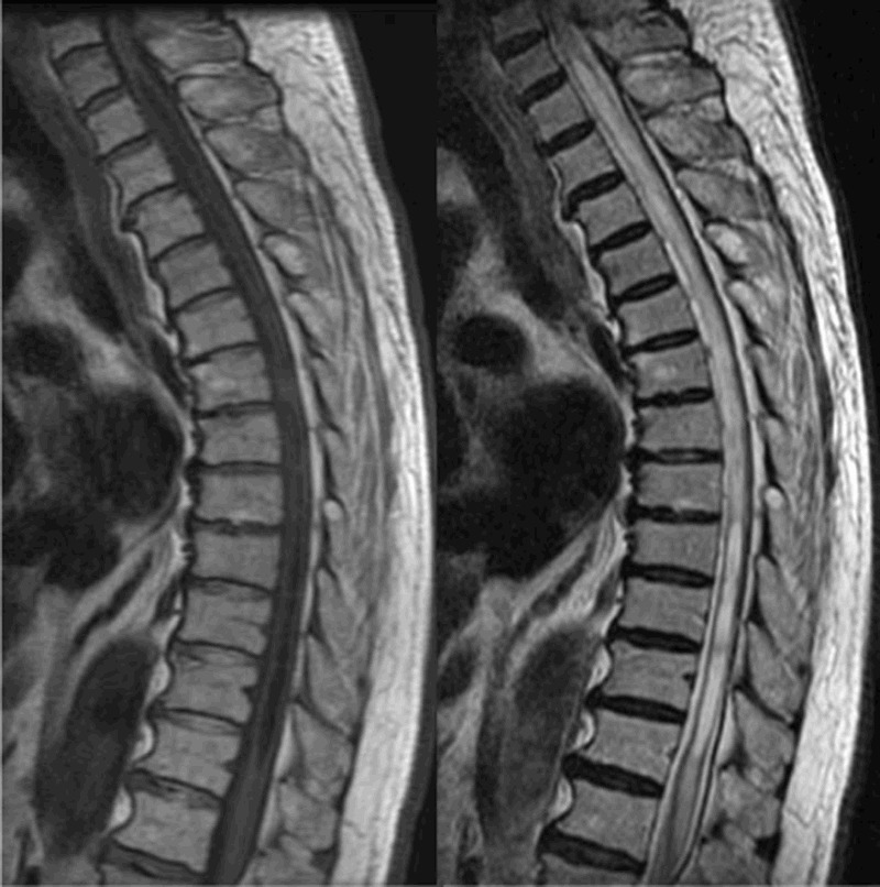
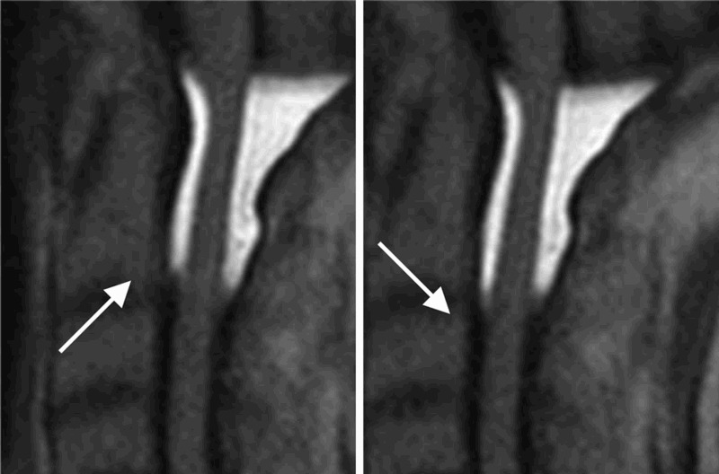
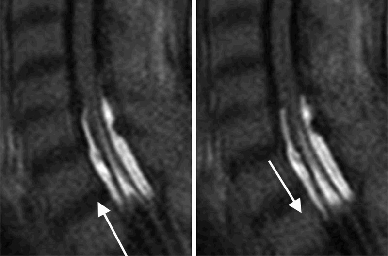
Findings: We investigated an 85-year-old male experiencing progressive gait disturbance, with leg numbness and muscle weakness. Conventional MRI revealed syringomyelia from C7 to T12, with multiple webs of cavities. We then applied the Time-SLIP approach to characterize CSF flow in the syringomyelic cavities. Time-SLIP detected several unique CSF flow patterns that could not be observed by conventional imaging. The basic CSF flow pattern in the subarachnoid space was pulsatile and was harmonious with the heartbeat. Several unique flow patterns, such as bubbles, jumping, and fast flow, were observed within syringomyelic cavities by Time-SLIP imaging. These patterns likely reflect the complex flow paths through the septum and/or webs of cavities.
Conclusion/clinical relevance: Time-SLIP permits observation of CSF motion over a long period of time and detects patterns of flow velocity and direction. Thus, this novel approach to CSF flow analysis can be used to gain a more extensive understanding of spinal disease pathology and to optimize surgical access in the treatment of spinal lesions. Additionally, Time-SLIP has broad applicability in the field of spinal research.
Keywords: Cerebrospinal fluid (CSF); Magnetic resonance imaging (MRI); Spinal disease; Syringomyelia; Time-spatial labeling inversion pulse (Time-SLIP).
Figures

Similar articles
-
Time-spatial Labeling Inversion Pulse (Time-SLIP) with Pencil Beam Pulse: A Selective Labeling Technique for Observing Cerebrospinal Fluid Flow Dynamics.Magn Reson Med Sci. 2018 Jul 10;17(3):259-264. doi: 10.2463/mrms.tn.2017-0032. Epub 2017 Aug 24. Magn Reson Med Sci. 2018. PMID: 28835572 Free PMC article.
-
Cerebrospinal fluid flow in small-breed dogs with idiopathic epilepsy observed using time-spatial labeling inversion pulse images: a preliminary study.J Vet Med Sci. 2024 Nov 15;86(11):1168-1176. doi: 10.1292/jvms.23-0305. Epub 2024 Oct 3. J Vet Med Sci. 2024. PMID: 39358237 Free PMC article.
-
[Optimal imaging parameters and the advantage of cerebrospinal fluid flow image using time-spatial labeling inversion pulse at 3 tesla magnetic resonance imaging: comparison of image quality for 1.5 tesla magnetic resonance imaging].Nihon Hoshasen Gijutsu Gakkai Zasshi. 2014 Dec;70(12):1439-44. doi: 10.6009/jjrt.2014_JSRT_70.12.1439. Nihon Hoshasen Gijutsu Gakkai Zasshi. 2014. PMID: 25672449 Japanese.
-
Cerebrospinal fluid physiology: visualization of cerebrospinal fluid dynamics using the magnetic resonance imaging Time-Spatial Inversion Pulse method.Croat Med J. 2014 Aug 28;55(4):337-46. doi: 10.3325/cmj.2014.55.337. Croat Med J. 2014. PMID: 25165048 Free PMC article. Review.
-
The pathogenesis of syringomyelia associated with lesions at the foramen magnum: a critical review of existing theories and proposal of a new hypothesis.J Neurol Sci. 2004 May 15;220(1-2):3-21. doi: 10.1016/j.jns.2004.01.014. J Neurol Sci. 2004. PMID: 15140600 Review.
Cited by
-
Bulat-Klarica-Oreskovic Hypothesis: A Comprehensive Review.Cureus. 2023 Sep 23;15(9):e45821. doi: 10.7759/cureus.45821. eCollection 2023 Sep. Cureus. 2023. PMID: 37876400 Free PMC article. Review.
-
Cerebrospinal fluid flow in normal beagle dogs analyzed using magnetic resonance imaging.J Vet Sci. 2021 Jan;22(1):e2. doi: 10.4142/jvs.2021.22.e2. J Vet Sci. 2021. PMID: 33522154 Free PMC article.
-
The significance of cerebrospinal fluid dynamics in adolescent idiopathic scoliosis using time-SLIP MRI.Sci Rep. 2024 May 28;14(1):12214. doi: 10.1038/s41598-024-63135-3. Sci Rep. 2024. PMID: 38806612 Free PMC article.
-
Application of time-spatial labeling inversion pulse magnetic resonance imaging in the diagnosis of spontaneous intracranial hypotension due to high-flow cerebrospinal fluid leakage at C1-2.Surg Neurol Int. 2016 Dec 26;7(Suppl 42):S1085-S1088. doi: 10.4103/2152-7806.196765. eCollection 2016. Surg Neurol Int. 2016. PMID: 28144490 Free PMC article.
References
-
- Satogami N, Okada T, Koyama T, Gotoh K, Kamae T, Togashi K.. Visualization of external carotid artery and its branches: Non-contrast-enhanced MR angiography using balanced steady-state free-precession sequence and a time-spatial labeling inversion pulse. J Magn Reson Imaging 2009;30(3):678–83. doi: 10.1002/jmri.21883 - DOI - PubMed
Publication types
MeSH terms
LinkOut - more resources
Full Text Sources
Other Literature Sources
Medical
Miscellaneous