Age-Associated Pathology in Rhesus Macaques (Macaca mulatta)
- PMID: 26864889
- PMCID: PMC5909965
- DOI: 10.1177/0300985815620628
Age-Associated Pathology in Rhesus Macaques (Macaca mulatta)
Abstract
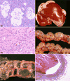
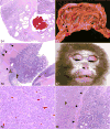
The rhesus macaque (Macaca mulatta) is one of the most extensively used nonhuman primate models for human diseases. This article presents a literature review focusing on major organ systems and age-associated conditions in humans and primates, combined with information from the Wisconsin National Primate Research Center Electronic Health Record database to highlight and contrast age-associated lesions in geriatric rhesus macaques with younger cohorts. Rhesus macaques are excellent models for age-associated conditions, including diabetes, osteoarthritis, endometriosis, visual accommodation, hypertension, osteoporosis, and amyloidosis. Adenocarcinoma of the large intestine (ileocecocolic junction, cecum, and colon) is the most common spontaneous neoplasm in the rhesus macaque. A combination of cross-sectional and longitudinal studies is required to truly define mechanisms of maturation, aging, and the pathology of age-associated conditions in macaques and thus humans. The rhesus macaque is and will continue to be an appropriate and valuable model for investigation of the mechanisms and treatment of age-associated diseases.
Keywords: Macaca mulatta; aging; amyloid; cross-sectional studies; diabetes; disease prevalence; diverticulosis; endometriosis; neoplasia; rhesus macaque.
© The Author(s) 2016.
Conflict of interest statement
The author(s) declared no potential conflicts of interest with respect to the research, authorship, and/or publication of this article.
Figures

Similar articles
-
The incidence of spontaneous neoplasia in two populations of captive rhesus macaques (Macaca mulatta).Antioxid Redox Signal. 2011 Jan 15;14(2):221-7. doi: 10.1089/ars.2010.3311. Epub 2010 Oct 28. Antioxid Redox Signal. 2011. PMID: 20524847 Free PMC article.
-
Genomic resources for rhesus macaques (Macaca mulatta).Mamm Genome. 2022 Mar;33(1):91-99. doi: 10.1007/s00335-021-09922-z. Epub 2022 Jan 9. Mamm Genome. 2022. PMID: 34999909 Free PMC article.
-
Postnatal development of the hippocampus in the Rhesus macaque (Macaca mulatta): a longitudinal magnetic resonance imaging study.Hippocampus. 2014 Jul;24(7):794-807. doi: 10.1002/hipo.22271. Epub 2014 Apr 1. Hippocampus. 2014. PMID: 24648155 Free PMC article.
-
Acute Radiation Effects, the H-ARS in the Non-human Primate: A Review and New Data for the Cynomolgus Macaque with Reference to the Rhesus Macaque.Health Phys. 2021 Oct 1;121(4):304-330. doi: 10.1097/HP.0000000000001442. Health Phys. 2021. PMID: 34546214 Review.
-
Common and Not-So-Common Pathologic Findings of the Gastrointestinal Tract of Rhesus and Cynomolgus Macaques.Toxicol Pathol. 2022 Jul;50(5):638-659. doi: 10.1177/01926233221084634. Epub 2022 Apr 1. Toxicol Pathol. 2022. PMID: 35363082 Free PMC article. Review.
Cited by
-
Cross-sectional comparison of health-span phenotypes in young versus geriatric marmosets.Am J Primatol. 2019 Feb;81(2):e22952. doi: 10.1002/ajp.22952. Epub 2019 Jan 21. Am J Primatol. 2019. PMID: 30664265 Free PMC article.
-
Considerations of Medical Preparedness to Assess and Treat Various Populations During a Radiation Public Health Emergency.Radiat Res. 2023 Mar 1;199(3):301-318. doi: 10.1667/RADE-22-00148.1. Radiat Res. 2023. PMID: 36656560 Free PMC article. Review.
-
Ketamine-induced neuromuscular reactivity is associated with aging in female rhesus macaques.PLoS One. 2020 Sep 21;15(9):e0236430. doi: 10.1371/journal.pone.0236430. eCollection 2020. PLoS One. 2020. PMID: 32956357 Free PMC article.
-
Plasma lipidomic signatures of spontaneous obese rhesus monkeys.Lipids Health Dis. 2019 Jan 8;18(1):8. doi: 10.1186/s12944-018-0952-9. Lipids Health Dis. 2019. PMID: 30621707 Free PMC article.
-
An intranasally administrated SARS-CoV-2 beta variant subunit booster vaccine prevents beta variant replication in rhesus macaques.PNAS Nexus. 2022 Jun 17;1(3):pgac091. doi: 10.1093/pnasnexus/pgac091. eCollection 2022 Jul. PNAS Nexus. 2022. PMID: 35873792 Free PMC article.
References
-
- Abbott DP, Majeed SK. A survey of parasitic lesions in wild-caught, laboratory-maintained primates (rhesus, cynomolgus, and baboon) Vet Pathol. 1984;21(2):198–207. - PubMed
-
- Alpers CE. The kidney. In: Kumar V, Abbas AK, Fausto N, editors. Robbins and Cotran Pathologic Basis of Disease. 7th. Philadelphia, PA: Elsevier/Saunders; 2005. pp. 966–968.
-
- Andrade MCR, Marchevsky RS. Histopathologic findings of pulmonary acariasis in a rhesus monkeys breeding unit. Revista Brasileira de Parasitologia Veterinária. 2007;16:229–234. - PubMed
-
- Anirban M, Abbas AK. The endocrine system. In: Kumar V, Abbas AK, Fausto N, editors. Robbins and Cotran Pathologic Basis of Disease. 7th. Philadelphia, PA: Elsevier/Saunders; 2005. pp. 1189–1206.
Publication types
MeSH terms
Grants and funding
LinkOut - more resources
Full Text Sources
Other Literature Sources
Medical

