

Novel motif in calcineurin catalytic subunit is required for septal localization of calcineurin in Aspergillus fumigatus
- PMID: 26864964
- PMCID: PMC4764407
- DOI: 10.1002/1873-3468.12075
Novel motif in calcineurin catalytic subunit is required for septal localization of calcineurin in Aspergillus fumigatus
Abstract
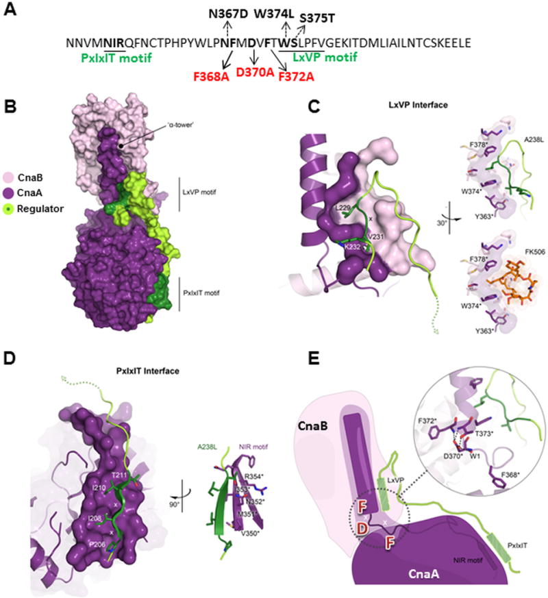
Calcineurin heterodimer, comprised of the catalytic (CnaA) and regulatory (CnaB) subunits, localizes at the hyphal tips and septa to direct growth, septation, and disease in the human pathogen Aspergillus fumigatus. Here we discovered a novel motif (FMDVF) required for this critical CnaA septal localization, including residues Phe368, Asp370 and Phe372 overlapping the cyclosporine A-cyclophilin A-binding domain, CnaB-binding helix and the FK506-FKBP12-binding pocket. Mutations in adjacent residues Asn367, Trp374, and Ser375 confer FK506 resistance without impacting CnaA septal localization. Modeling A. fumigatus CnaA confirmed that the FMDVF motif forms a bridge between the two known substrate-binding motifs, PxIxIT and LxVP, and concurrent mutations (F368A D370A; F368A F372A) in the FMDVF motif disrupt CnaA-substrate interaction at the septum.
Keywords: Aspergillus fumigatus; FK506; LxVP motif; PxIxIT motif; calcineurin; septum.
© 2016 Federation of European Biochemical Societies.
Figures


Similar articles
-
Phosphorylation of Calcineurin at a novel serine-proline rich region orchestrates hyphal growth and virulence in Aspergillus fumigatus.PLoS Pathog. 2013;9(8):e1003564. doi: 10.1371/journal.ppat.1003564. Epub 2013 Aug 22. PLoS Pathog. 2013. PMID: 23990785 Free PMC article.
-
Localization and activity of the calcineurin catalytic and regulatory subunit complex at the septum is essential for hyphal elongation and proper septation in Aspergillus fumigatus.Mol Microbiol. 2011 Dec;82(5):1235-59. doi: 10.1111/j.1365-2958.2011.07886.x. Epub 2011 Nov 8. Mol Microbiol. 2011. PMID: 22066998 Free PMC article.
-
Kin1 kinase localizes at the hyphal septum and is dephosphorylated by calcineurin but is dispensable for septation and virulence in the human pathogen Aspergillus fumigatus.Biochem Biophys Res Commun. 2018 Nov 2;505(3):740-746. doi: 10.1016/j.bbrc.2018.09.186. Epub 2018 Oct 4. Biochem Biophys Res Commun. 2018. PMID: 30292408 Free PMC article.
-
Calcineurin-mediated regulation of hyphal growth, septation, and virulence in Aspergillus fumigatus.Mycopathologia. 2014 Dec;178(5-6):341-8. doi: 10.1007/s11046-014-9794-9. Epub 2014 Aug 15. Mycopathologia. 2014. PMID: 25118871 Free PMC article. Review.
-
Calcineurin Orchestrates Hyphal Growth, Septation, Drug Resistance and Pathogenesis of Aspergillus fumigatus: Where Do We Go from Here?Pathogens. 2015 Dec 16;4(4):883-93. doi: 10.3390/pathogens4040883. Pathogens. 2015. PMID: 26694470 Free PMC article. Review.
Cited by
-
Disruption of calcineurin catalytic subunit (cnaA) in Epichloë festucae induces symbiotic defects and intrahyphal hyphae formation.Mol Plant Pathol. 2018 Jun;19(6):1414-1426. doi: 10.1111/mpp.12624. Epub 2018 Feb 9. Mol Plant Pathol. 2018. PMID: 28990722 Free PMC article.
-
Aspergillus fumigatus and Aspergillosis in 2019.Clin Microbiol Rev. 2019 Nov 13;33(1):e00140-18. doi: 10.1128/CMR.00140-18. Print 2019 Dec 18. Clin Microbiol Rev. 2019. PMID: 31722890 Free PMC article. Review.
-
Potential Antifungal Targets for Aspergillus sp. from the Calcineurin and Heat Shock Protein Pathways.Int J Mol Sci. 2022 Oct 19;23(20):12543. doi: 10.3390/ijms232012543. Int J Mol Sci. 2022. PMID: 36293395 Free PMC article. Review.
-
Calcineurin Inhibitor CN585 Exhibits Off-Target Effects in the Human Fungal Pathogen Aspergillus fumigatus.J Fungi (Basel). 2022 Dec 7;8(12):1281. doi: 10.3390/jof8121281. J Fungi (Basel). 2022. PMID: 36547614 Free PMC article.
-
Calcineurin in fungal virulence and drug resistance: Prospects for harnessing targeted inhibition of calcineurin for an antifungal therapeutic approach.Virulence. 2017 Feb 17;8(2):186-197. doi: 10.1080/21505594.2016.1201250. Epub 2016 Jun 20. Virulence. 2017. PMID: 27325145 Free PMC article. Review.
References
-
- Ferreira MEdS, et al. Functional characterization of the Aspergillus fumigatus calcineurin. Fungal Genetics and Biology. 2007;44:219–230. - PubMed
-
- Rusnak F, Mertz P. Calcineurin: Form and Function. Physiological Reviews. 2000;80:1483–1521. - PubMed
-
- Liu J, Farmer JD, Jr, Lane WS, Friedman J, Weissman I, Schreiber SL. Calcineurin is a common target of cyclophilin-cyclosporin A and FKBP-FK506 complexes. Cell. 1991;66:807–815. - PubMed
Publication types
MeSH terms
Substances
Grants and funding
LinkOut - more resources
Full Text Sources
Other Literature Sources
