. 2016 Apr 20;5(8):900-6.
doi: 10.1002/adhm.201500956. Epub 2016 Feb 10.

Modular Assembly Approach to Engineer Geometrically Precise Cardiovascular Tissue

Affiliations

Modular Assembly Approach to Engineer Geometrically Precise Cardiovascular Tissue

Benjamin W Lee et al. Adv Healthc Mater. .

Abstract

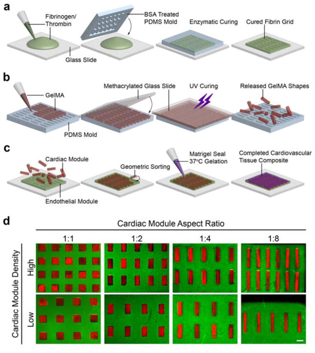
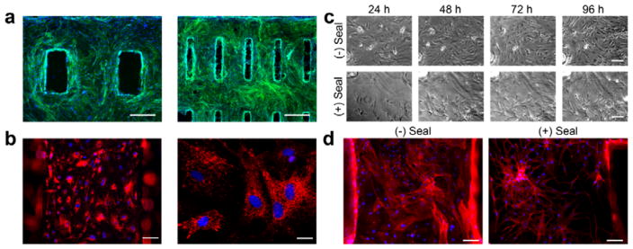
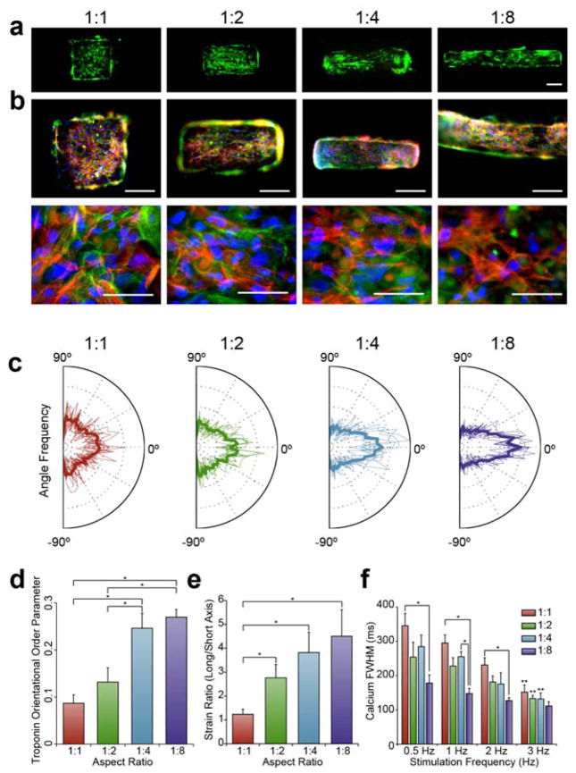
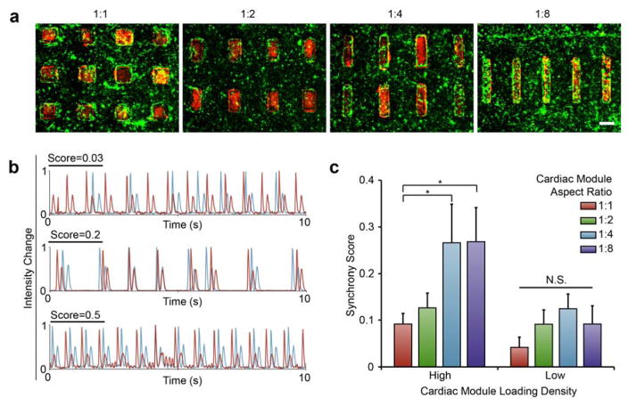
This modular assembly approach to microfabricate functional cardiovascular tissue composites enables quantitative assessment of the effects of microarchitecture on cellular function. Cardiac and endothelial modules are micromolded separately, designed to direct cardiomyocyte alignment and anisotropic contraction or vascular network formation. Assembled cardiovascular tissue composites contract synchronously, facilitating the use of this tissue-engineering platform to study structure-function relationships in the heart.

Keywords: anisotropy; cardiac tissue engineering; hydrogels; micropatterning; modular assembly.


Figures

Figure 1
Figure 1. Method for fabricating composite cardiovascular tissue
(a) Endothelial modules were fabricated by pipetting a fibrinogen thrombin mixture between a glass slide and a PDMS template mold that was allowed to cure for 5 min prior to endothelial cell seeding. (b) Cardiac modules were also fabricated by micromolding, using GelMA, a UV-light curable hydrogel, and a PDMS mold containing an array of any single type of rectangle. (c) To form tissues, cardiac modules were sorted within the endothelial module and sealed with a thin layer of Matrigel. (d) An array of cardiovascular tissue composite designs, where the cardiac modules are represented by red fluorescent beads encapsulated within hydrogels and the endothelial moduless are represented by green fluorescent beads encapsulated within hydrogels. Aspect ratio and loading density of the cardiac module were varied independently (scale: 500 μm).
Figure 2
Figure 2. Endothelial module supports vascular network formation
(a) Fluorescence micrographs of endothelial cells on fibrin hydrogels where green represents actin (Phalloidin) and blue represents nuclei (DAPI) (Scale = 500 μm). (b) Acetylated LDL uptake (red) in endothelial cells on fibrin hydrogels, nuclei counterstained with DAPI (blue). Low power (left, scale = 100 μm) and high power (right, scale = 25 μm). (c) Time course micrographs of endothelial cells grown on fibrin without or with the Matrigel seal over the course of 4 days, where the Matrigel promotes the formation of a network-like pattern (scale = 100 μm). (d) Actin staining with Phalloidin (red) at the end of the 4-day culture period further confirms the presence of dense endothelial networks in the sealed scaffolds (scale = 100 μm).
Figure 3
Figure 3. Cardiac module aspect ratio drives cardiomyocyte anisotropy and function
(a) Live (green) and dead (red) staining of cardiomyocyte-laden GelMA shapes demonstrates high fidelity of patterning and high density of live cardiomyocytes (scale = 200 μm). (b) Troponin (red) staining reveals that most cells on the GelMA are cardiomyocytes, counterstained with Phalloidin (green) and DAPI (blue). Troponin fibers are visibly disorganized in the 1:1 aspect ratio group, and become more aligned to the long axis as aspect ratio increases. Low power (top, scale: 200 μm) and high power (bottom, scale: 50 μm) views. (c) Distribution of calculated troponin fiber angles in individual analyzed images with the average in bold. A greater preponderance of angles near 0° occurred with cardiomyocytes on higher aspect ratios substrates. (d) The orientational order parameter (OOP, average Average ± SEM), measured from the distribution of angles, shows greater troponin alignment in the high aspect ratio groups (*p<0.05, n=8). (e) Long axis strain/short axis strain (strain ratio) (average ± SEM) generated in cardiac modules (*p<0.05, n≥14). (f) Full-width half maximum (FWHM) (average ± SEM) calcium transient all four aspect ratio groups stimulated at 0.5, 1, 2, and 3 Hz frequencies (*p<0.05, **p<0.05 between 3 Hz group and corresponding 0.5 Hz group with the same aspect ratio, n=3).
Figure 4
Figure 4. Composite tissue architecture defines cardiac beat synchrony
(a) Cardiac modules (red) within sorted within endothelial modules (green). (scale: 500 μm). (b) Traces of adjacent shapes from a range of synchrony scores where 0.03 shows no visible synchrony, and scores of 0.2 and 0.5 show high levels of synchrony between the shapes. (c) Synchrony score (average ± SEM) from all eight composite tissue designs, four shape aspect ratios in two loading densities. (*p<0.05, NS = not significant, n≥3).

References

    1. Gilbert SH, Benoist D, Benson AP, White E, Tanner SF, Holden AV, Dobrzynski H, Bernus O, Radjenovic A. Am J Physiol Heart Circ Physiol. 2012;302:H287. - PMC - PubMed
    2. Pope AJ, Sands GB, Smaill BH, LeGrice IJ. Am J Physiol Heart Circ Physiol. 2008;295:H1243. - PMC - PubMed
    1. Sutton MG, Sharpe N. Circulation. 2000;101:2981. - PubMed
    1. Mann DL, Bristow MR. Circulation. 2005;111:2837. - PubMed
    1. Jessup M, Brozena S. N Engl J Med. 2003;348:2007. - PubMed
    1. Kolewe ME, Park H, Gray C, Ye X, Langer R, Freed LE. Adv Mater. 2013;25:4459. - PMC - PubMed
    2. McCain ML, Sheehy SP, Grosberg A, Goss JA, Parker KK. Proc Natl Acad Sci USA. 2013;110:9770. - PMC - PubMed

Publication types

LinkOut - more resources