

Application of Flow Cytometry in the Evaluation of Primary Immunodeficiencies
- PMID: 26865168
- PMCID: PMC5007620
- DOI: 10.1007/s12098-015-2011-0
Application of Flow Cytometry in the Evaluation of Primary Immunodeficiencies
Abstract
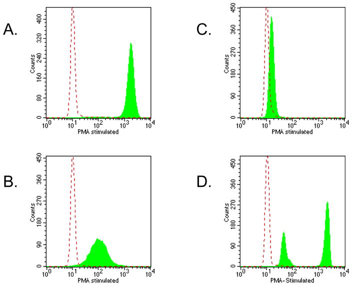
Primary immunodeficiency disorders (PIDDs) are a heterogeneous group of inherited disorders of the immune system. Currently more than 250 different PIDDs with a known genetic defect have been recognized. The diagnosis of many of these disorders is supported strongly by a wide variety of flow cytometry applications. Flow cytometry offers a rapid and sensitive tool for diagnosis and classification of PIDDs. It is applicable in the initial workup and subsequent management of several primary immunodeficiency diseases. As our understanding of the pathogenesis and management of these diseases increases, the majority of these tests can be easily established in the diagnostic laboratory. Thus, the focus of this article is on the application of flow cytometry in the diagnosis and/or evaluation of PIDDs.
Keywords: Diagnosis; Flow cytometry; Primary immunodeficiency.
Conflict of interest statement
Declaration on competing interests: Nil
Figures
Similar articles
-
Flow Cytometry, a Versatile Tool for Diagnosis and Monitoring of Primary Immunodeficiencies.Clin Vaccine Immunol. 2016 Apr 4;23(4):254-71. doi: 10.1128/CVI.00001-16. Print 2016 Apr. Clin Vaccine Immunol. 2016. PMID: 26912782 Free PMC article. Review.
-
Diagnostic Tools for Inborn Errors of Human Immunity (Primary Immunodeficiencies and Immune Dysregulatory Diseases).Curr Allergy Asthma Rep. 2018 Feb 22;18(3):19. doi: 10.1007/s11882-018-0770-1. Curr Allergy Asthma Rep. 2018. PMID: 29470720 Review.
-
Role of flow cytometry in the diagnosis and monitoring of primary immunodeficiency disease.Clin Lab Med. 2007 Sep;27(3):591-626, vii. doi: 10.1016/j.cll.2007.05.007. Clin Lab Med. 2007. PMID: 17658409 Review.
-
The utility of flow cytometry for the diagnosis of primary immunodeficiencies.Int J Lab Hematol. 2019 May;41 Suppl 1:63-72. doi: 10.1111/ijlh.13010. Int J Lab Hematol. 2019. PMID: 31069989 Review.
-
Current applications of flow cytometry in the diagnosis of primary immunodeficiency diseases.Arch Pathol Lab Med. 2004 Jan;128(1):23-31. doi: 10.5858/2004-128-23-CAOFCI. Arch Pathol Lab Med. 2004. PMID: 14692816 Review.
Cited by
-
Primary Immunodeficiency Diseases: Need for Awareness and Advocacy in India.Indian J Pediatr. 2016 Apr;83(4):328-30. doi: 10.1007/s12098-016-2070-x. Epub 2016 Feb 29. Indian J Pediatr. 2016. PMID: 26924652 No abstract available.
-
Coexistence of a Leaky SCID Phenotype With Hyperphenylalaninemia in an Adult Case.Case Reports Immunol. 2025 Mar 19;2025:9988821. doi: 10.1155/crii/9988821. eCollection 2025. Case Reports Immunol. 2025. PMID: 40151380 Free PMC article.
-
Changes in Treg and Breg cells in a healthy pediatric population.Front Immunol. 2023 Nov 21;14:1283981. doi: 10.3389/fimmu.2023.1283981. eCollection 2023. Front Immunol. 2023. PMID: 38077340 Free PMC article.
-
Inborn errors of immunity (primary immunodeficiencies).Allergy Asthma Clin Immunol. 2025 Jan 8;20(Suppl 3):76. doi: 10.1186/s13223-024-00938-z. Allergy Asthma Clin Immunol. 2025. PMID: 39780212 Free PMC article. Review.
-
Genetically confirmed chronic granulomatous disease in a Kenyan child: case report.Front Immunol. 2023 May 18;14:1172848. doi: 10.3389/fimmu.2023.1172848. eCollection 2023. Front Immunol. 2023. PMID: 37275907 Free PMC article.
References
-
- Oliveira JB, Fleisher TA. Molecular- and flow cytometry-based diagnosis of primary immunodeficiency disorders. Curr Allergy Asthma Rep. 2010;10(6):460–67. - PubMed
-
- Shearer WT, Rosenblatt HM, Gelman RS, et al. Pediatric AIDS Clinical Trials Group. Lymphocyte subsets in healthy children from birth through 18 years of age: the Pediatric AIDS Clinical Trials Group P1009 study. J Allergy Clin Immunol. 2003;112(5):973–80. - PubMed
-
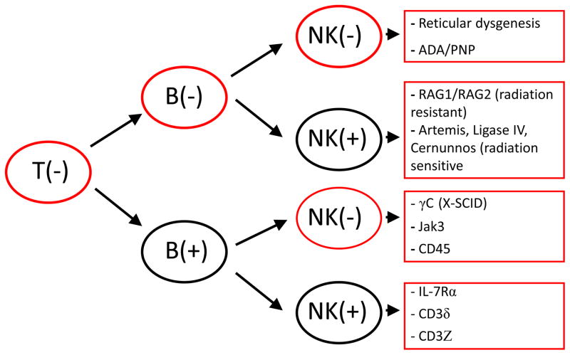
- Kalman L, Lindegren ML, Kobrynski L, Vogt R, Hannon H, Howard JT, Buckley R. Mutations in genes required for T-cell development: IL7R, CD45, IL2RG, JAK3, RAG1, RAG2, ARTEMIS, and ADA and severe combined immunodeficiency: HuGE review. Genet Med. 2004;6(1):16–26. - PubMed
Publication types
MeSH terms
Grants and funding
LinkOut - more resources
Full Text Sources
Other Literature Sources
Research Materials
