. 2016 Feb 11:6:20629.
doi: 10.1038/srep20629.

New insights into the mechanism of substrates trafficking in Glyoxylate/Hydroxypyruvate reductases

Affiliations

New insights into the mechanism of substrates trafficking in Glyoxylate/Hydroxypyruvate reductases

Louise Lassalle et al. Sci Rep. .

Erratum in

Abstract

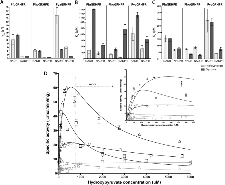
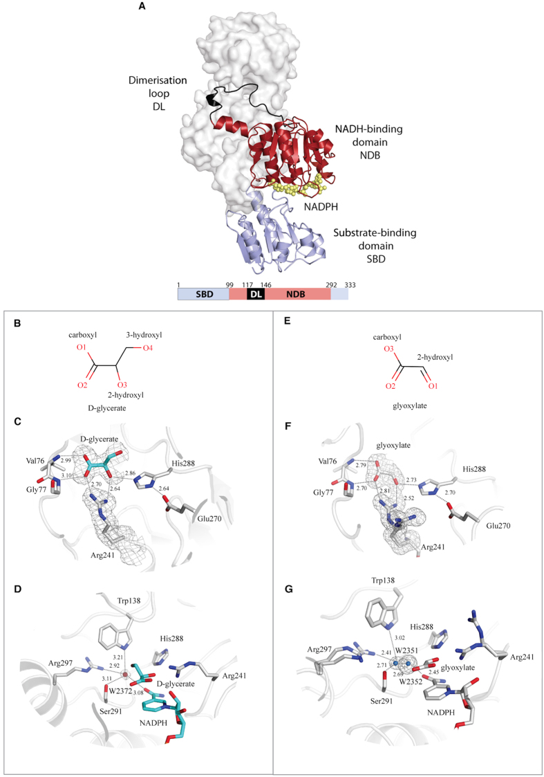
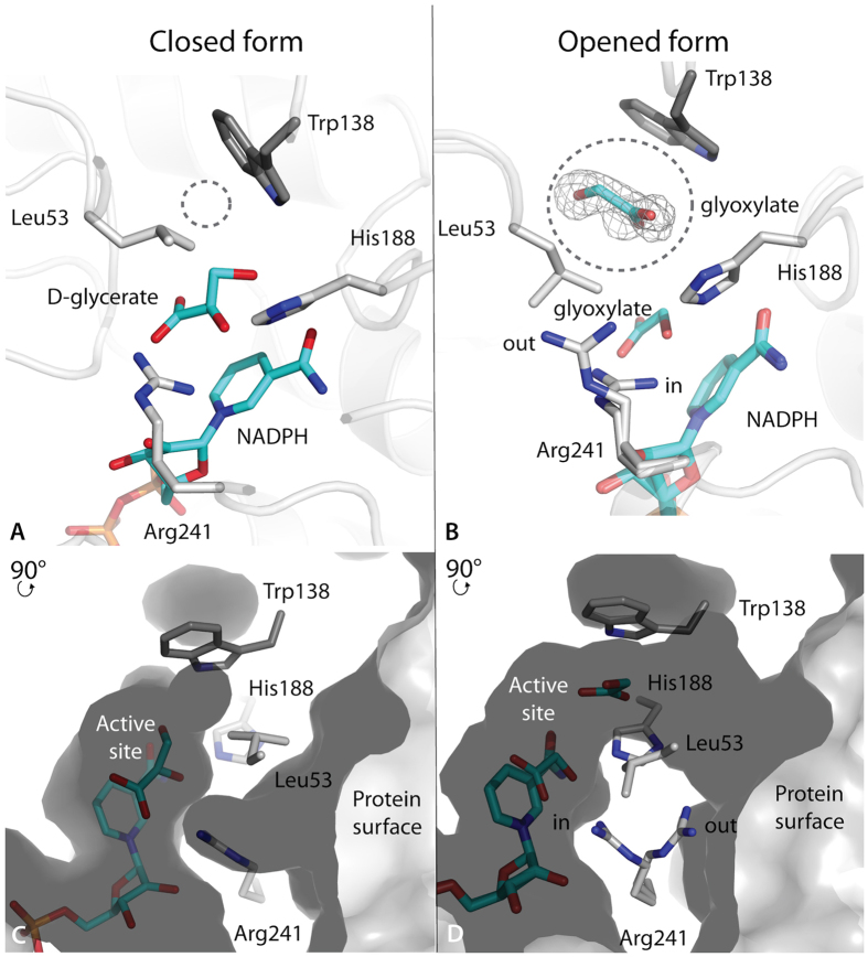
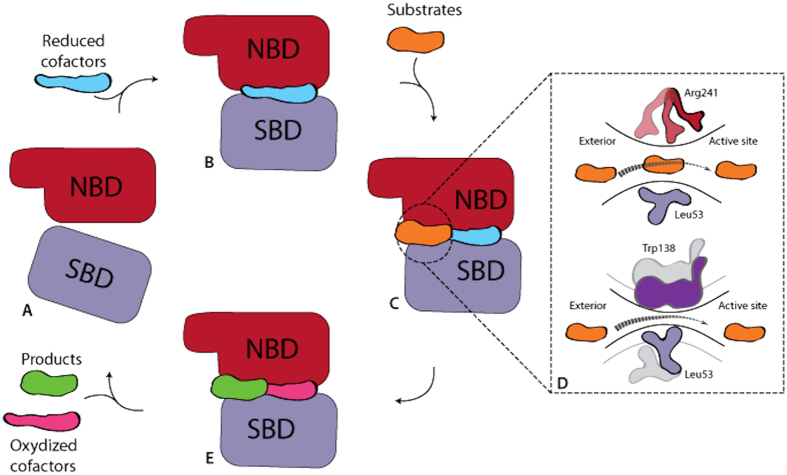
Glyoxylate accumulation within cells is highly toxic. In humans, it is associated with hyperoxaluria type 2 (PH2) leading to renal failure. The glyoxylate content within cells is regulated by the NADPH/NADH dependent glyoxylate/hydroxypyruvate reductases (GRHPR). These are highly conserved enzymes with a dual activity as they are able to reduce glyoxylate to glycolate and to convert hydroxypyruvate into D-glycerate. Despite the determination of high-resolution X-ray structures, the substrate recognition mode of this class of enzymes remains unclear. We determined the structure at 2.0 Å resolution of a thermostable GRHPR from Archaea as a ternary complex in the presence of D-glycerate and NADPH. This shows a binding mode conserved between human and archeal enzymes. We also determined the first structure of GRHPR in presence of glyoxylate at 1.40 Å resolution. This revealed the pivotal role of Leu53 and Trp138 in substrate trafficking. These residues act as gatekeepers at the entrance of a tunnel connecting the active site to protein surface. Taken together, these results allowed us to propose a general model for GRHPR mode of action.


Figures

Figure 1
Figure 1. Kinetics parameters of PfuGRHPR, PhoGRHPR and PyaGRHPR.
For all panels, measurements in presence of hydroxypyruvate and glyoxylate are represented in white and in black, respectively. (A) Catalytic activity of GRHPR enzymes for substrates. (B) Affinity values of GRHPR enzymes for substrates. (C) Affinity values of GRHPR enzymes for cofactors. (D) Specific activity (in mmol.min−1.mg−1) is represented as a function of hydroxypyruvate concentration (in mM) for PfuGRHPR (square), PhoGRHPR (circle) and PyaGRHPR (diamond) with NADH (black) and NADPH (gray) as cofactors. A close-up view of GRHPR activities at low hydroxypyruvate concentration (0–900 μM) is shown at right corner. Curve fit with Michaelis-Menten or substrate inhibition models are shown. Error bars are not visible when they are smaller than the font size used for the data point. Assays were performed as described under “Methods”.
Figure 2
Figure 2. Overall structure of GRHPR.
(A) GRHPRs are symmetrical homodimers with a large dimerization interface. One monomer is represented in cartoon while the adjacent subunit is shown as a molecular surface and white. NADPH is shown in sphere and yellow. The NADH-binding domain (residues 99–117 and 146–292), the substrate-binding domain (1–99 and 293–333) and the dimerisation loop (118–146) are represented in red, blue and black, respectively. (BD) Views of the active sites of PyaGRHPR in presence of D-glycerate: (B) Skeletal formula of D-glycerate, (C) PyaGRHPR active site in presence of D-glycerate, (D) Alternative view of PyaGRHPR active site in presence of D-glycerate showing a fragment of NADPH. (E–G) Views of the active sites of PfuGRHPR in presence of glyoxylate: (E) Skeletal formula of glyoxylate, (F) PfuGRHPR active site residues in presence of glyoxylate, (G) Alternative view of PfuGRHPR active site in presence of glyoxylate showing a fragment of NADPH. D-glycerate and glyoxylate are colored in cyan and white, respectively. Superimposed sigmaA-weighted Fo - Fc OMIT map contoured at 3.0 σ depicting substrate/product, key water molecules as well as Arg241. Distance are indicated in angstrom. For PyaGRHPR the indicated distances correspond to the average of the two molecules present in the asymmetric unit (Table 3).
Figure 3
Figure 3. Active site accessibility in PyaGRHPR and PfuGRHPR structures.
Top panel, details of the catalytic residues showing the cofactor as well as substrates, D-glycerate (A) and glyoxylate (B). The tunnel is indicated with a gray circle. In Panel (B), the second glyoxylate molecule is represented within the tunnel with the cage representing simulated-annealing sigmaA-weighted Fo - Fc OMIT map contoured at 3 sigma. Bottom panel, cross section of the surface representation showing the tunnel in closed form (C) and in opened form (D). Substrate and cofactor molecules are represented with sticks in cyan. Panels (A,C) are from PyaGRHPR structure with D-glycerate and panels (B,D) are from PfuGRHPR structure with glyoxylate.
Figure 4
Figure 4. Model of catalytic process in GRHPR.
(A) Schematic representation of GRHPR monomer with NBD domain in red and SBD domain in mauve. This corresponds to the apo form of the enzyme. (B) Binding of the cofactor induces a domains movement and formation of the tunnel. (C) Substrate binding. (D) Residue rearrangements associated with substrate trafficking. (E) Product release.

Similar articles

Cited by

References

    1. Yoshikawa S. et al.. Structure of archaeal glyoxylate reductase from Pyrococcus horikoshii OT3 complexed with nicotinamide adenine dinucleotide phosphate. Acta Crystallogr. D Biol. Crystallogr. 63, 357–365 (2007). - PubMed
    1. Rumsby G. & Cregeen D. P. Identification and expression of a cDNA for human hydroxypyruvate/glyoxylate reductase. Biochimica et Biophysica Acta 1446, 383–388 (1999). - PubMed
    1. Hullin R. P. Glyoxylate reductase, two forms from Pseudomonas. Meth. Enzymol. 41, 343–348 (1975). - PubMed
    1. Yoshida T. et al.. Cloning and expression of the gene for hydroxypyruvate reductase (D-glycerate dehydrogenase from an obligate methylotroph Hyphomicrobium methylovorum GM2. Eur. J. Biochem. 223, 727–732 (1994). - PubMed
    1. Nuñez M. F., Pellicer M. T., Badia J., Aguilar J. & Baldoma L. Biochemical characterization of the 2-ketoacid reductases encoded by ycdW and yiaE genes in Escherichia coli. Biochem. J. 354, 707–715 (2001). - PMC - PubMed

MeSH terms

LinkOut - more resources