. 2016 Feb 11:6:20643.
doi: 10.1038/srep20643.

Basic FGF or VEGF gene therapy corrects insufficiency in the intrinsic healing capacity of tendons

Affiliations

Basic FGF or VEGF gene therapy corrects insufficiency in the intrinsic healing capacity of tendons

Jin Bo Tang et al. Sci Rep. .

Abstract

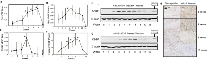
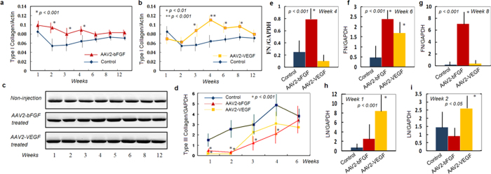
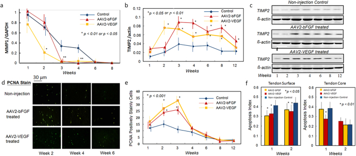
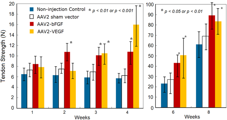
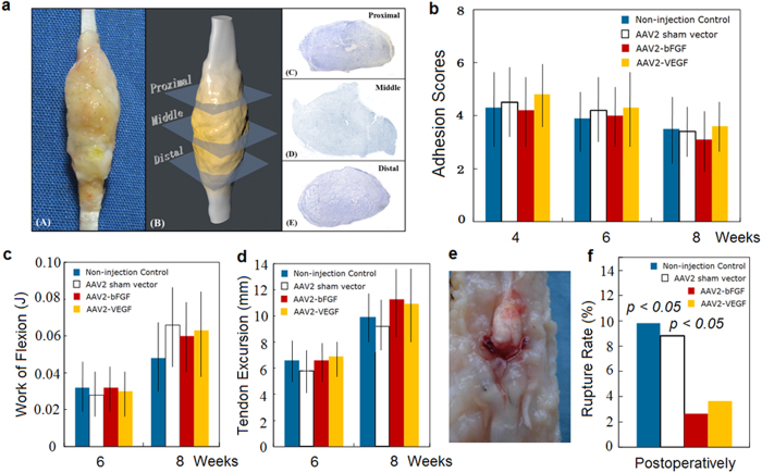
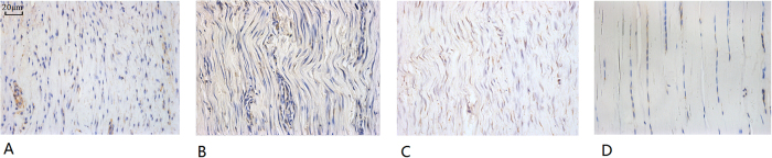
Tendon injury during limb motion is common. Damaged tendons heal poorly and frequently undergo unpredictable ruptures or impaired motion due to insufficient innate healing capacity. By basic fibroblast growth factor (bFGF) or vascular endothelial growth factor (VEGF) gene therapy via adeno-associated viral type-2 (AAV2) vector to produce supernormal amount of bFGF or VEGF intrinsically in the tendon, we effectively corrected the insufficiency of the tendon healing capacity. This therapeutic approach (1) resulted in substantial amelioration of the low growth factor activity with significant increases in bFGF or VEGF from weeks 4 to 6 in the treated tendons (p < 0.05 or p < 0.01), (2) significantly promoted production of type I collagen and other extracellular molecules (p < 0.01) and accelerated cellular proliferation, and (3) significantly increased tendon strength by 68-91% from week 2 after AAV2-bFGF treatment and by 82-210% from week 3 after AAV2-VEGF compared with that of the controls (p < 0.05 or p < 0.01). Moreover, the transgene expression dissipated after healing was complete. These findings show that the gene transfers provide an optimistic solution to the insufficiencies of the intrinsic healing capacity of the tendon and offers an effective therapeutic possibility for patients with tendon disunion.


Conflict of interest statement

The authors declare no competing financial interests.

Figures

Figure 1
Figure 1. Transgene and protein production in AAV2-bFGF or AAV2-VEGF injected tendons.
(a) Transgene (rat bFGF) expression in AAV2-bFGF injected tendon increased from weeks 1 to 3, peaked from weeks 4 to 8, dropped drastically after week 8, and was very low at week 12. *indicate the data significantly greater than that at other time-points (p < 0.05 or p < 0.01). (b) bFGF protein levels relative to beta-actin. *indicates the data significantly greater than that at weeks 1, 2, 12, 16 (p < 0.01 or p < 0.01).(c) Representative pictures of gel electrophoresis of western blot using mouse-anti-rat bFGF antibody. Rat bFGF was increased from weeks 2 to 4, peaked at weeks 4 and 5, and declined at weeks 6 to 12. The bFGF was not detectable at week 16. (d) immunohistochemistry showing the changes of the bFGF (chicken and rat origins) in the AAV2-bFGF injected and non-injection control tendons upto week 16. The bFGF was increased at weeks 2 and 4 in the AAV2-bFGF injected tendon. (e) Transgene (human VEGF) expression in the AAV2-VEGF injected tendon. Transgene expression peaked at week 4. The expression was minimal at week 6, 8, and 12. *indicates the data significantly greater than that at other time-points (p < 0.05 or p < 0.001). (f) Western blot analysis showed gradual increase in the expression of human VEGF from weeks 1 to 6. The VEGF peaked at week 6 and dropped thereafter. Expression of the VEGF relative to beta-actin is shown. *indicates the data significantly greater than that at week 1, 12, or 16 (p < 0.01 or p < 0.001). (g) gel pictures showing the changes in human VEGF. The VEGF was not present in the gel at week 16. The sample number (n) was 6 for analysis of gene expression and 4 for western blot analysis at each time point in each group.
Figure 2
Figure 2. Changes in expression of extracellular matrix after AAV2-bFGF or AAV2-VEGF injection to the tendons.
(a) Type I collagen were significantly increased at weeks 2, 3, and 4 in the AAV2-bFGF injected tendon compared with the non-injection controls (p < 0.001). (b) Type I collagen was significantly increased at weeks 3, 4, 6, and 8 in the AAV2-VEGF injected tendon (p < 0.01, or p < 0.001). (c) gel pictures showing the changes in protein levels of type I collagen. Note an earlier increase (weeks 2 to 5) of the collagen I after AAV2-bFGF injection, but a greater and more persistent increase (up to week 8) after AAV2-VEGF injection. (d) Changes in type III collagen gene expression of the AAV2-bFGF and AAV2-VEGF injected tendons compared with non-injection controls (p < 0.001, 1 to 4 weeks after AAV2-bFGF treatment, and 1 and 2 weeks after AAV2-VEGF treatment). (e–i) showing the real-time PCR analysis of changes in expression of the fibronectin (FN) at weeks 4, 6, and 8 and the laminin (LN) at weeks 1 and 2. Statistical significance is shown in the graph. * indicates the data of significant difference from those in the non-injection controls. Sample sizes at each time point in each group were 6 to 8 for gene expression analysis and 5 or 6 for western blot analysis.
Figure 3
Figure 3. Changes in regulators MMPs and TIMPs of metabolism in the AAV2-bFGF and AAV2-VEGF treated tendons.
(a,b) significant changes in the expression of the MMP1 and TIMP2 was found in the tendons after either AAV2-bFGF or AAV2-VEGF treatment (n = 6, in each group at each time point), typically from weeks 2 to 8 (*p < 0.05 or p < 0.01, compared with non-injection controls). (c) western blot gel pictures show that the TIMP2 was activated after the therapy from weeks 2 to 8 to inhibit collagen degradation. (d,e) PCNA staining showed significant increases in the positively-stained cells after injection of AAV2-bFGF or AAV2-VEGF at weeks 2 and 3. (d) the representative pictures (200 X magnification). (e). Data presented are from 6 fields of each of 6 tendon samples per group under 200 X magnification. *indicates data of significant difference from the non-injection controls at weeks 2 and 3.(f) apoptosis index of the AAV2-bFGF or AAV2-VEGF injected tendons and non-injection controls at weeks 1 and 2 (n = 6, each group at each time point, *p < 0.05 or p < 0.01). No significant difference was found in the number of the PCNA positively stained cells and apoptosis index in these groups at weeks 4, 6, 8, and 12 (data not shown). *indicates the data of significant difference from the non-injection controls. The data of sham vector controls (not shown) were not significantly different from the non-injection controls.
Figure 4
Figure 4. Tendon healing strengths (data of weeks 1, 2, 3, 4, 6, and 8 shown, n = 12, each group at each time point).
Compared with non-injection and sham vector controls, the strengths of the AAV2-bFGF injected tendon had significant increases from week 2 and lasted up to week 8 (p < 0.01 or p < 0.001). In contrast, AAV2-VEGF treatment brought more robust and significant increases at week 3 (p < 0.01) and week 4 (p < 0.001). The strengths of the tendons injected with AAV2-VEGF were significantly greater compared with non-injection controls or sham vector injection controls at weeks 6 and 8 (p < 0.05 or p < 0.01). No significant difference in the strengths between the sham vector and non-treatment controls (p > 0.05, statistical power > 0.80). Compared with the strengths of non-injection controls, the percent increases in the strength were 72%, 68% and 91% for the AAV2-bFGF treated tendons at weeks 2, 3, and 4, respectively, and the increases were 82% and 210% for the AAV2-VEGF treated tendons at week 3 and 4, respectively. *indicates the data of significant difference from those in the non-injection and sham vector controls at individual time points.
Figure 5
Figure 5. Effects of AAV2-bFGF and AAV2-VEGF injection to the tendon on adhesion formation and amplitude of tendon movement.
(a) a three-dimensional analysis method for quantification of adhesions around the tendon. The tendon is sectioned through 3 cross-sectional levels (0.5 cm apart, with the middle section at the site of tendon repair) and is stained histologically. The area of adhesions and the ratio of adhesions to the healing tendons are computed to obtain adhesion scores. (b) adhesion scores (n = 8, each group at each time point) are presented. No significant difference was found in the scores (shown in b) and area of adhesions (not shown). (c) work of flexion of the toes (n = 12, each group at each time point). (d) tendon excursions under 10 N load to the repaired FDP tendon (n = 12, each group at each time point). No significant differences were found in the work of flexion and tendon movement at week 6 and 8 (p > 0.05, statistical power > 0.85). (e) a picture shows a typical tendon rupture. (f) overall rate of tendon ruptures recorded during dissection in the samples for mechanical test at weeks 4, 5, 6, and 8 (48 toes at each group) after surgery. Significant differences in the rupture rate were noted between the AAV2-bFGF or AAV2-VEGF injection, sham vector and non-injection groups. P values shown are comparison of the non-injection and sham vector groups with the AAV2-bFGF or AAV2-VEGF injection groups.
Figure 6
Figure 6. Three sections of healing tendons at week 6 and the uninjured tendons shown.
(A) AAV2-bFGF treated tendon, (B) AAV2-VEGF treated tendon, (C) non-injection control tendon, and (D) uninjured tendon. Morphologically, the cellularity and collagen formation in AAV2-bFGF or AAV2-VEGF treated tendon (A,B) are greater than those in the non-treatment control (C) or uninjured tendon (D). This is at the beginning of the tendon remodeling, so cellularity in the tendon still much more robust in these healing tendons. The sections stained for immunohistochemistry were used for the observation (X400, magnification). Section shown in (A,C,D) was stained with mouse anti-rat bFGF antibody (05–118, Millipore Corp., Billerica, Mass.) and that shown in b was stained with mouse anti-human VEGF (Santa Cruz, Dallas, Texas).

Similar articles

Cited by

References

    1. de Jong J. P. et al. The incidence of acute traumatic tendon injuries in the hand and wrist: a 10-year population-based study. Clin Orthop Surg. 6, 196–202 (2014). - PMC - PubMed
    1. Clayton R. A. & Court-Brown C. M. The epidemiology of musculoskeletal tendinous and ligamentous injuries. Injury. 39, 1338–1344 (2008). - PubMed
    1. Evans C. H., Ghivizzani S. C. & Robbins P. D. Orthopedic gene therapy in 2008. Mol Ther. 17, 231–244 (2009). - PMC - PubMed
    1. Pennisi E. Tending tender tendons. Science. 295 (5557), 1011 (2002). - PubMed
    1. Beredjiklian P. K. Biological aspects of flexor tendon laceration and repair. J Bone Joint Surg (Am). 85, 539–550 (2003). - PubMed

Publication types

MeSH terms