. 2016 Mar;117(3):441-55.
doi: 10.1093/aob/mcv192. Epub 2016 Feb 9.

Pectic polysaccharides are attacked by hydroxyl radicals in ripening fruit: evidence from a fluorescent fingerprinting method

Affiliations

Pectic polysaccharides are attacked by hydroxyl radicals in ripening fruit: evidence from a fluorescent fingerprinting method

Othman B Airianah et al. Ann Bot. 2016 Mar.

Abstract

Background and aims: Many fruits soften during ripening, which is important commercially and in rendering the fruit attractive to seed-dispersing animals. Cell-wall polysaccharide hydrolases may contribute to softening, but sometimes appear to be absent. An alternative hypothesis is that hydroxyl radicals ((•)OH) non-enzymically cleave wall polysaccharides. We evaluated this hypothesis by using a new fluorescent labelling procedure to 'fingerprint' (•)OH-attacked polysaccharides.

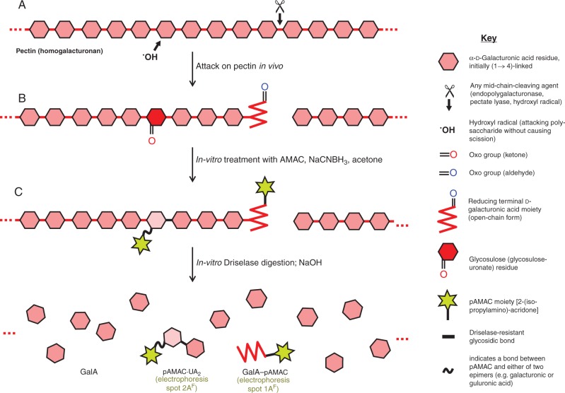
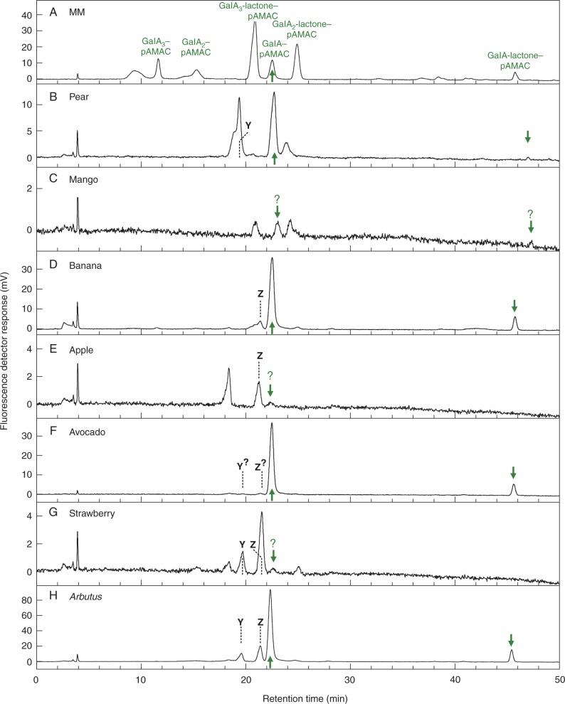
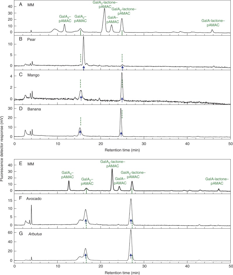
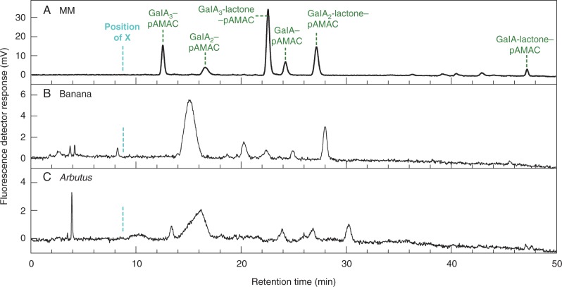
Methods: We tagged fruit polysaccharides with 2-(isopropylamino)-acridone (pAMAC) groups to detect (a) any mid-chain glycosulose residues formed in vivo during (•)OH action and (b) the conventional reducing termini. The pAMAC-labelled pectins were digested with Driselase, and the products resolved by high-voltage electrophoresis and high-pressure liquid chromatography.

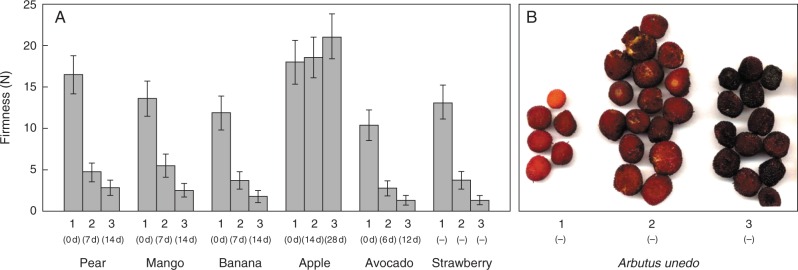
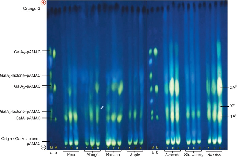
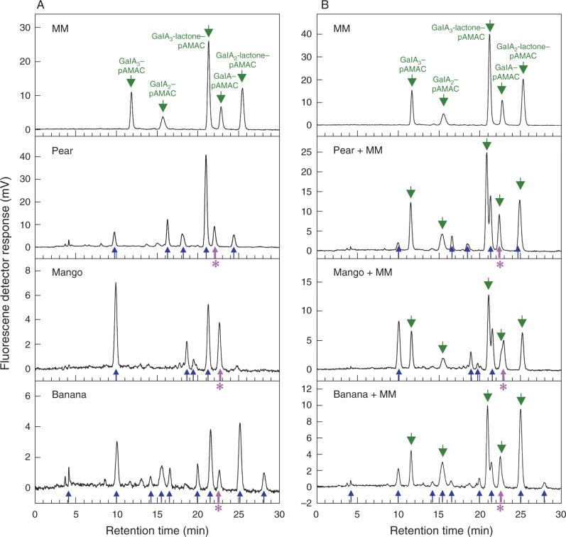
Key results: Strawberry, pear, mango, banana, apple, avocado, Arbutus unedo, plum and nectarine pectins all yielded several pAMAC-labelled products. GalA-pAMAC (monomeric galacturonate, labelled with pAMAC at carbon-1) was produced in all species, usually increasing during fruit softening. The six true fruits also gave pAMAC·UA-GalA disaccharides (where pAMAC·UA is an unspecified uronate, labelled at a position other than carbon-1), with yields increasing during softening. Among false fruits, apple and strawberry gave little pAMAC·UA-GalA; pear produced it transiently.

Conclusions: GalA-pAMAC arises from pectic reducing termini, formed by any of three proposed chain-cleaving agents ((•)OH, endopolygalacturonase and pectate lyase), any of which could cause its ripening-related increase. In contrast, pAMAC·UA-GalA conjugates are diagnostic of mid-chain oxidation of pectins by (•)OH. The evidence shows that (•)OH radicals do indeed attack fruit cell wall polysaccharides non-enzymically during softening in vivo. This applies much more prominently to drupes and berries (true fruits) than to false fruits (swollen receptacles). (•)OH radical attack on polysaccharides is thus predominantly a feature of ovary-wall tissue.

Keywords: Fruit; cell wall; fingerprint compounds; fluorescent labelling; hydroxyl radicals; non-enzymic scission; pectic polysaccharides; ripening.


Figures

F<sc>ig</sc>. 1.
Fig. 1.
Schematic view of in-vivo attack on pectins and strategies used to detect it. (A) Part of a pectin (homogalacturonan) chain in the wall of a living fruit cell may be attacked either non-enzymically by a hydroxyl radical (OH) or enzymically by endo-polygalacturonase or pectate lyase. Any of these three agents can cleave the backbone (e.g. at ✂), creating a new reducing terminus (shown in its non-cyclic form, and thus possessing an oxo group). In addition, OH can non-enzymically abstract an H atom (e.g. from C-2 or C-3 of a GalA residue) without causing chain scission; in an aerobic environment, this initial reaction leads to the formation of a relatively stable glycosulose residue possessing a mid-chain oxo group. (B) Wall material (AIR) is treated in vitro with AMAC, NaCNBH3 and acetone; oxo groups are reductively aminated to form yellow–green-fluorescing pAMAC conjugates. (C) The pAMAC-labelled homogalacturonan is then digested with Driselase, which hydrolyses all glycosidic bonds except any whose sugar residue carries a pAMAC group. The products tend to lactonize and are therefore briefly de-lactonized with NaOH before being fractionated. Further details of the reactions are given in figs 1 and 2 of Vreeburg et al. (2014).
F<sc>ig</sc>. 2.
Fig. 2.
Softening of fruits at three stages of ripening. (A) Firmness data were obtained by penetrometer at three stages of ripening (1–3). Values are means (n = 3) ± s.e. Stages of softening were at various days after purchase as stated in parentheses. Strawberry and Arbutus fruit were chosen based on their colour, the different stages being picked on the same day. (B) No firmness readings for the Arbutus berries are available as they were frozen immediately after picking; their appearance is illustrated here.
F<sc>ig</sc>. 3.
Fig. 3.
HVPE resolution of total Driselase digests of pAMAC-labelled AIR samples from seven fruit species. Fruit AIRs, each harvested at three stages of ripening (1–3; see Fig. 2), were successively treated with AMAC, acetone and Driselase (14 d); the pAMAC-labelled oligosaccharides generated were partially purified on a Supelco C18 cartridge column and de-lactonized in NaOH before electrophoresis. Each electrophoretogram loading was the products obtained from 20 mg f. wt of fruit tissue. Markers Ma and Mb are identical mixtures of acidic sugar–pAMAC conjugates before and after de-lactonization. Electrophoresis was at pH 6·5 and 4·0 kV for 45 min on Whatman No. 1 paper. Fluorescent spots were photographed under a 254-nm UV lamp. Orange G, loaded as a tracker between each fruit sample, shows up as a dark spot under UV. (+), anode; (–), cathode; –, blank loading.
F<sc>ig</sc>. 4.
Fig. 4.
HPLC of total Driselase digests of pAMAC-labelled cell walls from three fruit species. AIRs from pear, mango and banana fruit (stages 3, 1 and 1, respectively) were treated with AMAC, acetone, Driselase, Supelco C18 and NaOH, all as in Fig. 3. Total fluorescent products (which will include conjugates of both neutral and acidic carbohydrates) were analysed by HPLC (A) before and (B) after addition of a marker mixture containing acidic sugar–pAMAC conjugates. Fluorescence detection was with excitation at 442 nm and emission at 520 nm. MM, marker mixture containing authentic acidic sugar–pAMAC conjugates. Green arrows, authentic sugar–pAMACs (including those added as a ‘spike’); blue arrows, unidentified peaks from fruit cell wall digests; thick purple arrows with asterisk, putative GalA–pAMAC from fruit cell wall digests.
F<sc>ig</sc>. 5.
Fig. 5.
HPLC of the acidic monomer (1AF) spots from Driselase-digested pAMAC-labelled cell walls of seven fruit species. Each 1AF spot (pooled for all three stages of development for each fruit; de-lactonized) shown in Fig. 3 was eluted from the electrophoretogram and analysed by HPLC. MM, marker mixture containing authentic acidic sugar–pAMAC conjugates. Arrows, putative GalA–pAMAC (and its lactone, which partially re-formed during elution from the electrophoretogram) from fruit cell wall digests. Dashed lines, compounds Y and Z, discussed in the text.
F<sc>ig</sc>. 6.
Fig. 6.
HPLC of the acidic dimer (2AF) spots from Driselase-digested pAMAC-labelled cell walls of five fruit species. 2AF spots were eluted from a paper electrophoretogram (similar to that shown in Fig. 3 but derived from non-de-lactonized samples; all three ripening stages combined) and analysed by HPLC. MM, marker mixture containing authentic acidic sugar–pAMAC conjugates. Arrows, the proposed fingerprints for OH attack: pAMAC·UA-GalA and its lactone (rapidly re-formed during elution from the electrophoretogram) from fruit cell wall digests. Dashed lines, predicted position of authentic GalA2–pAMAC and GalA2-lactone–pAMAC, deduced from the marker run. The samples in the upper and lower graphs were run on different days, accounting for the slight discrepancy in marker retention times. Strawberry and apple were not included because they did not show any appreciable 2AF spot in Fig. 3.
F<sc>ig</sc>. 7.
Fig. 7.
HPLC of the acidic unknown (XF) spots from Driselase-digested pAMAC-labelled cell walls of banana and Arbutus. The XF spot (similar to that shown in Fig. 3 but from a non-de-lactonized sample) for stage-1 banana and Arbutus was eluted from an electrophoretogram and analysed by HPLC. MM, marker mixture containing authentic acidic sugar–pAMAC conjugates. The cyan dashed line indicates the approximate retention time of unknown ‘X’ (relative to the GalA3–pAMAC peak) seen in the products obtained from in-vitro OH-treated pectin (Vreeburg et al., 2014). Green dashed lines indicate the authentic markers.

References

    1. Albersheim P, Darvill A, Roberts K, Sederoff R, Staehelin A. 2010. Plant cell walls. From chemistry to biology. New York: Garland Science.
    1. de Alcântara PH, Martim L, Silva CO, Dietrich SM, Buckeridge MS. 2006. Purification of a β-galactosidase from cotyledons of Hymenaea courbaril L. (Leguminosae). Enzyme properties and biological function. Plant Physiology and Biochemistry 44: 619–627. - PubMed
    1. Ali ZM, Chin LH, Lazan H. 2004. A comparative study on wall degrading enzymes, pectin modifications and softening during ripening of selected tropical fruits. Plant Science 167: 317–327.
    1. Asthir B, Duffus CM, Smith RC, Spoor W. 2002. Diamine oxidase is involved in H2O2 production in the chalazal cells during barley grain filling. Journal of Experimental Botany 53: 677–682. - PubMed
    1. Basanta MF, Ponce NM, Salum ML, et al. 2014. Compositional changes in cell wall polysaccharides from five sweet cherry (Prunus avium L.) cultivars during on-tree ripening. Journal of Agricultural and Food Chemistry 62: 12418–12427. - PubMed

Publication types