

Ocular Findings in Infants With Microcephaly Associated With Presumed Zika Virus Congenital Infection in Salvador, Brazil
- PMID: 26865554
- PMCID: PMC5444996
- DOI: 10.1001/jamaophthalmol.2016.0267
Ocular Findings in Infants With Microcephaly Associated With Presumed Zika Virus Congenital Infection in Salvador, Brazil
Abstract
Importance: The Zika virus (ZIKV) has rapidly reached epidemic proportions, especially in northeastern Brazil, and has rapidly spread to other parts of the Americas. A recent increase in the prevalence of microcephaly in newborn infants and vision-threatening findings in these infants is likely associated with the rapid spread of ZIKV.
Objective: To evaluate the ocular findings in infants with microcephaly associated with presumed intrauterine ZIKV infection in Salvador, Bahia, Brazil.
Design, setting, and participants: Case series at a tertiary hospital. Twenty-nine infants with microcephaly (defined by a cephalic circumference of ≤32 cm) with a presumed diagnosis of congenital ZIKV were recruited through an active search and referrals from other hospitals and health unities. The study was conducted between December 1 and December 21, 2015.
Interventions: All infants and mothers underwent systemic and ophthalmic examinations from December 1 through December 21, 2015, in the Roberto Santos General Hospital, Salvador, Brazil. Anterior segment and retinal, choroidal, and optic nerve abnormalities were documented using a wide-field digital imaging system. The differential diagnosis included toxoplasmosis, rubella, cytomegalovirus, herpes simplex virus, syphilis, and human immunodeficiency virus, which were ruled out through serologic and clinical examinations.
Main outcomes and measures: Ocular abnormalities associated with ZIKV.
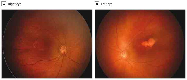
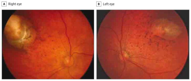
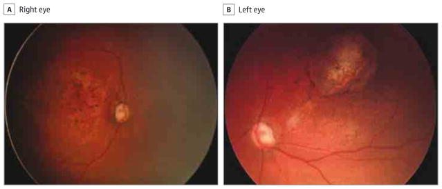
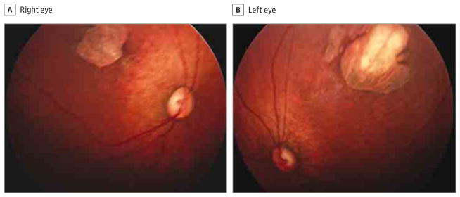
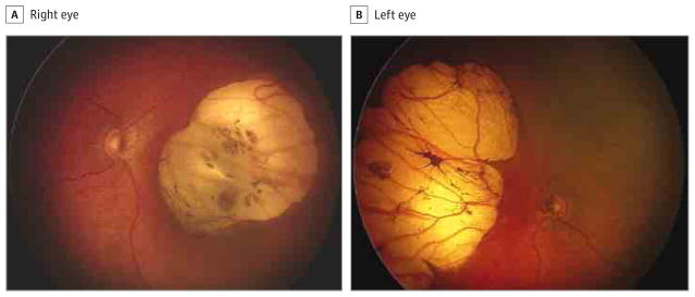
Results: Twenty-three of 29 mothers (79.3%) reported suspected ZIKV infection signs and symptoms during pregnancy, 18 in the first trimester, 4 in the second trimester, and 1 in the third trimester. Of the 29 infants (58 eyes) examined (18 [62.1%] female), ocular abnormalities were present in 17 eyes (29.3%) of 10 children (34.5%). Bilateral findings were found in 7 of 10 patients presenting with ocular lesions, the most common of which were focal pigment mottling of the retina and chorioretinal atrophy in 11 of the 17 eyes with abnormalities (64.7%), followed by optic nerve abnormalities in 8 eyes (47.1%), bilateral iris coloboma in 1 patient (2 eyes [11.8%]), and lens subluxation in 1 eye (5.9%).
Conclusions and relevance: Congenital infection due to presumed ZIKV exposure is associated with vision-threatening findings, which include bilateral macular and perimacular lesions as well as optic nerve abnormalities in most cases.
Conflict of interest statement
Figures
Comment in
-
Zika Virus, Microcephaly, and Ocular Findings.JAMA Ophthalmol. 2016 Aug 1;134(8):946. doi: 10.1001/jamaophthalmol.2016.1313. JAMA Ophthalmol. 2016. PMID: 27253205 No abstract available.
-
Zika Virus, Microcephaly, and Ocular Findings-Reply.JAMA Ophthalmol. 2016 Aug 1;134(8):946-7. doi: 10.1001/jamaophthalmol.2016.1305. JAMA Ophthalmol. 2016. PMID: 27253365 No abstract available.
-
Zika Virus, Microcephaly, and Ocular Findings-Reply.JAMA Ophthalmol. 2016 Aug 1;134(8):946. doi: 10.1001/jamaophthalmol.2016.1307. JAMA Ophthalmol. 2016. PMID: 27253432 No abstract available.
-
Zika Virus, Microcephaly, and Ocular Findings.JAMA Ophthalmol. 2016 Aug 1;134(8):945. doi: 10.1001/jamaophthalmol.2016.1303. JAMA Ophthalmol. 2016. PMID: 27254835 No abstract available.
-
Similarities in the Retinal Appearance of Patients With Zika Virus Compared With Cobalamin C Deficiency.JAMA Ophthalmol. 2016 Oct 1;134(10):1200-1201. doi: 10.1001/jamaophthalmol.2016.2398. JAMA Ophthalmol. 2016. PMID: 27490307 No abstract available.
-
Similarities in the Retinal Appearance of Patients With Zika Virus Compared With Cobalamin C Deficiency-Reply.JAMA Ophthalmol. 2016 Oct 1;134(10):1201. doi: 10.1001/jamaophthalmol.2016.2404. JAMA Ophthalmol. 2016. PMID: 27490445 No abstract available.
References
-
- Dick GW, Kitchen SF, Haddow AJ. Zika virus, I: isolations and serological specificity. Trans R Soc Trop Med Hyg. 1952;46(5):509–520. - PubMed
-
- Dick GW. Zika virus, II: pathogenicity and physical properties. Trans R Soc Trop Med Hyg. 1952;46(5):521–534. - PubMed
-
- Ioos S, Mallet HP, Leparc Goffart I, Gauthier V, Cardoso T, Herida M. Current Zika virus epidemiology and recent epidemics. Med Mal Infect. 2014;44(7):302–307. - PubMed
-
- Tan PC, Rajasingam G, Devi S, Omar SZ. Dengue infection in pregnancy: prevalence, vertical transmission, and pregnancy outcome. Obstet Gynecol. 2008;111(5):1111–1117. - PubMed
Grants and funding
LinkOut - more resources
Full Text Sources
Other Literature Sources
