. 2016 Feb 10;8(325):325ra18.
doi: 10.1126/scitranslmed.aad6833.

Filaggrin inhibits generation of CD1a neolipid antigens by house dust mite-derived phospholipase

Affiliations

Filaggrin inhibits generation of CD1a neolipid antigens by house dust mite-derived phospholipase

Rachael Jarrett et al. Sci Transl Med. .

Abstract

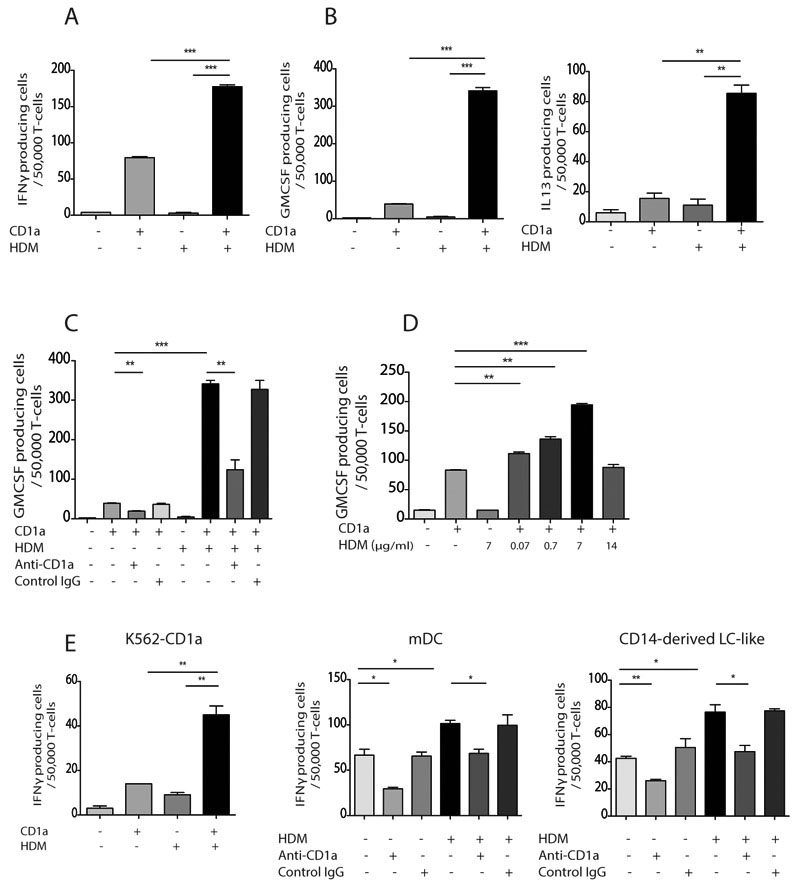
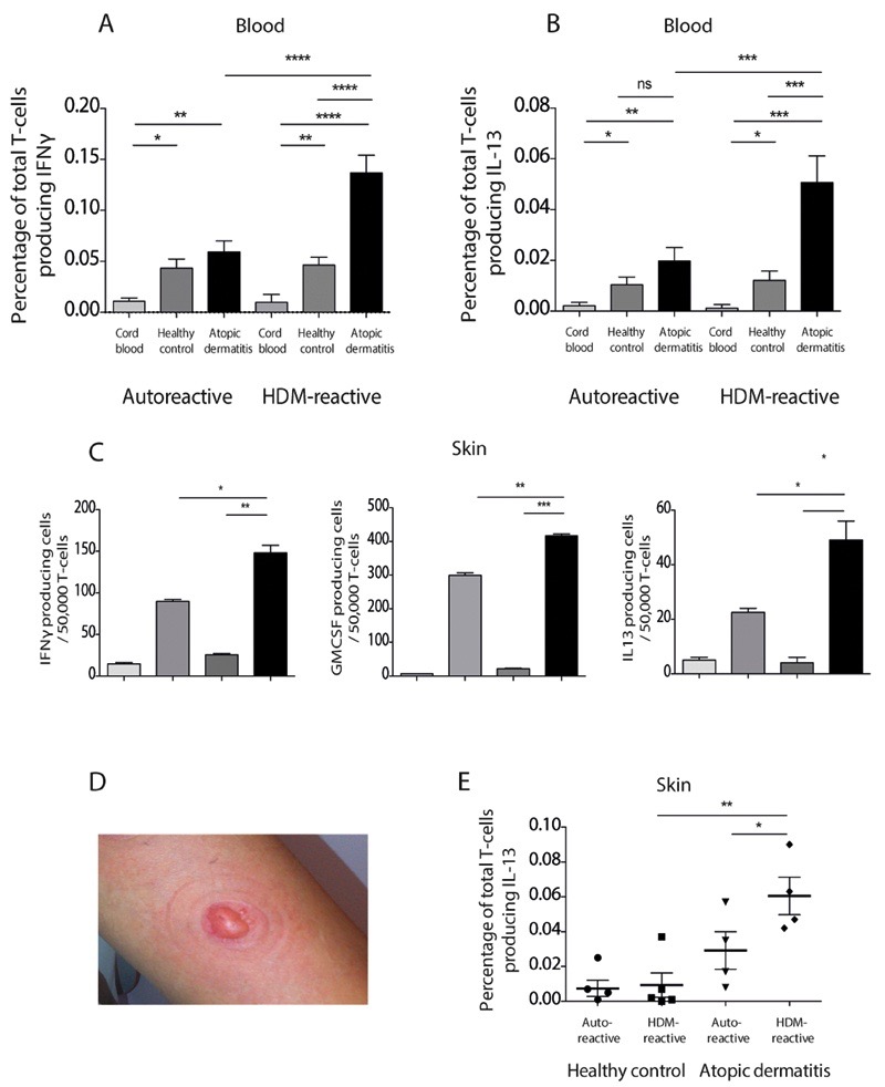
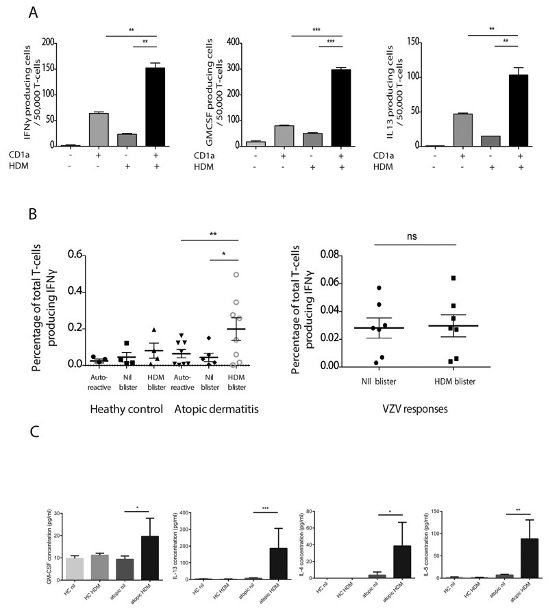
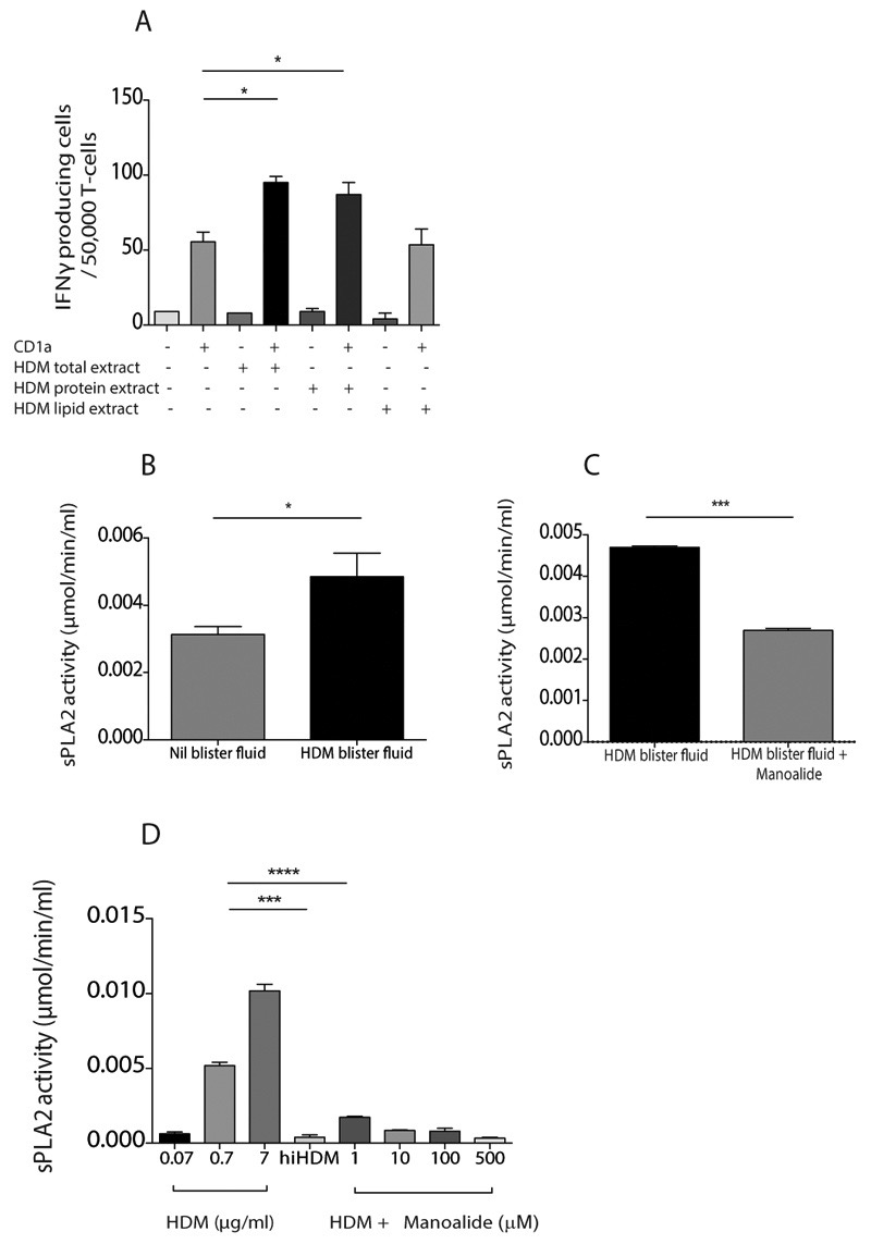
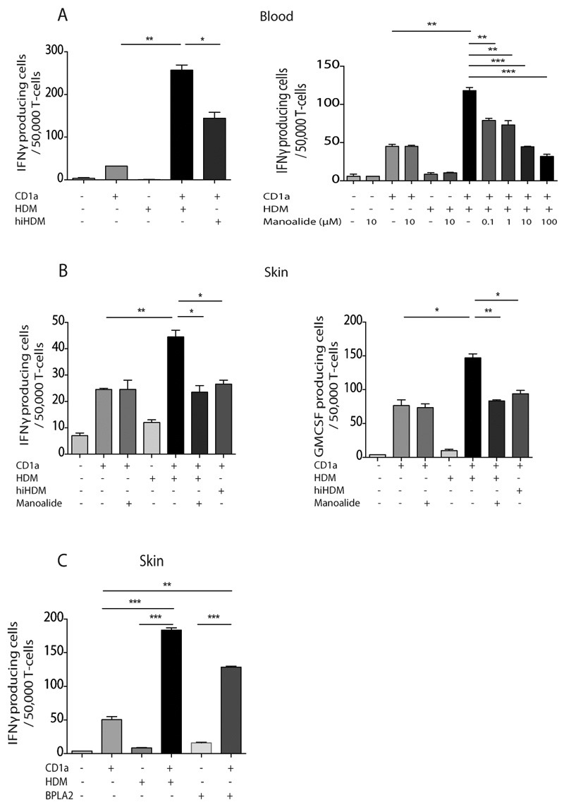
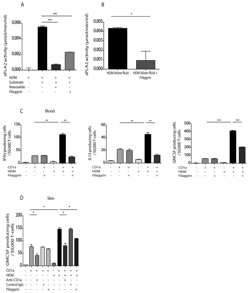
Atopic dermatitis is a common pruritic skin disease in which barrier dysfunction and cutaneous inflammation contribute to pathogenesis. Mechanisms underlying the associated inflammation are not fully understood, and although Langerhans cells expressing the nonclassical major histocompatibility complex (MHC) family member CD1a are known to be enriched within lesions, their role in clinical disease pathogenesis has not been studied. We observed that house dust mite (HDM) allergen generates neolipid antigens presented by CD1a to T cells in the blood and skin lesions of affected individuals. HDM-responsive CD1a-reactive T cells increased in frequency after birth in individuals with atopic dermatitis and showed rapid effector function, consistent with antigen-driven maturation. In HDM-challenged human skin, we observed phospholipase A2 (PLA2) activity in vivo. CD1a-reactive T cell activation was dependent on HDM-derived PLA2, and such cells infiltrated the skin after allergen challenge. Moreover, we observed that the skin barrier protein filaggrin, insufficiency of which is associated with atopic skin disease, inhibited PLA2 activity and decreased CD1a-reactive PLA2-generated neolipid-specific T cell activity from skin and blood. The most widely used classification schemes of hypersensitivity suggest that nonpeptide stimulants of T cells act as haptens that modify peptides or proteins; however, our results show that HDM proteins may also generate neolipid antigens that directly activate T cells. These data define PLA2 inhibition as a function of filaggrin, supporting PLA2 inhibition as a therapeutic approach.


Conflict of interest statement

Competing interests

The authors declare no competing interests.

Figures

Figure 1
Figure 1. Circulating CD1a-reactive house dust mite-responsive T cells produce IFNγ, GM-CSF and IL-13.
T cells were isolated by CD3 MACS beads from donor PBMC (R9500) and incubated overnight with CD1a-transfected K562 (CD1a) or untransfected K562 (EV) cells pulsed with HDM extract. IFN-γ (A), GM-CSF (B, left) and IL-13 (B, right) production were measured by ELISpot in the absence or presence of anti-CD1a antibody (C) and at different HDM concentrations (D). CD1a-expressing K562 (E, left), in vitro derived mDC (E, middle) or LC-like cells (E, right) were pulsed with HDM extract overnight and incubated with autologous peripheral blood T cells from donor R2. IFNγ production was measured by ELISpot in the presence or absence of anti-CD1a antibody. Data representative of at least three donors for each experiment are shown. Bars represent standard error. * P<0.05; ** P<0.01; ***P<0.001;****P<0.0001, t test.
Figure 2
Figure 2. HDM-responsive CD1a-reactive T cells are enriched in blood and skin of atopic dermatitis patients.
(A,B) T cells derived from the peripheral blood of healthy controls (HC), patients with atopic dermatitis (AD) or from cord blood were incubated with CD1a-transfected K562 or untransfected K562 cells in the presence or absence of HDM extract. IFNγ (A) and IL-13 (B) production were measured by ELISpot and expressed as percentage of responding T cells (A n=30 HC, 24 AD, 10 cord blood; B n=20 HC, 17 AD, 10 cord blood). Auto-reactive is the response to CD1a-K562 in the absence of HDM. (C) Skin blister T cells from donor R229 were incubated with CD1a-transfected K562 or untransfected K562 cells in the presence or absence of HDM extract. IFNγ (C, left), GM-CSF (C, middle) and IL-13 (C, right) production were measured by ELISpot. Data are representative of at least three separate donors for each experiment. (D) Example of skin suction blister raised after 60 minutes of 200mmHg negative pressure. (E) Skin blister T cells were isolated from unchallenged skin of healthy controls (n=5) or patients with atopic dermatitis (n=4) and incubated with CD1a-transfected or untransfected K562 cells in the presence or absence of HDM extract. IFNγ production was measured by ELISpot and expressed as percentage of CD1a-reactive T cells. Bars represent standard error. * P<0.05; ** P<0.01; ***P<0.001;****P<0.0001, t test .
Figure 3
Figure 3. HDM-responsive CD1a-reactive T cells infiltrate skin after HDM challenge.
Skin blister T cells were isolated from donor R4 24 hours after HDM skin challenge, expanded and incubated with CD1a-transfected or untransfected K562 cells in the presence or absence of HDM extract. IFNγ (A, left), GM-CSF (A, middle) and IL-13 (A, right) production by donor R4 cells were measured by ELISpot. The data are representative of at least three separate donors for each experiment. (B, left panel) Overall frequencies of HDM-responsive CD1a-reactive IFNγ-producing T cells infiltrating human skin after saline or HDM skin challenge were compared between healthy controls (n=4) and atopic dermatitis patients (n=8). Auto-reactive refers to responses to unpulsed K562-CD1a cells. (B, right panel) Skin blister cells derived from HDM-challenged or unchallenged skin were incubated with live attenuated varicella zoster virus and IFNγ production was measured by ELISpot. (C) Concentrations of type 2 cytokines were measured in skin blister fluid by multiplex bead array after saline (nil) or HDM skin challenge in healthy controls (HC, n=8) or atopic (n=16) individuals. Bars represent standard error. * P<0.05; ** P<0.01; ***P<0.001;****P<0.0001, t test.
Figure 4
Figure 4. HDM extract contains PLA2 activity in vivo and in vitro.
(A) T cells were isolated by CD3 MACS beads from healthy donor PBMC and incubated overnight with CD1a-transfected K562 (CD1a) or untransfected K562 (EV) cells pulsed with HDM total extract, aqueous protein phase or lipid phase. IFN-γ production was measured by ELISpot (A). PLA2 activity in saline or HDM challenged skin blister fluid was detected by measuring free thiol release in the presence of the diheptanoyl thio-PC substrate in the absence (B) or presence (C) of 10μM of the PLA2 inhibitor manoalide. (D) PLA2 activity in HDM extract in vitro was detected by measuring free thiol release in the presence of the diheptanoyl thio-PC substrate and manoalide and after heat inactivation (HDMhi). Data are representative of at least three separate experiments. Bars represent standard error. * P<0.05; ** P<0.01; ***P<0.001;****P<0.0001, t test.
Figure 5
Figure 5. HDM-derived PLA2 generates neolipid antigens for presentation by CD1a to blood and skin T cells.
T cells were isolated by CD3 MACS beads from healthy donor PBMC and incubated overnight with CD1a-transfected K562 (CD1a) or untransfected K562 (EV) cells pulsed with HDM total extract. IFN-γ production was measured by ELISpot after HDM heat inactivation (hiHDM) (A, left) or overnight incubation with a dose titration of the PLA2 inhibitor manoalide (A, right). Skin blister T cells were isolated 24 hours after HDM skin challenge, expanded and incubated with CD1a-transfected or untransfected K562 cells in the presence or absence of HDM extract that had been heat inactivated or incubated with manoalide. IFNγ (B, left) and GM-CSF (B, right) production were measured by ELISpot. (C) Skin blister T cells were isolated 24 hours after HDM skin challenge, expanded and incubated with CD1a-transfected or untransfected K562 cells in the presence or absence of HDM extract or 1μg/ml purified bee venom PLA2. IFNγ production was measured by ELISpot. Data are representative of at least three separate experiments. Bars represent standard error. * P<0.05; ** P<0.01; ***P<0.001;****P<0.0001, t test.
Figure 6
Figure 6. Filaggrin inhibits HDM PLA2 activity and inhibits responses of HDM-responsive CD1a-reactive T cells isolated from blood and skin.
(A) PLA2 activity in HDM extract in vitro was detected by measuring free thiol release in the presence of the diheptanoyl thio-PC substrate, 10μM of the PLA2 inhibitor manoalide and 1μg/ml human recombinant filaggrin. (B) PLA2 activity in HDM challenged skin blister fluid was detected by measuring free thiol release diheptanoyl thio-PC substrate in the absence or presence of filaggrin. (C) T cells were isolated by CD3 MACS beads from healthy donor PBMC and incubated overnight with CD1a-transfected K562 or untransfected K562 cells pulsed with HDM total extract. GM-CSF, IL-13 and IFNγ production were measured by ELISpot in the presence or absence of filaggrin. (D) Skin blister T cells were isolated 24 hours after HDM skin challenge and incubated with CD1a-transfected or untransfected K562 cells in the presence or absence of HDM extract, filaggrin or anti-CD1a antibody. Data are representative of at least three separate experiments. Bars represent standard error. * P<0.05; ** P<0.01; ***P<0.001;****P<0.0001, t test.

References

    1. Asher MI, Montefort S, Bjorksten B, Lai CK, Strachan DP, Weiland SK, Williams H. Worldwide time trends in the prevalence of symptoms of asthma, allergic rhinoconjunctivitis, and eczema in childhood: ISAAC Phases One and Three repeat multicountry cross-sectional surveys. Lancet. 2006;368:733–743. - PubMed
    1. Barnes KC. An update on the genetics of atopic dermatitis: scratching the surface in 2009. J Allergy Clin Immunol. 2010;125:16–29 e11-11. - PMC - PubMed
    1. Palmer CN, Irvine AD, Terron-Kwiatkowski A, Zhao Y, Liao H, Lee SP, Goudie DR, Sandilands A, Campbell LE, Smith FJ, O'Regan GM, et al. Common loss-of-function variants of the epidermal barrier protein filaggrin are a major predisposing factor for atopic dermatitis. Nat Genet. 2006;38:441–446. - PubMed
    1. Rodríguez E, Baurecht H, Herberich E, Wagenpfeil S, Brown SJ, Cordell HJ, Irvine AD, Weidinger S. Meta-analysis of filaggrin polymorphisms in eczema and asthma: Robust risk factors in atopic disease. Journal of Allergy and Clinical Immunology. 2009;123:1361–1370.e1367. - PubMed
    1. Sandford AJ, Shirakawa T, Moffatt MF, Daniels SE, Ra C, Faux JA, Young RP, Nakamura Y, Lathrop GM, Cookson WO, et al. Localisation of atopy and beta subunit of high-affinity IgE receptor (Fc epsilon RI) on chromosome 11q. Lancet. 1993;341:332–334. - PubMed

Publication types

MeSH terms