. 2016 Feb 10;36(6):1930-41.
doi: 10.1523/JNEUROSCI.0350-15.2016.

Aβ40 Reduces P-Glycoprotein at the Blood-Brain Barrier through the Ubiquitin-Proteasome Pathway

Affiliations

Aβ40 Reduces P-Glycoprotein at the Blood-Brain Barrier through the Ubiquitin-Proteasome Pathway

Anika M S Hartz et al. J Neurosci. .

Abstract

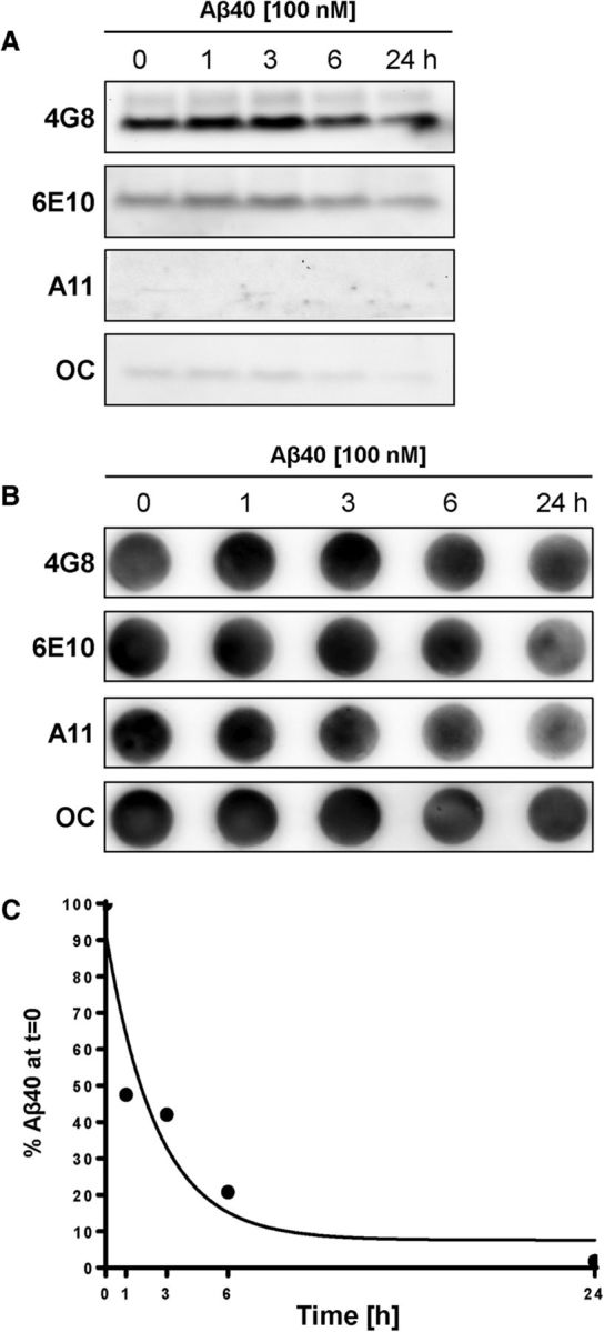
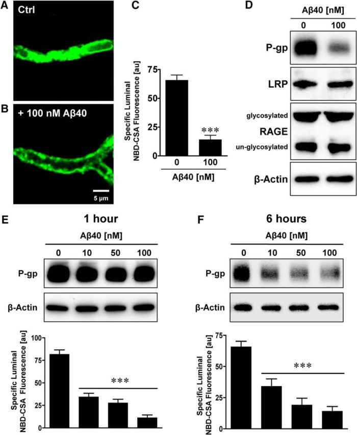
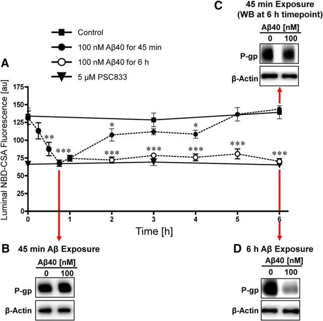
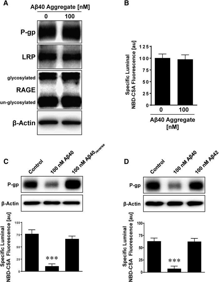
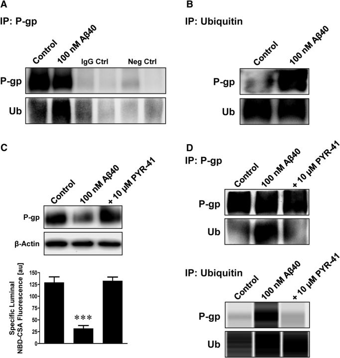
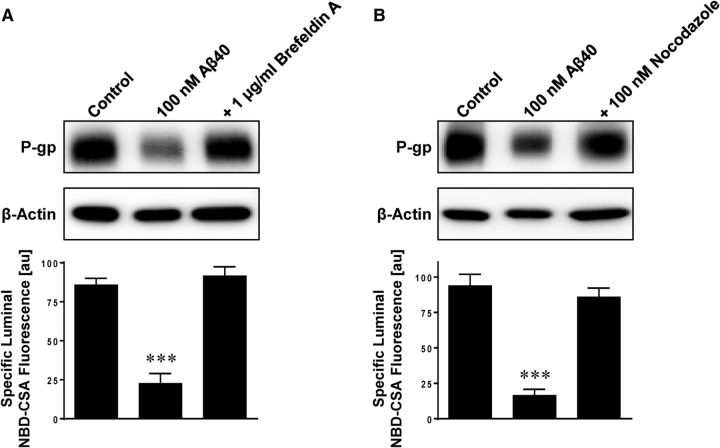
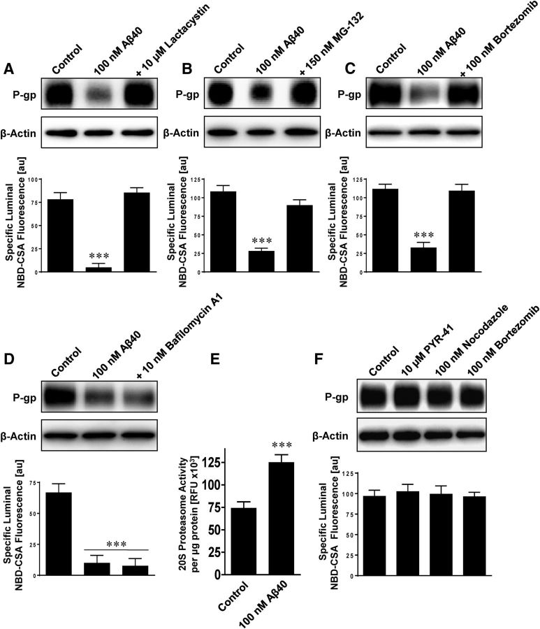
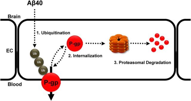
Failure to clear amyloid-β (Aβ) from the brain is in part responsible for Aβ brain accumulation in Alzheimer's disease (AD). A critical protein for clearing Aβ across the blood-brain barrier is the efflux transporter P-glycoprotein (P-gp) in the luminal plasma membrane of the brain capillary endothelium. P-gp is reduced at the blood-brain barrier in AD, which has been shown to be associated with Aβ brain accumulation. However, the mechanism responsible for P-gp reduction in AD is not well understood. Here we focused on identifying critical mechanistic steps involved in reducing P-gp in AD. We exposed isolated rat brain capillaries to 100 nm Aβ40, Aβ40, aggregated Aβ40, and Aβ42. We observed that only Aβ40 triggered reduction of P-gp protein expression and transport activity levels; this occurred in a dose- and time-dependent manner. To identify the steps involved in Aβ-mediated P-gp reduction, we inhibited protein ubiquitination, protein trafficking, and the ubiquitin-proteasome system, and monitored P-gp protein expression, transport activity, and P-gp-ubiquitin levels. Thus, exposing brain capillaries to Aβ40 triggers ubiquitination, internalization, and proteasomal degradation of P-gp. These findings may provide potential therapeutic targets within the blood-brain barrier to limit P-gp degradation in AD and improve Aβ brain clearance.

Significance statement: The mechanism reducing blood-brain barrier P-glycoprotein (P-gp) in Alzheimer's disease is poorly understood. In the present study, we focused on defining this mechanism. We demonstrate that Aβ40 drives P-gp ubiquitination, internalization, and proteasome-dependent degradation, reducing P-gp protein expression and transport activity in isolated brain capillaries. These findings may provide potential therapeutic avenues within the blood-brain barrier to limit P-gp degradation in Alzheimer's disease and improve Aβ brain clearance.

Keywords: Alzheimer's disease; P-glycoprotein; blood–brain barrier; transporter; ubiquitin–proteasome system.


Figures

Figure 1.
Figure 1.
Determination of Aβ40 forms. A, Western blot showing bands for Aβ40 at 4 kDa with 4G8 antibody (recognizes Aβ17–24), weak bands at 4 kDa with 6E10 antibody (recognizes Aβ1–17), and no bands with A11 antibody (detects amyloid oligomers) and faint bands with OC antibody (detects amyloid fibrils) 0, 1, 6, and 24 h after making the 100 nm Aβ40 working solution in PBS. B, Dot blots showing low Aβ40 levels with 4G8, 6E10, A11, and OC antibodies that are decreasing over the course of 24 h. C, Oligomer-specific configuration ELISA shows that the total amount of Aβ40 in solution decreases over time.
Figure 2.
Figure 2.
Aβ40 reduces P-gp protein expression and transport function. Representative confocal microscopy images of isolated brain capillaries that were exposed to 100 nm Aβ40 for 6 h and then exposed to the fluorescent P-gp substrate NBD-CSA (marker for P-gp transport activity). A, Representative image of an isolated brain capillary after 1 h of exposure to 2 μm NBD-CSA, showing steady-state NBD-CSA fluorescence. B, NBD-CSA fluorescence in the capillary lumen is reduced in capillaries that were exposed to Aβ40 for 6 h, indicating a decrease in P-gp transport activity. C, Specific NBD-CSA fluorescence in the capillary lumen was obtained through analysis of the confocal images and shows decreased luminal fluorescence in capillaries exposed to 100 nm Aβ40. D, Western blot showing that 100 nm Aβ40 reduces P-gp protein expression but has no effect on LRP or RAGE protein expression levels. E, Western blot showing that P-gp protein levels in capillaries exposed to 10–100 nm Aβ40 for 1 h remain at control levels. In contrast, P-gp transport activity levels decreased. F, Western blot showing that P-gp protein and transport activity levels decreased in a concentration-dependent manner after 6 h of Aβ40 exposure. β-Actin was used as protein loading control for Western blots. C, E, F, Data were obtained through analysis of the confocal images. Specific NBD-CSA fluorescence is the difference between total luminal fluorescence and fluorescence in the presence of the specific P-gp inhibitor PSC833, representing specific P-gp transport activity. Data are mean ± SEM (n = 10–15 capillaries per treatment group from one brain capillary isolation; pooled tissue from 10 rats). Units are arbitrary fluorescence units (scale, 0–255). ***p < 0.001, significantly lower than control.
Figure 3.
Figure 3.
Time course of Aβ40-mediated P-gp reduction. A, The effect of Aβ40 on P-gp transport activity is reversible. Capillaries were loaded for 60 min to steady state with 2 μm NBD-CSA. When 100 nm Aβ40 was added to the buffer (time 0 on graph), P-gp transport activity decreased rapidly; P-gp activity stayed decreased in capillaries exposed to Aβ40 for 6 h but recovered completely when Aβ40 was removed at time point 45 min. Western blot showing (B) no change of P-gp protein expression after 45 min exposure to Aβ40 and (C) no change of P-gp expression in capillaries that were first exposed to Aβ40 for 45 min followed by 5¼ h in Aβ40-free buffer (total experiment duration of 6 h). D, Consistent with Figure 2, Western blot showing reduced P-gp protein expression after 6 h exposure to Aβ40. β-Actin was used as a protein loading control for all Western blots. Data are mean ± SEM (n = 10–15 capillaries per treatment group from one brain capillary isolation; pooled tissue from 10 rats). Units are arbitrary fluorescence units (scale, 0–255). *p < 0.05, significantly lower than control. **p < 0.01, significantly lower than control. ***p < 0.001, significantly lower than control.
Figure 4.
Figure 4.
Aggregated Aβ40, reverse Aβ40, and Aβ42 have no effect on P-gp. Exposing capillaries to 100 nm Aβ40 decreases P-gp protein expression (Western blot) and transport activity (luminal NBD-CSA fluorescence). Aggregated Aβ40 (A, B), Aβ40reverse (reverse amino acid sequence of Aβ40) (C), and Aβ42 (D) had no effect on P-gp expression or activity. β-Actin was used as protein loading control for Western blots. Data are mean ± SEM (n = 10 capillaries per treatment group from one brain capillary isolation; pooled tissue from 10 rats). Units are arbitrary fluorescence units (scale, 0–255). ***p < 0.001, significantly lower than control.
Figure 5.
Figure 5.
Aβ40 triggers ubiquitination of P-gp. A, Isolated capillaries were exposed to 100 nm Aβ40. After 6 h, P-gp was immunoprecipitated and examined for P-gp and ubiquitin by Western blotting. Capillaries exposed to Aβ40 showed increased ubiquitination. IgG control: capillary lysate sample plus IgG antibody; negative control: capillary lysate sample but no primary antibody. B, Cross-experiment showing immunoprecipitation of ubiquitin followed by Western blot analysis of P-gp and ubiquitin. Increased ubiquitination of P-gp was found in capillaries exposed to Aβ40. C, The ubiquitin ligase inhibitor PYR-41 (10 μm) prevented Aβ40-mediated reduction of P-gp expression (Western blot, β-actin was used as protein loading control) and transport activity (luminal NBD-CSA fluorescence). D, P-gp and ubiquitin were immunoprecipitated and examined for P-gp and ubiquitin by Western blotting and simple Western assay using the Wes instrument. PYR-41 prevented P-gp ubiquitination in capillaries exposed to 100 nm Aβ40. Data are mean ± SEM (n = 10 capillaries per treatment group from one brain capillary isolation; pooled tissue from 10 rats). Units are arbitrary fluorescence units (scale, 0–255). ***p < 0.001, significantly lower than control.
Figure 6.
Figure 6.
Microtubule inhibitors prevent Aβ40-mediated P-gp reduction. The microtubule inhibitors brefeldin A (1 μg/ml) (A) and nocodazole (100 nm) (B) prevent Aβ40-mediated reduction of P-gp protein expression (Western blot) and transport activity (luminal NBD-CSA fluorescence). β-Actin was used as protein loading control for Western blots. Data are mean ± SEM (n = 10 capillaries per treatment group from one brain capillary isolation; pooled tissue from 10 rats). Units are arbitrary fluorescence units (scale, 0–255). ***p < 0.001, significantly lower than control.
Figure 7.
Figure 7.
Inhibition of the proteasome prevents Aβ40-mediated P-gp reduction. The proteasome inhibitors lactacystin (10 μm) (A), MG-132 (150 nm) (B), and bortezomib (100 nm) (C) abolish Aβ40-mediated reduction of P-gp expression (Western blot) and transport activity (luminal NBD-CSA fluorescence). D, The lysosomal inhibitor bafilomycin A1 (10 nm) did not prevent P-gp reduction triggered by Aβ40. E, The 20S proteasome activity was increased in isolated rat brain capillaries exposed to Aβ40 for 6 h. β-Actin was used as protein loading control for Western blots. F, The inhibitors PYR-41, nocodazole, and bortzomib had no effect on P-gp protein expression and transport activity. Data are mean ± SEM (n = 10 capillaries per treatment group from one brain capillary isolation; pooled tissue from 10 rats). Units are arbitrary fluorescence units (scale, 0–255). ***p < 0.001, significantly lower or higher than control.
Figure 8.
Figure 8.
Proposed signaling pathway. Based on the data presented here, we propose the following signaling pathway: Aβ40 triggers: (1) ubiquitination, (2) internalization, and (3) proteasomal degradation of blood–brain barrier P-gp, which results in reduced P-gp protein expression and transport function.

Similar articles

Cited by

References

    1. Aida K, Hayashi H, Inamura K, Mizuno T, Sugiyama Y. Differential roles of ubiquitination in the degradation mechanism of cell surface-resident bile salt export pump and multidrug resistance-associated protein 2. Mol Pharmacol. 2014;85:482–491. doi: 10.1124/mol.113.091090. - DOI - PubMed
    1. Akkaya BG, Zolnerciks JK, Ritchie TK, Bauer B, Hartz AMS, Sullivan JA, Linton KJ. The multidrug resistance pump ABCB1 is a substrate for the ubiquitin ligase Nedd4–1. Mol Membr Biol. 2015;32:39–45. doi: 10.3109/09687688.2015.1023378. - DOI - PMC - PubMed
    1. Bauer B, Yang X, Hartz AM, Olson ER, Zhao R, Kalvass JC, Pollack GM, Miller DS. In vivo activation of human pregnane X receptor tightens the blood–brain barrier to methadone through P-glycoprotein up-regulation. Mol Pharmacol. 2006;70:1212–1219. doi: 10.1124/mol.106.023796. - DOI - PubMed
    1. Beccano-Kelly DA, Kuhlmann N, Tatarnikov I, Volta M, Munsie LN, Chou P, Cao LP, Han H, Tapia L, Farrer MJ, Milnerwood AJ. Synaptic function is modulated by LRRK2 and glutamate release is increased in cortical neurons of G2019S LRRK2 knock-in mice. Front Cell Neurosci. 2014;8:301. doi: 10.3389/fncel.2014.00301. - DOI - PMC - PubMed
    1. Brenn A, Grube M, Peters M, Fischer A, Jedlitschky G, Kroemer HK, Warzok RW, Vogelgesang S. Beta-amyloid downregulates MDR1-P-glycoprotein (Abcb1) expression at the blood–brain barrier in mice. Int J Alzheimers Dis. 2011;2011:690121. doi: 10.4061/2011/690121. - DOI - PMC - PubMed

Publication types

MeSH terms

Substances

LinkOut - more resources