. 2016 Feb 10;36(6):2044-56.
doi: 10.1523/JNEUROSCI.3550-15.2016.

Evidence That the Laminar Fate of LGE/CGE-Derived Neocortical Interneurons Is Dependent on Their Progenitor Domains

Affiliations

Evidence That the Laminar Fate of LGE/CGE-Derived Neocortical Interneurons Is Dependent on Their Progenitor Domains

Makio Torigoe et al. J Neurosci. .

Abstract

Neocortical interneurons show tremendous diversity in terms of their neurochemical marker expressions, morphology, electrophysiological properties, and laminar fate. Allocation of interneurons to their appropriate regions and layers in the neocortex is thought to play important roles for the emergence of higher functions of the neocortex. Neocortical interneurons mainly originate from the medial ganglionic eminence (MGE) and the caudal ganglionic eminence (CGE). The diversity and the laminar fate of MGE-derived interneurons depend on the location of their birth and birthdate, respectively. However, this relationship does not hold for CGE-derived interneurons. Here, using the method of in utero electroporation, which causes arbitrary occurrence of labeled progenitor domains, we tracked all descendants of the lateral ganglionic eminence (LGE)/CGE progenitors in mice. We provide evidence that neocortical interneurons with distinct laminar fate originate from distinct progenitor domains within the LGE/CGE. We find layer I interneurons are predominantly labeled in a set of animals, whereas other upper layer neurons are predominantly labeled in another set. We also find distinct subcortical structures labeled between the two sets. Further, interneurons labeled in layer I show distinct neurochemical properties from those in other layers. Together, these results suggest that the laminar fate of LGE/CGE-derived interneurons depends on their spatial origin.

Significance statement: Diverse types of neocortical interneurons have distinct laminar fate, neurochemical marker expression, morphology, and electrophysiological properties. Although the specifications and laminar fate of medial ganglionic eminence-derived neocortical interneurons depend on their location of embryonic origin and birthdate, no similar causality of lateral/caudal ganglionic eminence (LGE/CGE)-derived neocortical interneurons is known. Here, we performed in utero electroporation on mouse LGE/CGE and found two groups of animals, one with preferential labeling of layer I and the other with preferential labeling of other layers. Interneurons labeled in these two groups show distinct neurochemical properties and morphologies and are associated with labeling of distinct subcortical structures. These findings suggest that the laminar fate of LGE/CGE-derived neocortical interneurons depends on their spatial origin.

Keywords: caudal ganglionic eminence; cortex; in utero electroporation; interneuron; laminar fate.


Figures

Figure 1.
Figure 1.
Neocortical interneurons labeled by IUE. A, Low-magnification view of the labeled neocortex. Neurons in the upper layers are labeled preferentially. B, C, Many neurons with radially oriented processes were labeled. Neurons with horizontally oriented cell bodies (D) and multipolar shapes (E) were also labeled. F, Laminar distribution of the labeled cells shown in A. The cortical wall was subdivided into 10 equal bins (A) and the number of interneurons in each bin was counted. G, Laminar distribution of IUE-labeled cells analyzed in this study (56 animals). A majority of labeled cells are localized in the upper cortical wall. The digits in the ordinate represent bin number and the abscissa indicates the proportion of labeled cells in each bin. Error bars indicate SD. Scale bars: A, 500 μm; B, C, E, 50 μm; D, 30 μm.
Figure 2.
Figure 2.
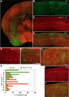
Extra-MGE/POA origin of IUE-labeled neocortical interneurons. AF, IUE was performed on an Nkx2.1Cre; Ai9 mouse. A, Low-magnification view of an IUE-labeled mouse. Note that, whereas a remnant of Nkx2.1 activity occurs in the ventral ventricular zone of the lateral ventricle (arrow), an EGFP signal can be seen at the ventricular surface in the dorsolateral corner of the lateral ventricle (arrowhead). BC, Laminar distribution of IUE-labeled interneurons (B) and Nkx2.1 fate-mapped cells (C) show IUE-labeled interneurons are more superficially distributed (D). D, Merged view of B and C. E, F, Higher-magnification view of Nkx2.1 fate-mapped cells (red) and IUE-labeled cells (green). There is no overlap of green and red signals. G, Laminar distribution of Nkx2.1 fate-mapped cells (red bars) and IUE-labeled cells (green bars) in the animal shown in AF. Although Nkx2.1 fate-mapped cells are localized preferentially to the deep zone (n = 4225 cells), IUE-labeled cells are localized preferentially to the upper layers (n = 74). H, IUE-labeled cells in a wild-type animal were immunostained for SOX6. No IUE-labeled cell expresses SOX6. I, J, IUE-labeled interneurons express SP8, a LGE/CGE marker. I, SP8 expression. J, Merged view of IUE-labeled interneurons and SP8 immunoreactivity. The positions of IUE-labeled interneurons are indicated by arrows in I. Scale bars: A, 500 μm; D, 200 μm; F, J, 50 μm; H, 100 μm.
Figure 3.
Figure 3.
Preferential labeling of interneurons in upper layers other than layer I in a Group 1 animal. A, Distribution of labeled interneurons in the neocortex. Labeled interneurons are preferentially localized in the upper layers, but layer I is almost devoid of labeled cells. The section was counterstained with ToPro3 (pseudocolored red). The graph on the left of B shows the laminar distribution of the interneurons in the animal shown in A and that on the right shows the average distribution of 16 animals in which the number of labeled cells in bin 1 was <1/3 those in bin 2. Ordinate indicates bin number. Abscissa indicate the proportion of labeled cells. Error bars indicate SD. C, IUE-labeled cells (green) and vasoactive intestinal peptide (VIP) immunoreactivity (red) in the same section show many VIP+ IUE-labeled interneurons (arrows) in the lamina below layer I. The dashed line represents the approximate border of layer I and layers II/III. D, Similar to C but for an NPY-immunostained preparation. IUE-labeled interneurons (green) immunoreactive for NPY (red; arrows) are seen below layer I (yellow). Scale bars: A, 500 μm; C, D, 100 μm.
Figure 4.
Figure 4.
Preferential labeling of layer I neocortical interneurons in a Group 2 animal. A, Low-magnification view of IUE-labeled interneurons in the neocortex. Counter-stained with ToPro3 (pseudocolored red). Inset shows a high-magnification view of layer I interneurons. Interneurons with horizontally oriented processes are labeled. B, Laminar distribution of interneurons in the animal shown in A. C, Average laminar distribution of interneurons in animals in which bin 1 interneurons outnumber the other bins (n = 5). Error bars indicate SD. D, IUE-labeled neurons in Group 2 (green) do not express SOX6 (red). SOX6 immunostaining was performed on an IUE-labeled section. E, F, Expression of RLN and VIP in IUE-labeled cells (green) of a Group 2 animal. Double immunostaining against RLN (blue) and VIP (red). IUE-labeled cells were stained for RLN (arrows) or VIP (double arrowhead). The dashed line in E shows the approximate border of layer I and layers II/III. Note that two layer I interneurons near the upper left corner express RLN. Scale bars: A, 300 μm; A inset, 50 μm; D, 100 μm; F, 50 μm.
Figure 5.
Figure 5.
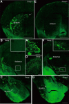
Labeled cells in subcortical structures in Group 1 and 2 animals. AD, Labeled cells in Group 1 animals. A, B, Low-magnification view at the level that includes the rostral striatum (A) and the hypothalamus (B). The striatum was densely labeled (A), but no cells were labeled in the hypothalamus (B and B inset). In these animals, cells facing the lateral ventricle were often observed at the dorsolateral corner, possibly a remnant of IUE-labeled progenitors (arrow in A). An enlarged view is shown in A′. The BLA/LA were almost devoid of labeled cells, although axonal and dendritic processes of unidentified cells can be recognized (C, arrow). Olfactory granule cells were also labeled (D). EG, Labeled cells in Group 2 animals. E, Low-magnification view at the level that includes the rostral striatum. In the neocortex, interneurons near the cortical surface were predominantly labeled. Only a small number of cells were labeled in the striatum (E). F, At a more caudal level, cells were labeled in the hypothalamus (F and F inset). G, In the amygdala, the BLA/LA were labeled (arrow). Insets in B and F are high-magnification views of boxed areas in each. All panels show images of coronal sections. Scale bars: A, B, E, F, 1 mm; C, G, 500 μm; D, 250 μm; A′, 50 μm.
Figure 6.
Figure 6.
Proportion of neocortical interneurons expressing neurochemical markers. A, Proportion of layer I interneurons expressing RLN (n = 3 animals), VIP (n = 2 animals), or NPY (n = 3 animals) (all Group 2 animals). The expression of RLN is outstanding. B, Proportion of layer II/III-VI interneurons expressing RLN (6 animals), VIP (6 animals) or NPY (4 animals) (all Group 1 animals). Comparable proportions of interneurons express each neurochemical marker. Error bars indicate SD.
Figure 7.
Figure 7.
Diversity of labeled interneurons in layers II-VI in Group 1 animals. A, B, Two types of labeled interneurons with distinct morphological features. In some Group 1 animals, interneurons having radially extending processes were prominent (A), whereas those with multipolar interneurons were dominant in others (B). C, IUE-labeled interneurons (left) immunostained for VIP (middle). In the merged view shown on the right, VIP+ interneurons with radially oriented processes are indicated by arrows, whereas a VIP interneuron with multipolar shape is indicated by an arrowhead. D, IUE-labeled interneurons (left) immunostained for NPY (middle). In the merged view shown on the right, NPY+ interneurons with multipolar shape are indicated by arrows, whereas an NPY interneuron extending radially oriented processes is indicated by an arrowhead. E, F, Laminar distribution of radially oriented and multipolar interneurons observed in bins of layers II/III-VI. Although both types of cells are preferentially localized to bins 2–3, radially oriented cells are almost absent in bins 7–10. Shown is a summary of 12 samples. Error bars indicate SD. Scale bars: B, 300 μm; C, 30 μm; D, 50 μm in D. The scale bar in B also applies to A.
Figure 8.
Figure 8.
Summary diagram showing that distinct PDs within the LGE/CGE generate subsets of neocortical interneurons with distinct laminar distribution and molecular and morphological characteristics. Progenitors possibly located within the LGE and/or rostral part of the CGE give rise to interneurons mainly in layers II/III. A subset of the interneurons express VIP and are radially oriented. NPY-expressing neurons are also generated. The same PD or a PD nearby may give rise to striatal cells and olfactory granule cells. Progenitors possibly located within the caudal part of the CGE give rise to interneurons in layer I. These interneurons, which exhibit multipolar shapes or horizontally oriented processes, express RLN. The same PD or a PD nearby may also give rise to the BLA/LA of the amygdala. Interneurons with radially oriented processes and multipolar cells might originate from distinct sub-PDs located within the PD that generates layers II/III interneurons.

Similar articles

Cited by

References

    1. Altman J, Bayer SA. Time of origin of neurons of the rat superior colliculus in relation to other components of the visual and visuomotor pathways. Exp Brain Res. 1981;42:424–434. - PubMed
    1. Altman J, Bayer SA. Development of the precerebellar nuclei in the rat: IV. The anterior precerebellar extramural migratory stream and the nucleus reticularis tegmenti pontis and the basal pontine gray. J Comp Neurol. 1987;257:529–552. doi: 10.1002/cne.902570405. - DOI - PubMed
    1. Anderson SA, Eisenstat DD, Shi L, Rubenstein JL. Interneuron migration from basal forebrain to neoneocortex: dependence on Dlx genes. Science. 1997;278:474–476. doi: 10.1126/science.278.5337.474. - DOI - PubMed
    1. Anderson SA, Marín O, Horn C, Jennings K, Rubenstein JL. Distinct cortical migrations from the medial and lateral ganglionic eminences. Development. 2001;128:353–363. - PubMed
    1. Angevine JB, Jr, Sidman RL. Autoradiographic study of cell migration during histogenesis of cerebral cortex in the mouse. Nature. 1961;192:766–768. - PubMed

Publication types