. 2016 Mar 25;291(13):7004-16.
doi: 10.1074/jbc.M115.693655. Epub 2016 Feb 10.

Aryl Hydrocarbon Receptor Plays Protective Roles against High Fat Diet (HFD)-induced Hepatic Steatosis and the Subsequent Lipotoxicity via Direct Transcriptional Regulation of Socs3 Gene Expression

Affiliations

Aryl Hydrocarbon Receptor Plays Protective Roles against High Fat Diet (HFD)-induced Hepatic Steatosis and the Subsequent Lipotoxicity via Direct Transcriptional Regulation of Socs3 Gene Expression

Taira Wada et al. J Biol Chem. .

Abstract

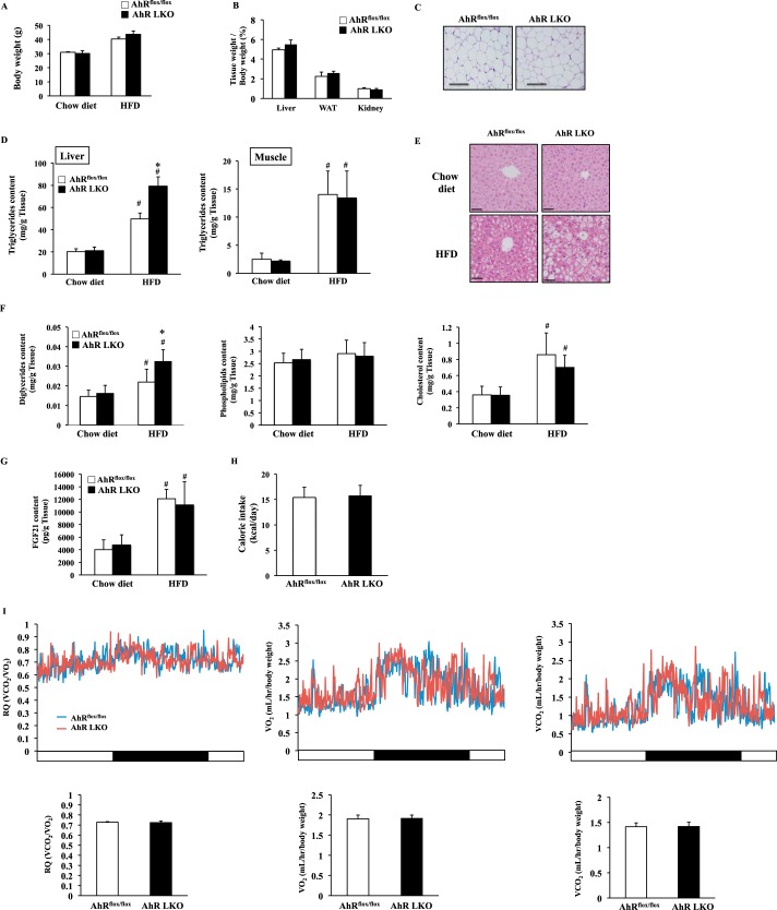
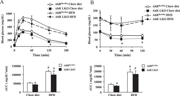
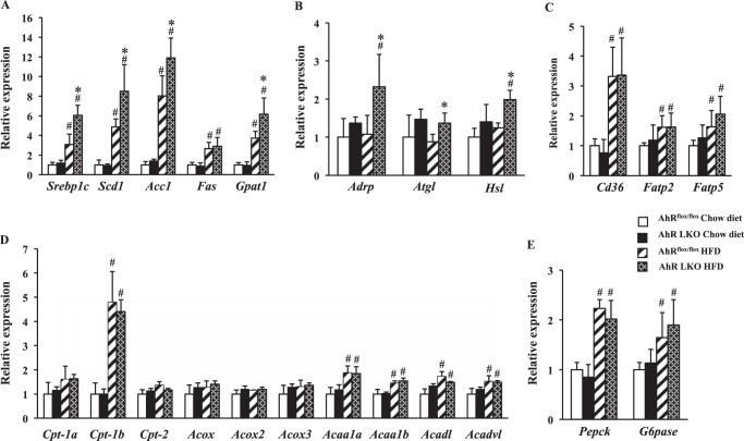
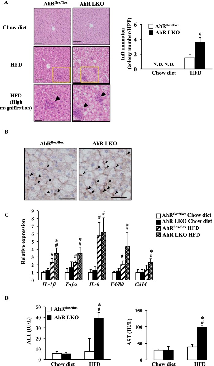
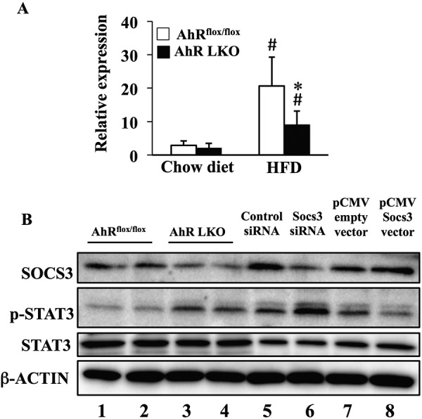
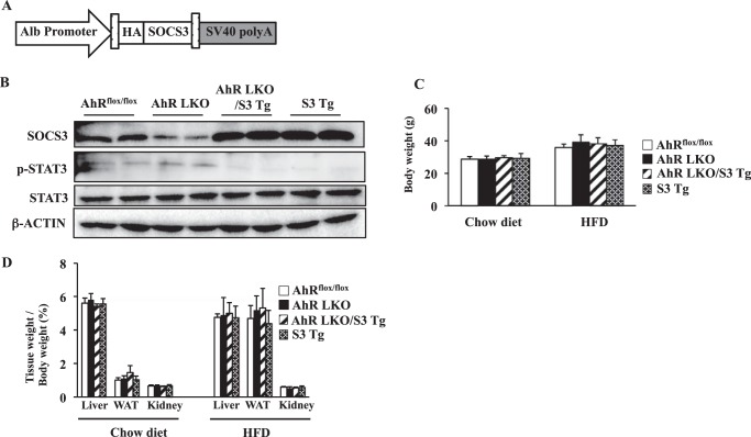
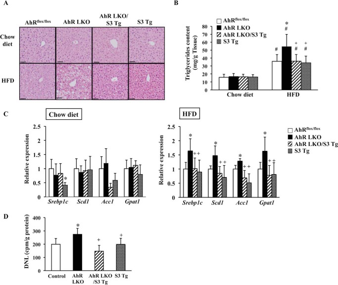
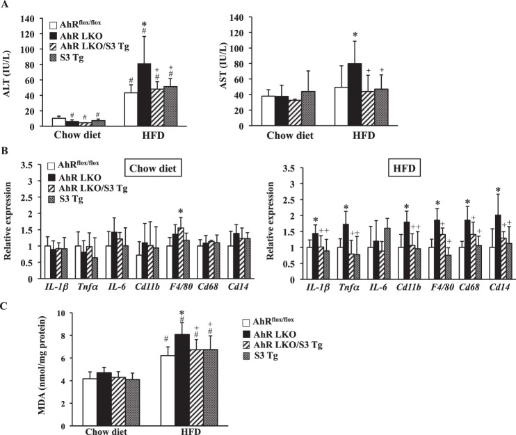
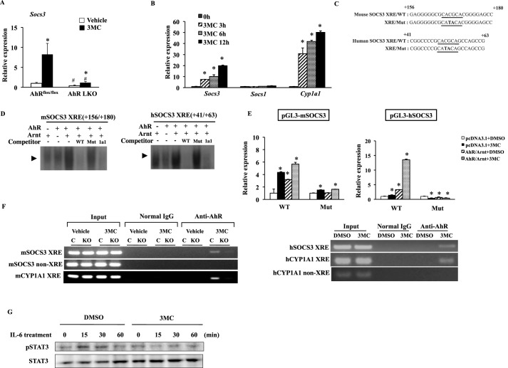
Aryl hydrocarbon receptor (AhR) is a ligand-activated transcription factor regulating the expression of genes involved in xenobiotic response. Recent studies have suggested that AhR plays essential roles not only in xenobiotic detoxification but also energy metabolism. Thus, in this study, we studied the roles of AhR in lipid metabolism. Under high fat diet (HFD) challenge, liver-specific AhR knock-out (AhR LKO) mice exhibited severe steatosis, inflammation, and injury in the liver. Gene expression analysis and biochemical study revealed thatde novolipogenesis activity was significantly increased in AhR LKO mice. In contrast, induction of suppressor of cytokine signal 3 (Socs3) expression by HFD was attenuated in the livers of AhR LKO mice. Rescue of theSocs3gene in the liver of AhR LKO mice cancelled the HFD-induced hepatic lipotoxicities. Promoter analysis established Socs3 as novel transcriptional target of AhR. These results indicated that AhR plays a protective role against HFD-induced hepatic steatosis and the subsequent lipotoxicity effects, such as inflammation, and that the mechanism of protection involves the direct transcriptional regulation ofSocs3expression by AhR.

Keywords: aryl hydrocarbon receptor (AhR); inflammation; lipid metabolism; liver; liver injury; liver metabolism; suppressor of cytokine signal 3 (Socs3).


Figures

FIGURE 1.
FIGURE 1.
Deletion of the Ahr gene in the liver accelerates HFD-induced hepatic steatosis. AhRflox/flox mice and AhR LKO mice were fed a chow diet or HFD for 12 weeks before being analyzed (n = 5–6). A, body weight. B, tissue weight of the liver, the epididymal white adipose tissue (WAT), and the kidney (n = 5–6). C, representative hematoxylin and eosin (H&E) staining of epididymal white adipose tissue. Magnification, ×40. Scale bar, 200 μm. D, triglyceride content in the liver and skeletal muscle. E, representative H&E staining of a liver section around the central vein. Magnification, ×100. Scale bar, 50 μm. F, content of diglyceride, phospholipid, and cholesterol in the liver (n = 5–6). G, FGF21 content in the liver (n = 5–6). H, food intake (kcal per day). I, top panel, representative daily changes of oxygen consumption (VO2), carbon dioxide production (VCO2), and RQ. Bottom panel, daily average of VO2, VCO2, and RQ (n = 5–6). #, p < 0.05 relative to AhRflox/flox mice fed a chow diet. *, p < 0.05 relative to AhRflox/flox mice fed a HFD.
FIGURE 2.
FIGURE 2.
HFD feeding attenuates the effects of deletion of the hepatic Ahr gene on insulin sensitivity. AhRflox/flox mice and AhR LKO mice were fed a chow diet or HFD for 12 weeks before being analyzed (n = 6). A, glucose tolerance test. B, insulin tolerance test. The area under the curve (AUC) was calculated for respective group. #, p < 0.05 relative to AhRflox/flox mice fed a chow diet. *, p < 0.05 relative to AhRflox/flox mice on the same diet.
FIGURE 3.
FIGURE 3.
Deletion of the Ahr gene in the liver increases expression of lipogenesis-related genes under the HFD condition. AhRflox/flox mice and AhR LKO mice were fed a chow diet or HFD for 12 weeks before being analyzed (n = 5–6). Gene expression in the mouse liver was analyzed by qRT-PCR. A, expression of genes involved in lipogenesis. B, expression of genes related to lipid droplet or lipolysis. C, expression of genes involved in fatty acid uptake. D, expression of genes involved in β-oxidation. E, expression of genes involved in gluconeogenesis. #, p < 0.05 relative to AhRflox/flox mice fed a chow diet. *, p < 0.05 relative to AhRflox/flox mice fed a HFD.
FIGURE 4.
FIGURE 4.
Deletion of the Ahr gene in the liver accelerates HFD-induced inflammation. AhRflox/flox mice and AhR LKO mice were fed a chow diet or HFD for 12 weeks before being analyzed (n = 5–6). A, left panel, representative H&E staining of a liver section around the central vein with arrows indicating inflammatory cell infiltration. Magnification, ×100 (top and middle) and ×200 (bottom). Scale bar, 50 μm (top and middle) and 100 μm (bottom). Right panel, the colony number of inflammatory cells per high power field. B, representative immunohistochemistry to detect F4/80 in the liver of mice fed a HFD (magnification, ×200. Scale bar, 50 μm). C, qRT-PCR analysis of inflammatory-related gene expression. D, activity of ALT and AST in serum. #, p < 0.05 relative to AhRflox/flox mice fed a chow diet. *, p < 0.05 relative to AhRflox/flox mice fed a HFD. N.D., not detected.
FIGURE 5.
FIGURE 5.
Deletion of the Ahr gene in the liver attenuates the HFD-dependent induction of Socs3 expression. AhRflox/flox mice and AhR LKO mice were fed a chow diet or HFD for 12 weeks before being analyzed (n = 5). A, qRT-PCR analysis of Socs3 gene expression in the liver. B, representative Western blot of SOCS3, total STAT3, and phosphorylated STAT3 in the liver extract of mice fed a HFD. β-Actin was used as a loading control. Lanes 1 and 2 were run using the liver extract prepared from two distinct AhRflox/flox mice, and lanes 3 and 4 were run using the liver extract prepared from two distinct AhR LKO mice. Lane 6 was run using a cell extract prepared from Socs3 siRNA-transfected RAW264.7 cells as negative control. Lane 8 was run using a cell extract prepared from Socs3 expression vector-transfected RAW264.7 cells as positive control. Lanes 5 and 7 were transfection control for Socs3 siRNA-transfected cells (lane 6) and Socs3 expression vector-transfected cells (lane 8), respectively. #, p < 0.05 relative to AhRflox/flox mice fed a chow diet. *, p < 0.05 relative to AhRflox/flox mice fed a HFD.
FIGURE 6.
FIGURE 6.
Generation of AhR LKO mice expressing transgene of the Socs3. A, schematic representations of the transgene construct. The transgene is under the control of the mouse albumin promoter. B, representative Western blot of SOCS3, phosphorylated STAT3, total STAT3, and β-actin in the liver extract. Each lane was run using samples from two different male mice. AhRflox/flox mice, AhR LKO mice, AhR LKO/S3 Tg mice, and S3 Tg mice were fed a HFD. C, body weight of mice fed a chow diet or HFD for 10 weeks (n = 5–7). D, tissue weight of mice fed a chow diet or HFD for 10 weeks (n = 5–7).
FIGURE 7.
FIGURE 7.
Rescue of Socs3 expression in AhR LKO mice attenuates HFD-induced steatosis. AhRflox/flox mice, AhR LKO mice, AhR LKO/S3 Tg mice, and S3 Tg mice were fed a chow or HFD for 10 weeks (n = 5–7). A, representative H&E staining of the liver section (magnification, ×100. Scale bar, 50 μm). B, triglyceride content in the liver. C, qRT-PCR analysis of the expression of lipogenic genes. D, de novo lipogenesis activity in the mouse liver of mice fed a HFD. #, p < 0.05 relative to AhRflox/flox mice fed a chow diet. *, p < 0.05 relative to AhRflox/flox mice fed a HFD (in B and D) or AhRflox/flox mice fed a same diet (C). +, p < 0.05 relative to AhR LKO mice fed a HFD.
FIGURE 8.
FIGURE 8.
Rescue of Socs3 expression in AhR LKO mice attenuates HFD-induced lipotoxicity. AhRflox/flox mice, AhR LKO mice, AhR LKO/S3 Tg mice, and S3 Tg mice were fed a chow diet or HFD for 10 weeks (n = 5–7). A, activity of ALT and AST in serum. B, qRT-PCR analysis of the expression of inflammation-related genes (n = 5–7). C, TBARS level in the liver. #, p < 0.05 relative to AhRflox/flox mice fed a chow diet. *, p < 0.05 relative to AhRflox/flox mice fed a HFD. +, p < 0.05 relative to AhR LKO mice fed a HFD.
FIGURE 9.
FIGURE 9.
Socs3 is a novel transcriptional target of AhR. A, qRT-PCR analysis of Socs3 expression in the livers of AhRflox/flox mice and AhR LKO mice treated with an AhR agonist, 3MC (20 mg/kg), or corn oil for 3 consecutive days under a regular diet (n = 3–4). *, p < 0.05 relative to same genotype treated with corn oil (vehicle). +, p < 0.05 relative to AhRflox/flox mice treated with 3MC. B, qRT-PCR analysis of SOCS3, SOCS1, and CYP1A1 expression in HepG2 cells treated with 3MC (3 μm) for the indicated period of time (n = 3). *, p < 0.05 relative to 0 h (untreated cells). C, partial DNA sequences of the mouse and human Socs3 gene promoters. The putative Socs3/XRE sequences are underlined, and the mutated nucleotides are in capital letters. D, specific binding of the in vitro-synthesized AhR/aryl hydrocarbon receptor nuclear translocator heterodimer to the Socs3/XRE and the Cyp1a1/XRE in EMSA. In the competition lanes, unlabeled probes were present in 100-fold molar excess relative to the radiolabeled probe. E, luciferase activity in DMSO-treated or 3MC (3 μm)-treated HepG2 cells transfected with the reporter plasmids containing the mouse or human Socs3 promoter and its mutant variants in the presence of AhR, Arnt, or pcDNA 3.1 empty vector. The value of pcDNA3.1-transfected cells treated with DMSO was normalized to 1. The data represent the average and standard deviation from three independent experiments. *, p < 0.05 relative to DMSO-treated cells transfected with same plasmids. +, p < 0.05 relative to wild type (WT) reporter gene-transfected cells treated with same compound (DMSO or 3MC). F, ChIP analysis of the interaction between AhR and the region containing the Socs3/XRE in mice treated with corn oil (vehicle) or 3MC for 3 consecutive days (left) and HepG2 cells treated with 3MC (3 μm) for 16 h (right). C, AhRflox/flox mice; KO, AhR LKO mice. G, representative Western blot of total STAT3 and phosphorylated STAT3 in HepG2 cells exposed to IL-6 (10 ng/ml) for the indicated period of time after treatment with DMSO or 3MC (3 μm) for 16 h.

Similar articles

Cited by

References

    1. Ema M., Sogawa K., Watanabe N., Chujoh Y., Matsushita N., Gotoh O., Funae Y., and Fujii-Kuriyama Y. (1992) cDNA cloning and structure of mouse putative Ah receptor. Biochem. Biophys. Res. Commun. 184, 246–253 - PubMed
    1. Burbach K. M., Poland A., and Bradfield C. A. (1992) Cloning of the Ah-receptor cDNA reveals a distinctive ligand-activated transcription factor. Proc. Natl. Acad. Sci. U.S.A. 89, 8185–8189 - PMC - PubMed
    1. Poland A., and Knutson J. C. (1982) 2,3,7,8-Tetrachlorodibenzo-p-dioxin and related halogenated aromatic hydrocarbons: examination of the mechanism of toxicity. Annu. Rev. Pharmacol. Toxicol. 22, 517–554 - PubMed
    1. Reyes H., Reisz-Porszasz S., and Hankinson O. (1992) Identification of the Ah receptor nuclear translocator protein (Arnt) as a component of the DNA binding form of the Ah receptor. Science 256, 1193–1195 - PubMed
    1. Hankinson O. (1995) The arylhydrocarbon receptor complex. Annu. Rev. Pharmacol. Toxicol. 35, 307–340 - PubMed

Publication types

MeSH terms

Substances

LinkOut - more resources