. 2016 Apr 1;291(14):7527-40.
doi: 10.1074/jbc.M115.688796. Epub 2016 Feb 10.

Structural Insights into the Carbohydrate Binding Ability of an α-(1→2) Branching Sucrase from Glycoside Hydrolase Family 70

Affiliations

Structural Insights into the Carbohydrate Binding Ability of an α-(1→2) Branching Sucrase from Glycoside Hydrolase Family 70

Yoann Brison et al. J Biol Chem. .

Abstract

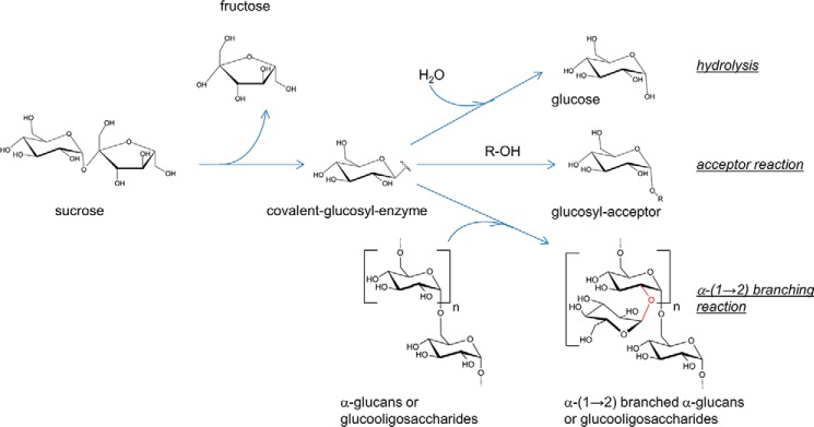
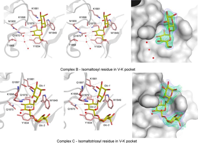
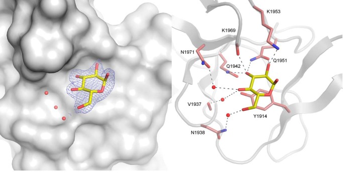
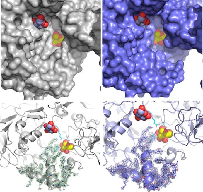
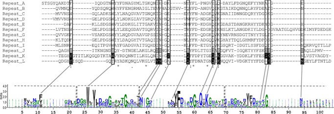
The α-(1→2) branching sucrase ΔN123-GBD-CD2 is a transglucosylase belonging to glycoside hydrolase family 70 (GH70) that catalyzes the transfer ofd-glucosyl units from sucroseto dextrans or gluco-oligosaccharides via the formation of α-(1→2) glucosidic linkages. The first structures of ΔN123-GBD-CD2 in complex withd-glucose, isomaltosyl, or isomaltotriosyl residues were solved. The glucose complex revealed three glucose-binding sites in the catalytic gorge and six additional binding sites at the surface of domains B, IV, and V. Soaking with isomaltotriose or gluco-oligosaccharides led to structures in which isomaltosyl or isomaltotriosyl residues were found in glucan binding pockets located in domain V. One aromatic residue is systematically identified at the bottom of these pockets in stacking interaction with one glucosyl moiety. The carbohydrate is also maintained by a network of hydrogen bonds and van der Waals interactions. The sequence of these binding pockets is conserved and repeatedly present in domain V of several GH70 glucansucrases known to bind α-glucans. These findings provide the first structural evidence of the molecular interaction occurring between isomalto-oligosaccharides and domain V of the GH70 enzymes.

Keywords: alpha-1,2 branching sucrase; carbohydrate-binding protein; crystal structure; enzyme; family GH70; glucan-binding domain; glucansucrase; glycoside hydrolase; oligosaccharide; α-glucan.


Figures

FIGURE 1.
FIGURE 1.
The three types of reactions catalyzed by the α-(1→2) branching sucrase ΔN123-GBD-CD2.
FIGURE 2.
FIGURE 2.
Affinity gel electrophoresis of ΔN123-GBD-CD2 in gels containing variable amounts of dextrans. Left panel, affinity gels for which the content in dextran is expressed in % (w/v). Lanes 1 correspond to BSA, lanes 2 correspond to ΔN123-GBD-CD2. Right panel, double-reciprocal plots of the inverse of the relative electrophoretic mobility versus the inverse of dextran concentrations. The relative mobility was determined from the migration distances observed on the gels. * estimated average molecular weight.
FIGURE 3.
FIGURE 3.
Structure of ΔN123-GBD-CD2 in complex with carbohydrates. A, structure of ΔN123-GBD-CD2 in complex with d-glucose (complex A). Nine glucose-binding sites have been identified at the surface of the enzyme. Four sites in domain A (A-1, A2, A3, and A4), two sites in domain IV (IV1 and IV2), one site in domain V (named binding pocket V-L), and two sites spanning over domains B, IV, and V (site B-IV-V and site IV-V). Domains A, B, C, IV, and V are colored in blue, green, magenta, yellow, and fire-brick, respectively. Glucose molecules are shown as spheres (with carbon and oxygen atoms in gray and red, respectively). B, grooves at the surface of the enzyme in complex B. Grooves were searched using CAVER starting in the active site from subsite −1. Glucose molecules from the complex A have been superimposed. C, electrostatic potential surface color-coded from red (−5 kBT/e) to blue (+5 kBT/e) for complex B computed at pH 5.75; white is neutral. The nine glucose molecules were superimposed and figured as yellow and red spheres.
FIGURE 4.
FIGURE 4.
Glucose-binding sites found over the ΔN123-GBD-CD2 enzyme. The simulated annealing omit (FoFc) electron density maps (in green) around the nine glucose molecules bound to ΔN123-GBD-CD2 were contoured at 2σ. ΔN123-GBD-CD2 residues in the neighborhood of each glucose molecule are shown as lines and colored by atom types. Stereo view of subsites A-1, A2, A3, A4, B-IV-V, IV1, IV2, IV-V, and VL are shown on panels A–I, respectively.
FIGURE 5.
FIGURE 5.
Glucose-binding sites A2 (left panel) and A3 (right panel). The simulated annealing omit (FoFc) electron density maps around glucose were contoured at 3σ or 2.3σ for A2 and A3, respectively. Interacting water molecules are depicted as red spheres. Residues of domains A or B are represented as blue or green sticks, respectively. For residues that are involved in sugar binding through their backbone atoms, side chains are omitted. In the left panel, residues forming the catalytic triad (Asp-2210, Glu-2248, and Asp-2322) are shown as turquoise sticks.
FIGURE 6.
FIGURE 6.
Binding pocket V-K found in the GBD of ΔN123-GBD-CD2 in complexes B (upper panels) and C (lower panels). For this pocket, in complexes B or C, an isomaltosyl or isomaltotriosyl residue was observed and shown as yellow sticks. The network of hydrogen bonding is shown on the right panels, whereas the electron density map around carbohydrates is displayed on left panels. The residues and water molecules involved in binding are represented as pale red sticks and red spheres, respectively (right panels). The simulated annealing omit (FoFc) electron density maps (in green) around carbohydrates were contoured at 3σ or 2σ for isomaltosyl and isomaltotriosyl, respectively. The σA weighted 2FoFc electron density maps in blue were contoured at 0.9 σ.
FIGURE 7.
FIGURE 7.
View of the V-L binding pocket in the glucan-binding domain of ΔN123-GBD-CD2 with gluco-oligosaccharide. For gluco-oligosaccharide or isomaltotriose ligands, a glucosyl residue, shown as yellow stick, was visible in the electron density map. Residues and water molecules involved in binding are represented as pale red sticks and red spheres, respectively. The σA weighted 2FoFc electron density map was contoured at 0.9 σ.
FIGURE 8.
FIGURE 8.
Loop rearrangement in the catalytic gorge of ΔN123-GBD-CD2. The left panels show the catalytic gorge conformation in the apo form. The right panels show the same region for the structure of the complex B. The upper panels represent the protein surface, whereas the schematic representation has been used in the lower panels. The σA weighted 2FoFc electron density maps around loop 7 of domain A were contoured at 1σ. To figure the active site, the d-glucose molecule bound in subsite −1, as found in the structure of the complex with glucose, is superimposed and represented as yellow and red spheres. The d-glucose molecule found in the binding site A2 is also represented (light blue and red spheres). Residues forming the catalytic triad are represented as turquoise sticks.
FIGURE 9.
FIGURE 9.
Comparison of the sugar binding pockets found in the glucan-binding domains of ΔN123-GBD-CD2 (complex C), and putative sugar binding pockets of GTF180-ΔN (PDB entry 3KLK) and GTFA-ΔN (PDB entry 4AMC) glucansucrases. Secondary structure elements are shown in pale red in agreement with Fig. 9. Molecular surfaces are displayed in light gray. Residues delineating the binding pockets are shown as pale red sticks. Stacked sugars in the binding pockets of ΔN123-GBD-CD2 are represented in yellow lines.
FIGURE 10.
FIGURE 10.
Sequence alignment and LOGO sequence of the GBD repeats proved to bind α-glucans of GH70 glucansucrases. GBD1A, GBD4RS, and GBD5 bind dextrans with Kd values of 0.6 μm, 2.5 μm, and 11.1 nm, respectively. Sequences are GBD1A_GTFI_rp# (repeats 1 to 4 of the GBD1A of GTFI from S. downei), GBD-4RS_GTFI_rp# (repeats 1 to 4 of the GBD-4RS of GFTI from S. sobrinus), GBD5_DSR-S_rp# (repeats 1 to 3 of the GBD5 of DSR-S from L. mesenteroides B-512F), GBD_GTF180_rp1 (repeat 1 of the GBD of GTF180-ΔN from Lactobacillus reuteri), GBD_GTFA_rp1 (repeat 1 of the GBD of GTFA-ΔN from L. reuteri 121), and GBD_DSR-E_rp# (repeats J to L of the GBD of ΔN123-GBD-CD2 from L. citreum NRRL-1299). Residues highlighted in red are well conserved, whereas those tending toward blue are not. Residues framed in gray delineate the sugar binding pockets and are pointed out in the LOGO sequence. Residues framed in dashed line on the LOGO sequence correspond to YG repeats. In the repeats of GBD1A, residues circled in white, were mutated by Shah et al. (25) (see ”Discussion“). Secondary structure elements, as determined by DSSP from PDB entries 3TTQ, 3KLK, and 4AMC, are displayed below the LOGO sequence. β-Strands, numbered according to Fig. 8, linked by dashed lines represent either β-hairpins or three-stranded β-sheets.
FIGURE 11.
FIGURE 11.
Sequence alignment of the 12 repeats identified in the GBD of DSR-E. Black highlighted residues are involved in sugar binding in repeats K and L, whereas gray highlighted residues are proposed to play the same role in repeat J. Framed residues would participate in sugar binding for repeats A to I. A LOGO sequence based on this alignment is shown. The YG repeats are framed in dashed lines on the LOGO sequence.

Similar articles

Cited by

References

    1. Cantarel B. L., Coutinho P. M., Rancurel C., Bernard T., Lombard V., and Henrissat B. (2009) The Carbohydrate-Active EnZymes database (CAZy): an expert resource for Glycogenomics. Nucleic Acids Res. 37, D233–238 - PMC - PubMed
    1. Leemhuis H., Pijning T., Dobruchowska J. M., van Leeuwen S. S., Kralj S., Dijkstra B. W., and Dijkhuizen L. (2013) Glucansucrases: three-dimensional structures, reactions, mechanism, α-glucan analysis and their implications in biotechnology and food applications. J. Biotechnol. 163, 250–272 - PubMed
    1. Leemhuis H., Dijkman W. P., Dobruchowska J. M., Pijning T., Grijpstra P., Kralj S., Kamerling J. P., and Dijkhuizen L. (2013) 4,6-α-Glucanotransferase activity occurs more widespread in Lactobacillus strains and constitutes a separate GH70 subfamily. Appl. Microbiol. Biotechnol. 97, 181–193 - PMC - PubMed
    1. Côté G. L., and Leathers T. D. (2005) A method for surveying and classifying Leuconostoc spp. glucansucrases according to strain-dependent acceptor product patterns. J. Ind. Microbiol. Biotechnol. 32, 53–60 - PubMed
    1. Naessens M., Cerdobbel A., Soetart W., and Vandamme E. J. (2005) Leuconostoc dextransucrase and dextran: production, properties and applications. J. Chem. Technol. Biotechnol. 80, 845–860

Publication types

MeSH terms

LinkOut - more resources