

Development of a Highly Specific IgM Enzyme-Linked Immunosorbent Assay for Bartonella henselae Using Refined N-Lauroyl-Sarcosine-Insoluble Proteins for Serodiagnosis of Cat Scratch Disease
- PMID: 26865692
- PMCID: PMC4809944
- DOI: 10.1128/JCM.03009-15
Development of a Highly Specific IgM Enzyme-Linked Immunosorbent Assay for Bartonella henselae Using Refined N-Lauroyl-Sarcosine-Insoluble Proteins for Serodiagnosis of Cat Scratch Disease
Abstract
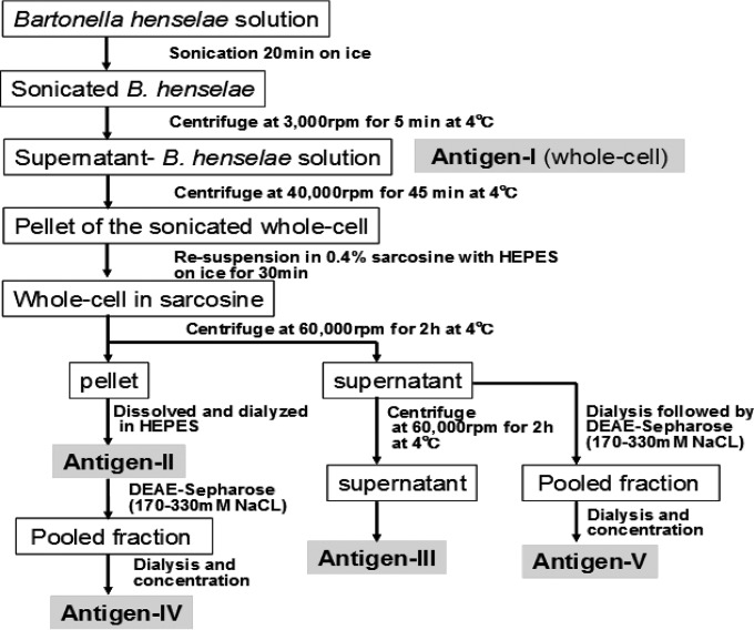
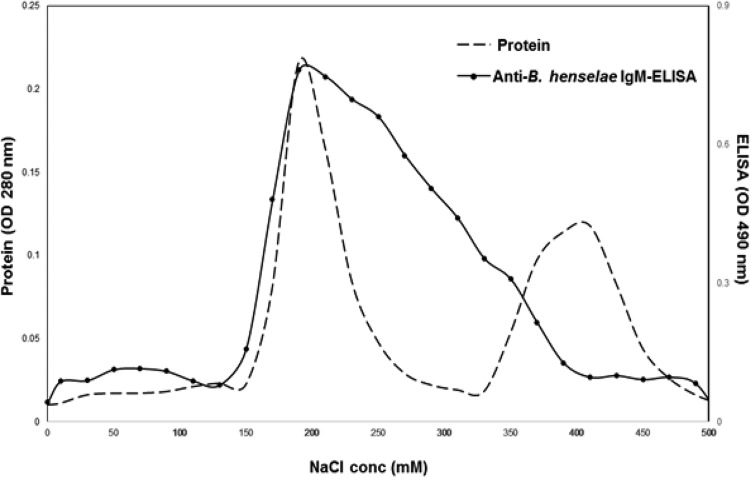
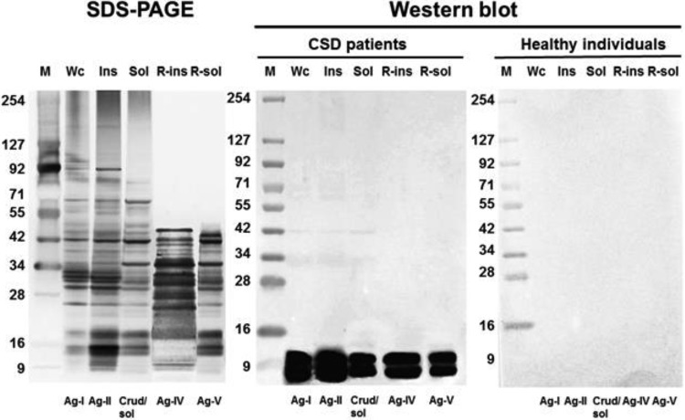
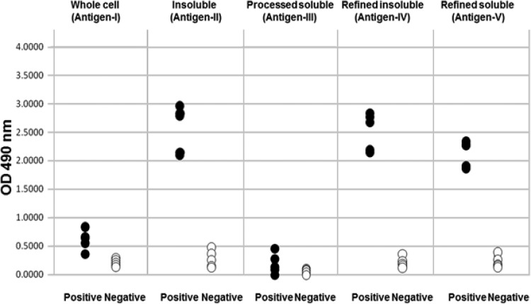
The conventional anti-Bartonella henselaeIgM enzyme-linked immunosorbent assay (IgM-ELISA) methods for diagnosing cat scratch disease (CSD) remain poor in both sensitivity and specificity. We sought to develop an IgM-ELISA with improved accuracy in the serodiagnosis of CSD by exploring the antigens that are most suitable for an ELISA. We prepared 5 different protein antigens: antigen I (sonicatedB. henselaewhole-cell antigen), antigen II (N-lauroyl-sarcosine-insoluble antigen), antigen III (processed sarcosine-soluble antigen), and antigen IV and antigen V (sarcosine-insoluble and sarcosine-soluble antigens refined by DEAE-Sepharose Fast Flow ion-exchange chromatography). The IgM antibodies in the sera of 47 patients with clinically suspected CSD (24 definite, 23 suspected) and of 85 healthy individuals were examined by ELISAs using the 5 antigens, and the results were compared with those of an IgM indirect fluorescent antibody assay (IgM-IFA). In a reference panel, which consisted of 5 positive and 5 negative sera, antigen I and antigen III failed to distinguish between the two statuses, whereas the other three antigens succeeded in distinguishing between them. When the cutoff value was set at the 98th percentile of the ELISA index for healthy individuals, the sensitivity of IgM-IFA for the 24 cases of definite CSD was 54%, whereas the sensitivities of the IgM-ELISAs with antigen II, IV, and V were 75%, 83%, and 75%, respectively. The sensitivities of these three IgM-ELISAs for all 47 of the clinically suspected cases were 49%, 64%, and 51%, respectively. In contrast, the sensitivity of IgM-IFA was 28%. These results indicate that the refined sarcosine-insoluble proteins (antigen IV), which possessed the highest specificity among the 5 antigens, are the most appropriate for developing an IgM-ELISA for the highly specific serodiagnosis of CSD.
Copyright © 2016, American Society for Microbiology. All Rights Reserved.
Figures
References
-
- Tsuneoka H, Fujii R, Yamamoto K, Fujisawa K, Iino H, Matsuda M, Tsukahara M. 1998. Determination of anti-Bartonella henselae antibody by indirect fluorescence antibody test—comparison of two types of antigen: non-cocultivated B. henselae and cocultivated B. henselae with Vero cells. Kansenshogaku Zasshi 72:801–807. (In Japanese.) doi: 10.11150/kansenshogakuzasshi1970.72.801. - DOI - PubMed
Publication types
MeSH terms
Substances
LinkOut - more resources
Full Text Sources
Other Literature Sources
Medical
Molecular Biology Databases
Miscellaneous
