. 2016 Apr 7;6(4):1031-47.
doi: 10.1534/g3.115.026815.

The Stress Granule RNA-Binding Protein TIAR-1 Protects Female Germ Cells from Heat Shock in Caenorhabditis elegans

Affiliations

The Stress Granule RNA-Binding Protein TIAR-1 Protects Female Germ Cells from Heat Shock in Caenorhabditis elegans

Gabriela Huelgas-Morales et al. G3 (Bethesda). .

Abstract

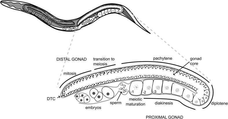
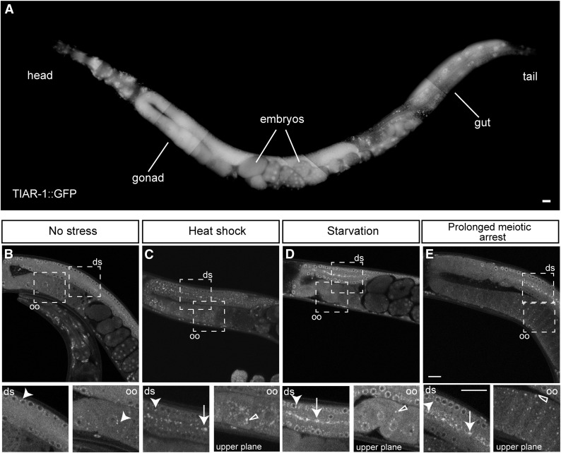
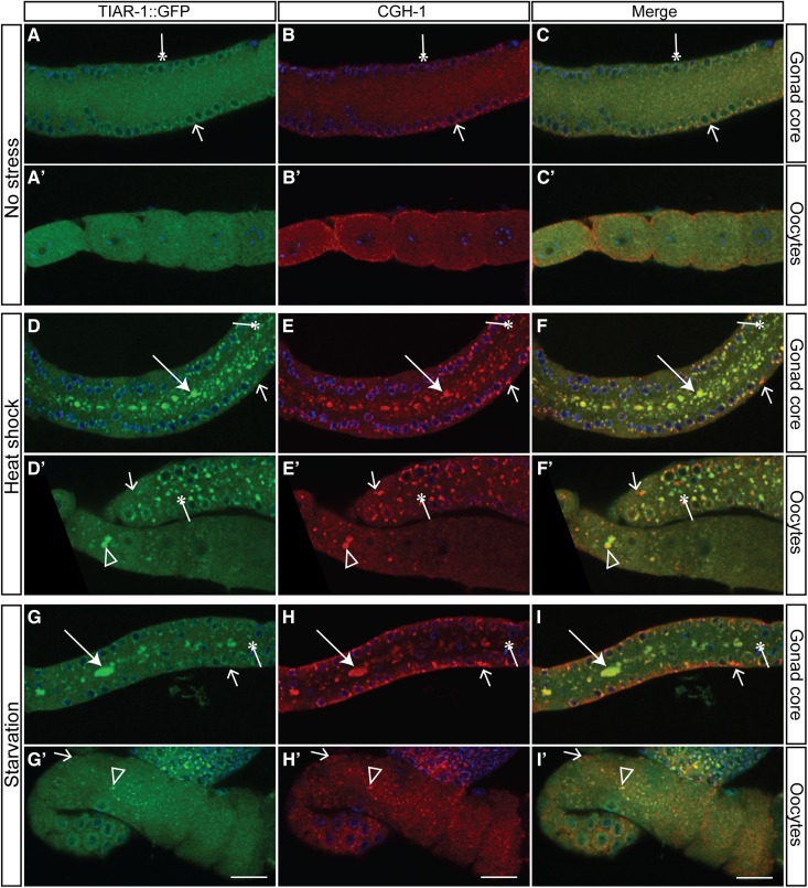
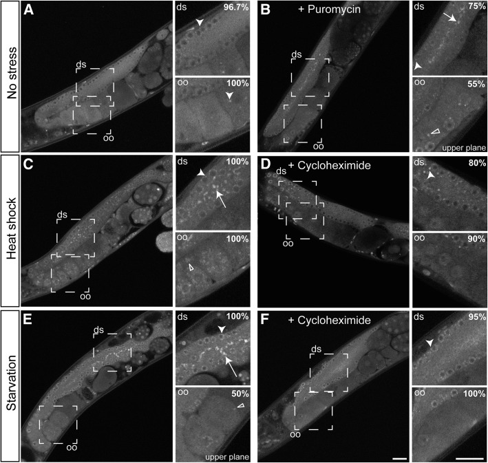
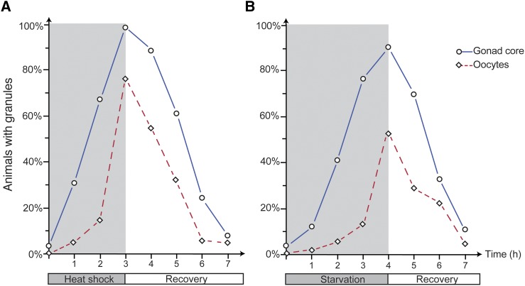
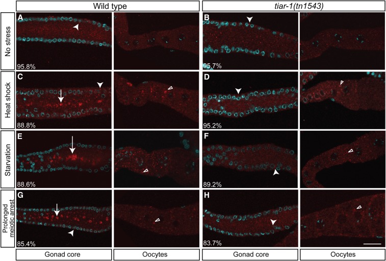
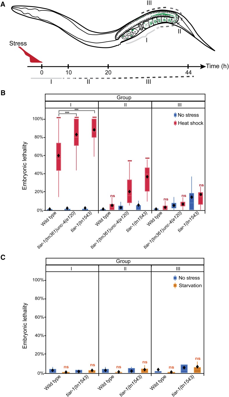
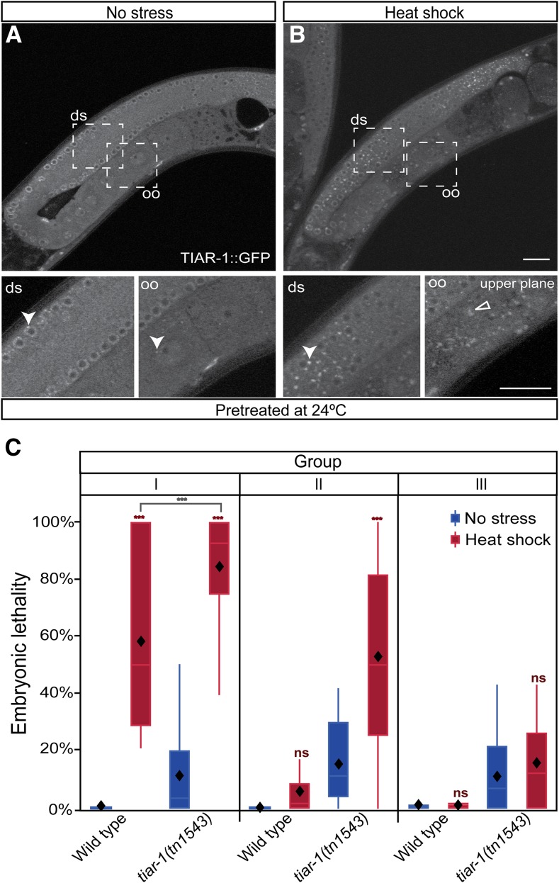
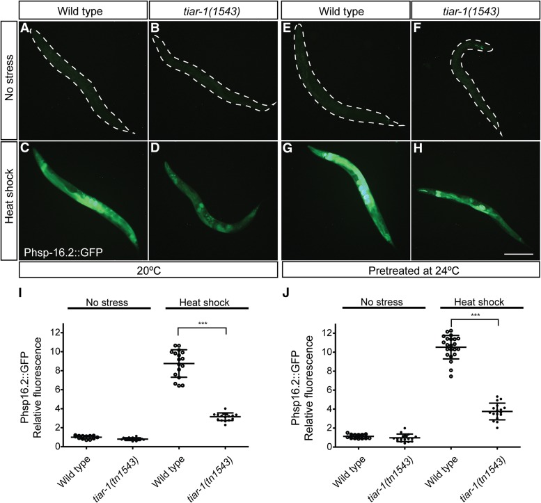
In response to stressful conditions, eukaryotic cells launch an arsenal of regulatory programs to protect the proteome. One major protective response involves the arrest of protein translation and the formation of stress granules, cytoplasmic ribonucleoprotein complexes containing the conserved RNA-binding proteins TIA-1 and TIAR. The stress granule response is thought to preserve mRNA for translation when conditions improve. For cells of the germline-the immortal cell lineage required for sexual reproduction-protection from stress is critically important for perpetuation of the species, yet how stress granule regulatory mechanisms are deployed in animal reproduction is incompletely understood. Here, we show that the stress granule protein TIAR-1 protects the Caenorhabditis elegans germline from the adverse effects of heat shock. Animals containing strong loss-of-function mutations in tiar-1 exhibit significantly reduced fertility compared to the wild type following heat shock. Analysis of a heat-shock protein promoter indicates that tiar-1 mutants display an impaired heat-shock response. We observed that TIAR-1 was associated with granules in the gonad core and oocytes during several stressful conditions. Both gonad core and oocyte granules are dynamic structures that depend on translation; protein synthesis inhibitors altered their formation. Nonetheless, tiar-1 was required for the formation of gonad core granules only. Interestingly, the gonad core granules did not seem to be needed for the germ cells to develop viable embryos after heat shock. This suggests that TIAR-1 is able to protect the germline from heat stress independently of these structures.

Keywords: C. elegans; TIA-1/TIAR; germ cells; stress; stress granules.


Figures

Figure 1
Figure 1
C. elegans adult hermaphrodite gonad. This model represents in detail the morphology of one of the two arms that compose the C. elegans adult hermaphrodite gonad. The upper picture represents a complete animal where the gonad and intestine (dark gray) are depicted. The bottom picture depicts one arm of the gonad zoomed in. Distal tip cell (DTC).
Figure 2
Figure 2
Alleles used to study the function of tiar genes in C. elegans. (A) The tiar-1 wild-type allele comprises six exons (gray) and five introns (thin peaks). The tiar-1(tm361) allele is an out-of-frame deletion of 581 bp (dotted line). We generated a deletion and a GFP fusion allele using CRISPR-Cas9 genome editing. The strain tiar-1(tn1543) results from a deletion of the tiar-1 open reading frame. In this strain, tiar-1 sequences were replaced with the C. briggsae unc-119 gene as a positive selection marker (blue). The strain tiar-1(tn1545) has an in-frame insertion of the tag S::TEV::GFP at the TIAR-1 C- terminus (green). (B) The tiar-2 wild-type allele comprises eight exons and eight introns. The tiar-2(tm2923) is a deletion of 336 bp (dotted line). (C) The tiar-3 wild-type allele comprises 10 exons and nine introns. The tiar-3(ok144) is a deletion of 1531 bp (dotted line).
Figure 3
Figure 3
TIAR-1 associates with cytoplasmic granules during stress in the gonad. TIAR-1::GFP expression and localization were assessed in tiar-1(tn1545). (A) 1-d-old tiar-1::gfp hermaphrodites were anesthetized and observed with the fluorescence microscope. The subcellular localization of TIAR-1::GFP in the gonad was observed under normal growth conditions (B), after exposure to heat shock (3 hr at 31°) (C), and after starvation (4 hr) (D). TIAR-1::GFP subcellular localization was also observed in tiar-1(tn1545); fog-2(q71) unmated females (E). These animals were imaged using confocal microscopy. Dotted squares indicate the zoomed-in areas. ds, distal gonad; oo, oocytes. Arrowheads indicate likely P granules, arrows gonad core granules, and empty triangles oocytes granules. Scale bars, 10 µm.
Figure 4
Figure 4
TIAR-1 and CGH-1 associate with large stress-induced granules in the gonad. tiar-1(tn1545) 1-d-old hermaphrodites were grown in normal conditions (no stress) (A–C’), exposed to heat shock (3 hr at 31°) (D–F’), or starved (4 hr without food) (G– I’). After the treatments, the animals were fixed and coimmunostained with anti-GFP, anti-CGH-1 (red), and DAPI (cyan) to visualize granules and DNA, respectively. The preparations were imaged using confocal microscopy. Note, TIAR-1 and CGH-1 associate with large stress-induced granules in the gonad core (panels A–I) and oocytes (panels A’–I’). However, some small granules appear to contain only TIAR-1 or CGH-1. Thin arrows indicate granules that appear to contain CGH-1 but not TIAR-1. Asterisks indicate granules that appear to contain TIAR-1 but not CGH-1. Arrows and empty triangles indicate granules in the gonad core and oocytes, respectively. Scale bar, 10 µm.
Figure 5
Figure 5
Cycloheximide treatment during stress impairs gonad granule formation. 1-d-old tiar-1::gfp hermaphrodites were grown at 20° in the absence of stress (A, B), or exposed to either heat shock (3 hr at 31°) (C) or starvation (4 hr) in liquid medium (see Materials and Methods) (E). tiar-1::gfp hermaphrodites were also incubated with 30 mM cycloheximide while being exposed to the heat shock (D) or starvation (F). Additionally, tiar-1::gfp hermaphrodites were treated with 15 mM puromycin in the absence of stress (B). After the incubation periods, the animals were mounted and imaged using confocal microscopy. Note, puromycin promotes granule formation in most of the nonstressed animals, while cycloheximide impairs granule formation in most of the stressed animals. Dotted squares indicate the zoomed-in areas; “upper plane” refers to a different confocal plane. ds, distal gonad; oo, oocytes. Arrowheads indicate likely P granules, arrows gonad core granules, and empty triangles oocytes granules. The percentage of animals with the depicted phenotype is shown. Scale bars, 10 µm.
Figure 6
Figure 6
Gonad granules dissociate after stress recovery. tiar-1::gfp 1-d-old hermaphrodites were exposed to heat shock (3 hr at 31°) in agar plates (A) or starvation (4 hr with no bacteria) in liquid medium (B). Then they were recovered from stress (20° with bacteria). The animals were mounted, observed under the fluorescence microscope, and scored for presence of TIAR-1::GFP granules in the gonad core (blue solid line, circles) and in oocytes (red dotted line, diamonds) every hr during stress exposure and recovery. At least two independent experiments were conducted with n ≥ 50 for each condition and time point. The average percentage of animals with visible granules is depicted in the graphs.
Figure 7
Figure 7
tiar-1 is required for gonad core granule formation. Wild-type and tiar-1(tn1543) 1-d-old mutant hermaphrodites were grown in normal conditions (no stress) (A and B), exposed to heat shock (3 hr at 31°) (C and D), or starved (4 hr without food) (E and F). For prolonged meiotic arrest experiments, 1-d-old fog-2(q71) and tiar-1(tn1543); fog-2(q71) unmated females were scored (G and H). After the treatments, the animals were fixed and immunostained with anti-CGH-1 (red) and DAPI (cyan) to visualize granules and DNA, respectively. The preparations were imaged using confocal microscopy. The average percentage of animals with the depicted phenotype was calculated from data obtained in at least three independent experiments. Note, granules are formed in the gonad core and oocytes of most wild-type animals. However, most of the tiar-1 null mutants do not form granules in the gonad core, but do in the oocytes. Arrowheads indicate likely P granules, arrows gonad core granules, and empty triangles oocytes granules. Scale bar, 10 µm.
Figure 8
Figure 8
tiar-1 protects embryos and germ cells from heat shock. Wild-type and tiar-1(0) young adult hermaphrodites were exposed to heat shock (31° for 3 hr), starved (4 hr), or kept at 20° on seeded plates as control (no stress). After stress, these animals were recovered to 20° on a seeded plate. Then their progeny were sorted into three groups: I) embryos that had been fertilized before or during stress, II) embryos that were produced shortly after stress, and III) embryos that were produced long after stress. (A) Scheme representing how the groups of embryos were assigned (see Materials and Methods). Embryonic lethality was quantified for all groups after heat shock (B) and starvation (C). Embryos not hatching within 24 hr of being laid were considered inviable. The graphs show the data obtained from three independent replicates. The boxes represent the 25–75% interquartile range, the diamonds the mean value, and the whiskers extend from the upper to the lower values. The *** (P < 0.001) and ns (nonsignificant) symbols placed on top of the boxes are comparisons between no-stress and stress groups, whereas the ones placed above them are comparisons between the stress groups. Least square means, Tukey HSD test.
Figure 9
Figure 9
tiar-1 protects female germ cells and embryos from heat shock independently of gonad core granules. Pretreated tiar-1::gfp animals (grown at 24°) were imaged using confocal microscopy. Representative images show gonads of animals (A) not exposed to stress, and (B) exposed to heat shock (31° for 3 hr). The photomicrograph in (B) is representative of animals that formed very small granules in the core gonad and normal granules in oocytes. However, most of the pretreated animals did not form core granules after the heat shock (Table 4). Arrowheads indicate likely P granules and empty triangles oocytes granules. Scale bar, 10 µm. (C) Pretreated wild-type and tiar-1(tn1543) hermaphrodites were exposed to heat shock (3 hr at 31°) or kept at 20° as a control. After, their progeny were sorted into three groups (as in Figure 7A). Embryonic lethality was quantified for all the groups (C). Embryos not hatching within 24 hr after being laid were considered inviable. The graphs show the data obtained from three independent replicates. The tiar-1(tn1543) animals included in this experiment were representative of the total population (see Table 4); we did not prescreen or exclude the small fraction of animals containing small granules in the gonad core. The boxes represent the 25–75% interquartile range, the diamonds the mean value, and the whiskers extend from the upper to the lower values. The *** (P < 0.001) and ns (nonsignificant) symbols placed on top of the boxes are comparisons between no-stress and stress groups, whereas the ones placed above them are comparisons between the stress groups. Least square means, Tukey HSD test.
Figure 10
Figure 10
tiar-1(tn1543) mutant animals respond weakly to heat shock. Expression of hsp-16.2::gfp was assessed in tiar-1(1543) animals and normal controls. Strains carrying the gpIs1[hsp-16.2::gfp] insertion were grown either at 20° or pretreated at 24°. Then these animals were cultured under normal conditions (no stress) (A, B and E, F) or exposed to heat shock (3 hr at 31°) (C, D and G, H). Afterward, the animals were mounted and imaged using fluorescence microscopy. Photomicrographs of representative animals for each strain and condition were taken using the same exposure settings. The fluorescence of GFP (arbitrary units) was quantified for each of the genotypes and conditions. The values were normalized relative to the fluorescence of wild-type animals, grown at 20° in normal conditions. The data of one of three independent replicates with similar results are shown. One-way ANOVA *** P < 0.001.

Similar articles

Cited by

References

    1. Angelo G., Van Gilst M. R., 2009. Starvation protects germline stem cells and extends reproductive longevity in C. elegans. Science 326: 954–958. - PubMed
    1. Anderson P., Kedersha N., 2008. Stress granules: the Tao of RNA triage. Trends Biochem. Sci. 33: 141–150. - PubMed
    1. Arribere J. A., Doudna J. A., Gilbert W. V., 2011. Reconsidering movement of eukaryotic mRNAs between polysomes and P bodies. Mol. Cell 44: 745–758. - PMC - PubMed
    1. Beck A. R. P., Miller I. J., Anderson P., Streuli M., 1998. RNA-binding protein TIAR is essential for primordial germ cell development. Proc. Natl. Acad. Sci. USA 95: 2331–2336. - PMC - PubMed
    1. Beshore E. L., McEwen T. J., Jud M. C., Marshall J. K., Schisa J. A., et al. , 2011. C. elegans Dicer interacts with the P-granule component GLH-1 and both regulate germline RNPs. Dev. Biol. 350: 370–381. - PMC - PubMed

Publication types

MeSH terms

Substances