. 2016 Mar 28;90(8):4232-4242.
doi: 10.1128/JVI.02804-15. Print 2016 Apr.

Distinct Entry Mechanisms for Nonenveloped and Quasi-Enveloped Hepatitis E Viruses

Affiliations

Distinct Entry Mechanisms for Nonenveloped and Quasi-Enveloped Hepatitis E Viruses

Xin Yin et al. J Virol. .

Abstract

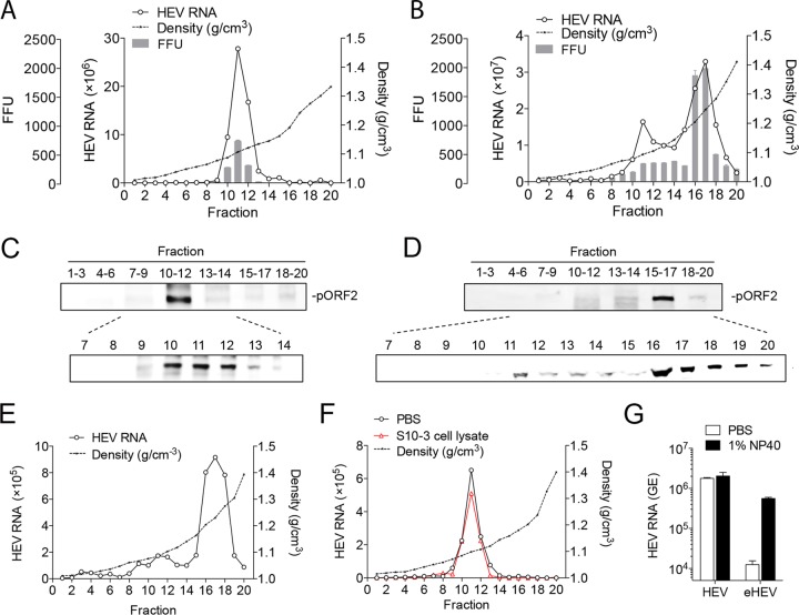
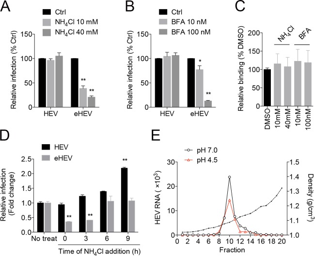
The hepatitis E virus (HEV) sheds into feces as nonenveloped virions but circulates in the blood in a membrane-associated, quasi-enveloped form (eHEV). Since the eHEV virions lack viral proteins on the surface, we investigated the entry mechanism for eHEV. We found that compared to nonenveloped HEV virions, eHEV attachment to the cell was much less efficient, requiring a longer inoculation time to reach its maximal infectivity. A survey of cellular internalization pathways identified clathrin-mediated endocytosis as the main route for eHEV entry. Unlike nonenveloped HEV virions, eHEV entry requires Rab5 and Rab7, small GTPases involved in endosomal trafficking, and blocking endosomal acidification abrogated eHEV infectivity. However, low pH alone was not sufficient for eHEV uncoating, suggesting that additional steps are required for entry. Supporting this concept, eHEV infectivity was substantially reduced in cells depleted of Niemann-Pick disease type C1, a lysosomal protein required for cholesterol extraction from lipid, or in cells treated with an inhibitor of lysosomal acid lipase. These data support a model in which the quasi-envelope is degraded within the lysosome prior to virus uncoating, a potentially novel mechanism for virus entry.

Importance: The recent discovery of quasi-enveloped viruses has shifted the paradigm of virus-host interactions. The impact of quasi-envelopment in the virus life cycle and pathogenesis is largely unknown. HEV is a highly relevant model to study these questions. HEV circulates as quasi-enveloped virions in the blood that are hidden from neutralizing antibodies. eHEV particles most likely are responsible for the cell-to-cell spread of the virus. Given the increasing concerns about persistent HEV infection and its potential for transmission via the blood supply, understanding how eHEV infects cells is important for understanding its pathogenesis and developing therapies. Our data provide evidence that eHEV uses a potentially novel mechanism for cellular entry. Several steps critical to eHEV entry were identified and may provide a basis for developing treatments for hepatitis E. Because quasi-enveloped viruses resemble exosomes, these data also may provide insights into the exosome-mediated intercellular communications.


Figures

FIG 1
FIG 1
Characterization of extracellular and intracellular HEV virions. (A and B) Buoyant density of HEV virions released into supernatant fluids (A) or in cell lysates (B) of Huh-7 cells transfected with HEV RNA. HEV RNA in fractions was determined by qRT-PCR. HEV infectivity was determined by IFA in HepG2 cells. (C and D) Distribution of the HEV capsid protein pORF2 in gradient fractions of panels A and B. (E) Buoyant density of HEV in a fecal sample of a rhesus macaque experimentally infected with HEV. (F) Buoyant density of eHEV after incubation with either PBS or uninfected Huh-7 cell lysates. (G) HEV and eHEV were either treated with 1% NP-40 or left untreated and immunoprecipitated with rabbit anti-pORF2 antibody. Bound virus was quantified by HEV-specific qRT-PCR. The results show the means ± SEM from 2 independent experiments. FFU, focus-forming units.
FIG 2
FIG 2
Cell attachment and entry kinetics of HEV and eHEV. (A) Immunofluorescence images showing the expression of the HEV pORF2 protein in HepG2 cells 5 days after inoculation with equal amounts of HEV and eHEV (3 × 10⁷ HEV GE). (B) Virus yields in wells inoculated with HEV and eHEV for 5 days. Shown are the means ± SEM from 2 independent experiments each performed in duplicate. *, P < 0.05; **, P < 0.01. (C) HEV RNA in supernatants of HEV-infected HepG2 culture. HEV RNA was detected by qRT-PCR. The results show the means ± SEM from 2 independent experiments performed in duplicate. *, P < 0.05; **, P < 0.01. (D) HepG2 cells were prechilled on ice for 15 min prior to inoculation with equal amounts of HEV and eHEV (3 × 10⁷ GE) at 4°C for 1 h. Cells were washed with cold PBS three times. Total RNA was extracted and subjected to HEV-specific qRT-PCR analysis. The results show means ± SEM from 2 independent experiments performed in duplicate. *, P < 0.05; **, P < 0.01. (E) HEV and eHEV (3 × 10⁷ GE) were incubated with heparan sulfate (HS) at the indicated concentrations for 1 h prior to inoculation. Virus yields were determined by IFA after 5 days. The results show the means ± SEM from 2 independent experiments performed in duplicate. *, P < 0.05; **, P < 0.01. (F) HepG2 cells were inoculated with HEV and eHEV (3 × 10⁷ GE). The inoculum was removed at different times. Cells were washed extensively with PBS, and fresh medium was added. Virus yields were determined by IFA after 5 days. Shown are representative results from two independent experiments.
FIG 3
FIG 3
Role of clathrin-mediated endocytosis, caveola-mediated endocytosis, and macropinocytosis in HEV and eHEV entry. (A and B) HepG2 cells were pretreated with the indicated inhibitors for 1 h prior to inoculation with HEV or eHEV. Inoculum was removed after 6 h, and culture was continued for 5 days. Virus yields were determined by IFA after 5 days. Data are represented as a percentage of the DMSO-treated control. The results show the means ± SEM from at least 2 independent experiments in duplicate. *, P < 0.05; **, P < 0.01. (C) HepG2 cells were treated with inhibitors at the indicated concentrations for 20 h, and cell viability was measured by an MTT assay. The results show the mean ± SEM results from triplicate wells. (D) HepG2 cells were treated with CPZ (10 μg/ml) or Dynasore (80 μM) for 1 h prior to incubation with Alexa-488-labeled transferrin (25 μg/ml). After 15 min of incubation, cells were fixed and analyzed by confocal microscopy. Scale bar, 10 μm. (E) HepG2 cells transfected with plasmids carrying GFP or CAV1-GFP for 48 h were treated with filipin (3 μg/ml) for 1 h and incubated with Alexa-594-labeled cholera toxin subunit B (CTxB; 20 μg/ml). After 1 h of incubation, cells were fixed and analyzed by confocal microscopy. Scale bar, 10 μm. (F) HepG2 cells were pretreated with EIPA or LY294002 for 1 h prior to inoculation with vaccinia virus (MVA-T7 strain) for 1 h. mRNA levels of the vaccinia DNA-directed RNA polymerase were determined 12 h after inoculation by qRT-PCR. Data are represented as a percentage of the DMSO-treated control. The data represent the means ± SEM from 2 independent experiments, each in duplicate. *, P < 0.05; **, P < 0.01. (G and H) HepG2 cells were transfected with nontargeting siRNA (siCtrl) or siRNA targeting the clathrin-1 heavy chain (CLTC) (G) or dynamin-2 (DNM2) (H). Thirty-six hours posttransfection, cells were inoculated with HEV or eHEV. (Upper) The knockdown efficiency was determined by immunoblotting. Virus yields were determined by IFA 5 days after inoculation. (Lower) Relative infection (percentage of control siRNA-transfected culture) was calculated based on FFU. Shown are the means ± SEM from 3 independent experiments, each in duplicate. *, P < 0.05; **, P < 0.01. (I, upper) Western blot showing the expression of GFP and CAV1-GFP in HepG2 cells transfected with the corresponding plasmids. Unmodified HepG2 cells were included as a control. Cells stably expressing GFP or CAV1-GFP were inoculated with HEV or eHEV in the presence or absence of filipin (in the case of cells expressing CAV1-GFP). (Lower) Virus yields were determined by IFA 5 days after inoculation. Shown are representative results from two independent experiments.
FIG 4
FIG 4
Role of Rab5 and Rab7 during HEV and eHEV entry. (A) Confocal images showing the localization of the HEV capsid protein pORF2 (green) in HepG2 cells previously transfected with mRFP-Rab5 or DsRed-Rab7 (red). Cells were inoculated with HEV or eHEV (1 × 10⁴ GE per cell) for 1 h and processed for confocal imaging analysis using rabbit anti-pORF2 antibody. Cells were counterstained with DAPI (blue). Scale bar, 10 μm. (B and C) HepG2 cells were transfected with control siRNA or siRNA targeting Rab5A or Rab7A. Thirty-six hours posttransfection, cells were inoculated with HEV or eHEV. (B) Immunoblots showing the knockdown efficiency 2 days after transfection. (C) Virus yields determined by IFA 5 days after inoculation. Data are represented as a percentage of the nontargeting siRNA (siCtrl)-treated control. The results represent the means ± SEM from 2 independent experiments, each in duplicate. *, P < 0.05; **, P < 0.01.
FIG 5
FIG 5
Effect of lysosomotropic agents on HEV and eHEV entry. (A and B) HepG2 cells were pretreated with NH4Cl (A) or bafilomycin A1 (BFA) (B) for 1 h prior to inoculation with HEV or eHEV. Virus yields were determined 5 days after inoculation. Data are represented as a percentage relative to the nontreated control (Ctrl). The results represent the means ± SEM from 3 independent experiments, each in duplicate. *, P < 0.05; **, P < 0.01. (C) HepG2 cells were inoculated with eHEV (3 × 10⁷ GE) in the presence or absence of NH4Cl or bafilomycin A1 (BFA) at 4°C for 1 h. Cells were washed with cold PBS three times. Total RNA was extracted and subjected to HEV-specific qRT-PCR analysis. Data represent the means ± SEM from 2 independent experiments, each in duplicate. (D) HepG2 cells were inoculated with HEV or eHEV. At different times postinoculation, NH4Cl was added and infection was continued in the presence of the drug. Virus yields were determined as indicated above. Data are represented as fold changes relative to the nontreated control. Data represent the means ± SEM from 2 independent experiments, each in duplicate. (E) Buoyant density of eHEV after incubation with solution of low (pH 4.5) or neutral (pH 7.0) pH for 30 min. HEV RNA in gradient fractions was measured by qRT-PCR.
FIG 6
FIG 6
Role of lysosomal lipid degradation in HEV and eHEV entry. (A and B) HepG2 cells were transfected with control siRNA or siRNA targeting NPC1. Thirty-six hours after transfection, cells were inoculated with HEV or eHEV. (A) Immunoblots showing the knockdown efficiency of NPC1. (B) Virus yields (FFU) 5 days after inoculation. Data are represented as a percentage relative to a nontargeting siRNA-treated control (siCtrl). Shown are the means ± SEM from 2 independent experiments, each in duplicate. *, P < 0.05; **, P < 0.01. (C) HepG2 cells were pretreated with Lalistat 2 at the indicated concentrations for 1 h prior to virus inoculation. Data are represented as a percentage of the DMSO-treated control. The result represents the means ± SEM from 2 independent experiments, each in duplicate. *, P < 0.05; **, P < 0.01.

Similar articles

Cited by

References

    1. Purcell RH, Emerson SU. 2008. Hepatitis E: an emerging awareness of an old disease. J Hepatol 48:494–503. doi: 10.1016/j.jhep.2007.12.008. - DOI - PubMed
    1. Smith DB, Simmonds P, Jameel S, Emerson SU, Harrison TJ, Meng XJ, Okamoto H, Van der Poel WH, Purdy MA. 2014. Consensus proposals for classification of the family Hepeviridae. J Gen Virol 95:2223–2232. doi: 10.1099/vir.0.068429-0. - DOI - PMC - PubMed
    1. Dalton HR, Bendall R, Ijaz S, Banks M. 2008. Hepatitis E: an emerging infection in developed countries. Lancet Infect Dis 8:698–709. doi: 10.1016/S1473-3099(08)70255-X. - DOI - PubMed
    1. Pavio N, Meng XJ, Doceul V. 2015. Zoonotic origin of hepatitis E. Curr Opin Virol 10:34–41. doi: 10.1016/j.coviro.2014.12.006. - DOI - PubMed
    1. Meng XJ. 2013. Zoonotic and foodborne transmission of hepatitis E virus. Semin Liver Dis 33:41–49. doi: 10.1055/s-0033-1338113. - DOI - PubMed

Publication types