. 2016 Apr 14;90(9):4278-4288.
doi: 10.1128/JVI.00039-16. Print 2016 May.

Transient CD4+ T Cell Depletion Results in Delayed Development of Functional Vaccine-Elicited Antibody Responses

Affiliations

Transient CD4+ T Cell Depletion Results in Delayed Development of Functional Vaccine-Elicited Antibody Responses

Nicholas M Provine et al. J Virol. .

Abstract

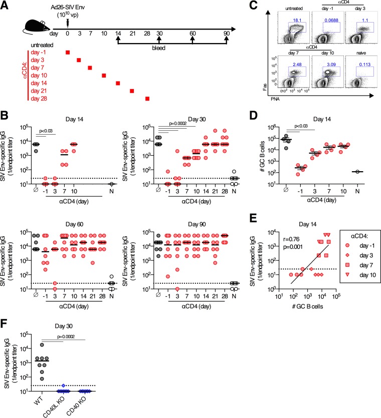
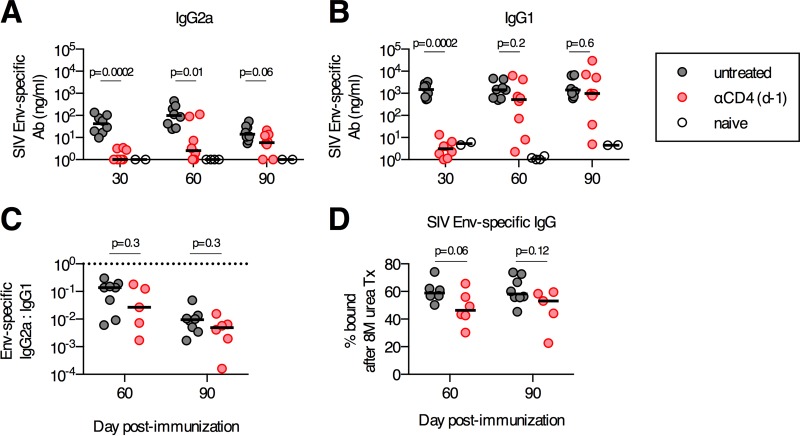
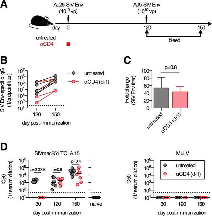
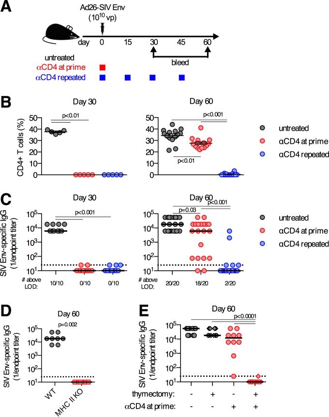
We have recently demonstrated that CD4(+)T cell help is required at the time of adenovirus (Ad) vector immunization for the development of functional CD8(+)T cell responses, but the temporal requirement for CD4(+)T cell help for the induction of antibody responses remains unclear. Here we demonstrate that induction of antibody responses in C57BL/6 mice can occur at a time displaced from the time of Ad vector immunization by depletion of CD4(+)T cells. Transient depletion of CD4(+)T cells at the time of immunization delays the development of antigen-specific antibody responses but does not permanently impair their development or induce tolerance against the transgene. Upon CD4(+)T cell recovery, transgene-specific serum IgG antibody titers develop and reach a concentration equivalent to that in undepleted control animals. These delayed antibody responses exhibit no functional defects with regard to isotype, functional avidity, expansion after boosting immunization, or the capacity to neutralize a simian immunodeficiency virus (SIV) Env-expressing pseudovirus. The development of this delayed transgene-specific antibody response is temporally linked to the expansion of de novo antigen-specific CD4(+)T cell responses, which develop after transient depletion of CD4(+)T cells. These data demonstrate that functional vaccine-elicited antibody responses can be induced even if CD4(+)T cell help is provided at a time markedly separated from the time of vaccination.

Importance: CD4(+)T cells have a critical role in providing positive help signals to B cells, which promote robust antibody responses. The paradigm is that helper signals must be provided immediately upon antigen exposure, and their absence results in tolerance against the antigen. Here we demonstrate that, in contrast to the current model that the absence of CD4(+)T cell help at priming results in long-term antibody nonresponsiveness, antibody responses can be induced by adenovirus vector immunization or alum-adjuvanted protein immunization even if CD4(+)T cell help is not provided until >1 month after immunization. These data demonstrate that the time when CD4(+)T cell help signals must be provided is more dynamic and flexible than previously appreciated. These data suggest that augmentation of CD4(+)T cell helper function even after the time of vaccination can enhance vaccine-elicited antibody responses and thereby potentially enhance the immunogenicity of vaccines in immunocompromised individuals.


Figures

FIG 1
FIG 1
Delayed development of antibody responses following depletion of CD4+ T cells. (A) C57BL/6 mice were depleted of CD4+ T cells at the indicated days or left untreated and immunized intramuscularly with 1010 vp of Ad26-SIV Env. (B) SIV Env-specific serum-binding antibody titers at the indicated days postimmunization. Gray, no anti-CD4 treatment; red, anti-CD4 given on the indicated day; white, naive animals. (C and D) Representative flow cytometry plots (C) and absolute numbers (D) of germinal center (GC) B cell responses in the iliac LNs on day 14 postimmunization, with gating on CD3ε CD19+ cells. (E) Spearman correlation analysis of the number of germinal center B cells in iliac LNs and serum SIV Env-specific binding antibody titers on day 14 postimmunization in mice treated with anti-CD4 at the days indicated. (F) SIV Env-specific serum-binding antibody titers in wild-type (WT), CD40L KO, or CD40 KO mice immunized with 109 vp of Ad26-SIV Env. Each dot represents an individual mouse, and the line is the median (B and F) or the mean ± the standard error of the mean (D). The horizontal dotted line denotes the limit of detection of the assay (for panel B, n = 4/group from one experiment [day 14] or n = 8/group pooled from two experiments [days 30, 60, and 90]; for panel D, n = 4/group from one experiment; for panel F, n = 8/group pooled from two experiments).
FIG 2
FIG 2
Transient depletion of CD4+ T cells at priming does not alter the isotype distribution or the antigen-binding avidity of delayed antibody (Ab) responses. C57BL/6 mice were depleted of CD4+ T cells or left untreated and immunized intramuscularly with 1010 vp of Ad26-SIV Env. (A and B) Concentrations of serum SIV Env-specific IgG2a (A) and IgG1 (B). (C) Ratios of SIV Env-specific IgG2a to IgG1 serum antibody titers. The horizontal dotted line denotes a ratio of 1. (D) Avidity of SIV Env-specific serum antibodies as determined by a urea disruption assay. Each dot represents an individual mouse, and the medians are indicated by the line (n = 5 to 8/group pooled from two experiments).
FIG 3
FIG 3
Boosting capacity and functional neutralization capacity of delayed antibody responses that develop following transient CD4+ T cell depletion. (A) C57BL/6 mice were depleted of CD4+ T cells or left untreated, immunized intramuscularly with 1010 vp of Ad26-SIV Env, and boosted 4 months postprime with 1010 vp of Ad5-SIV Env. (B) SIV Env-specific antibody titers prior to and following boosting immunization. (C) Fold change in SIV Env-specific antibody responses pre- to postboost. (D) Serum 50% neutralization capacity (IC50) of tier 1A SIVmac251.TCLA.15 Env-expressing pseudoviruses or a MuLV negative-control pseudovirus. Each dot represents an individual mouse, and the line indicates the mean ± the standard error of the mean (C) or the median (D). The horizontal dotted line denotes the limit of detection for the assay (n = 6 to 8/group pooled from two experiments).
FIG 4
FIG 4
Repeated depletion of CD4+ T cells prevents development of delayed antibody responses. (A) C57BL/6 mice were depleted of CD4+ T cells a single time (anti-CD4 at prime), depleted of CD4+ T cells repeatedly (anti-CD4 repeated), or left untreated and immunized intramuscularly with 1010 vp of Ad26-SIV Env. (B) Frequency of CD4 T cells in iliac (draining) LNs. (C) SIV Env-specific serum-binding antibody titers. (D) Serum SIV Env-specific antibody titers on day 60 postimmunization from wild-type or MHC-II KO mice immunized with 1010 vp of Ad26-SIV Env. (E) Serum SIV Env-specific antibody titers on day 60 postimmunization from mice that underwent adult thymectomy, or not, and were treated with anti-CD4 antibody, or not, as indicated. Each dot represents an individual mouse, and the line indicates the mean ± the standard error of the mean (B) or median (C to E). The horizontal dotted line denotes the limit of detection (LOD) for the assay (for panel B, n = 5/group from one experiment [day 30] or n = 20/group pooled from four experiments [day 60]; for panel C, n = 10/group pooled from two experiments [day 30] or n = 20/group pooled from four experiments [day 60]; for panel D, n = 8/group pooled from two experiments; for panel E, n = 10/group pooled from two experiments).
FIG 5
FIG 5
Germinal center B cell responses develop following transient depletion of CD4+ T cells. C57BL/6 mice were depleted of CD4 T cells a single time (anti-CD4 at prime) (red circles), depleted of CD4 T cells repeatedly (anti-CD4 repeated) (blue circles), or left untreated (gray circles) and immunized intramuscularly with 1010 vp of Ad26-SIV Env. (A to C) Representative flow cytometry plots (A), group average percentages (B), and absolute numbers (C) of germinal center B cells in the iliac (draining) LNs on day 60 postimmunization. (D) Expression of IgM and IgD on germinal center B cells on day 60 postimmunization. Each dot represents an individual mouse, and the means ± standard errors of the means are indicated by a line (n = 15/group pooled from three experiments).
FIG 6
FIG 6
Development of antigen-specific CD4+ T cell responses after transient depletion of CD4+ T cells. (A) Gating scheme for characterization of antigen-specific CD4+ T cells by intracellular cytokine staining. SSC, side scatter; FSC, forward scatter. (B) C57BL/6 mice were depleted of CD4+ T cells at immunization or left untreated and immunized intramuscularly with 1010 vp of Ad26-SIV Env. Spleens and iliac (draining) LNs were collected on day 60 postimmunization, and intracellular cytokine staining was performed to detect the production of IL-21, IFN-γ, and IL-2 by CD44+ CD4+ T cells following stimulation with an overlapping SIV Env peptide pool. Means ± standard errors of the means are shown. The horizontal dotted line denotes the limit of detection for the assay (n = 10/group pooled from two experiments).
FIG 7
FIG 7
Delayed antibody responses develop following immunization with an adjuvanted soluble protein formulated in Adju-Phos but not following immunization with a replication-incompetent poxvirus vector. SIV Env-specific serum antibody titers in C57BL/6 mice immunized intramuscularly with 50 μg of SIV Env gp140 plus Adju-Phos and depleted of CD4+ T cells at the time of immunization (anti-CD4 at prime) or continuously depleted of CD4+ T cells (anti-CD4 repeated) or mice left untreated were determined. Each dot represents an individual mouse, and the line is the median. The horizontal dotted line denotes the limit of detection for the assay (n = 4 to 5/group from one experiment).

Similar articles

Cited by

References

    1. Swain SL, McKinstry KK, Strutt TM. 2012. Expanding roles for CD4+ T cells in immunity to viruses. Nat Rev Immunol 12:136–148. doi:10.1038/nri3152. - DOI - PMC - PubMed
    1. Fulcher DA, Lyons AB, Korn SL, Cook MC, Koleda C, Parish C, Fazekas de St Groth B, Basten A. 1996. The fate of self-reactive B cells depends primarily on the degree of antigen receptor engagement and availability of T cell help. J Exp Med 183:2313–2328. doi:10.1084/jem.183.5.2313. - DOI - PMC - PubMed
    1. Victora GD, Nussenzweig MC. 2012. Germinal centers. Annu Rev Immunol 30:429–457. doi:10.1146/annurev-immunol-020711-075032. - DOI - PubMed
    1. Benjamin RJ, Waldmann H. 1986. Induction of tolerance by monoclonal antibody therapy. Nature 320:449–451. doi:10.1038/320449a0. - DOI - PubMed
    1. Goronzy J, Weyand CM, Fathman CG. 1986. Long-term humoral unresponsiveness in vivo, induced by treatment with monoclonal antibody against L3T4. J Exp Med 164:911–925. doi:10.1084/jem.164.3.911. - DOI - PMC - PubMed

Publication types

MeSH terms