. 2016 Apr 14;90(9):4289-97.
doi: 10.1128/JVI.00018-16. Print 2016 May.

Role of TSPAN9 in Alphavirus Entry and Early Endosomes

Affiliations

Role of TSPAN9 in Alphavirus Entry and Early Endosomes

Katie M Stiles et al. J Virol. .

Abstract

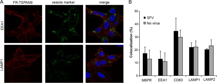
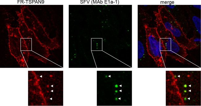
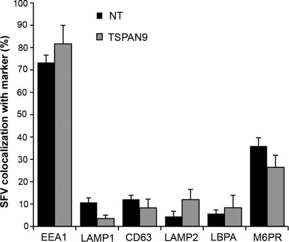
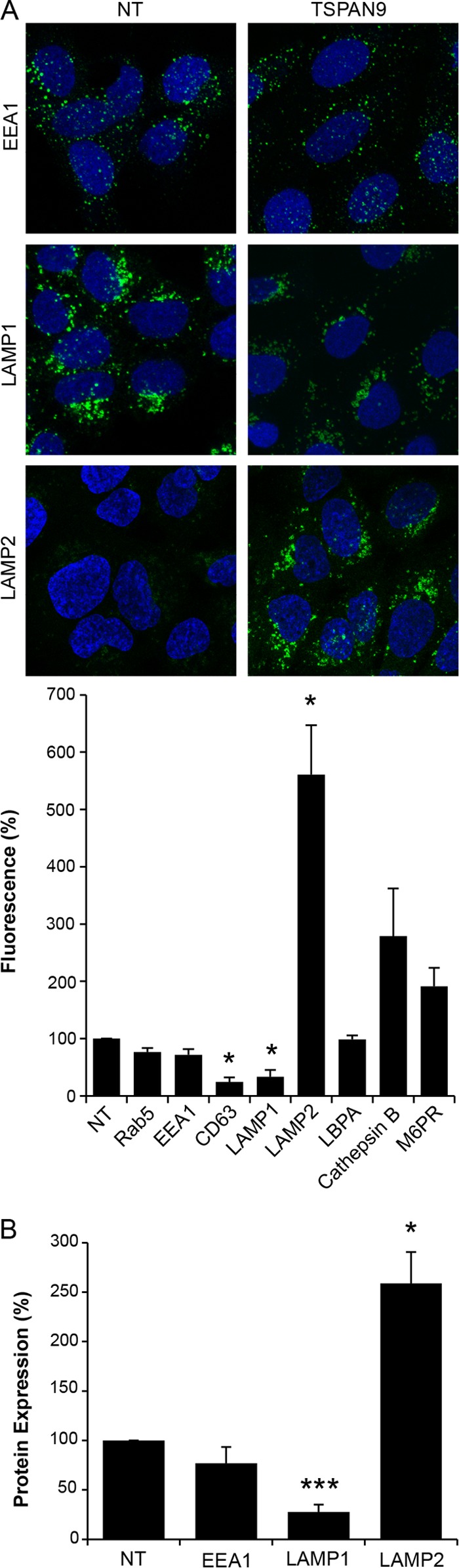
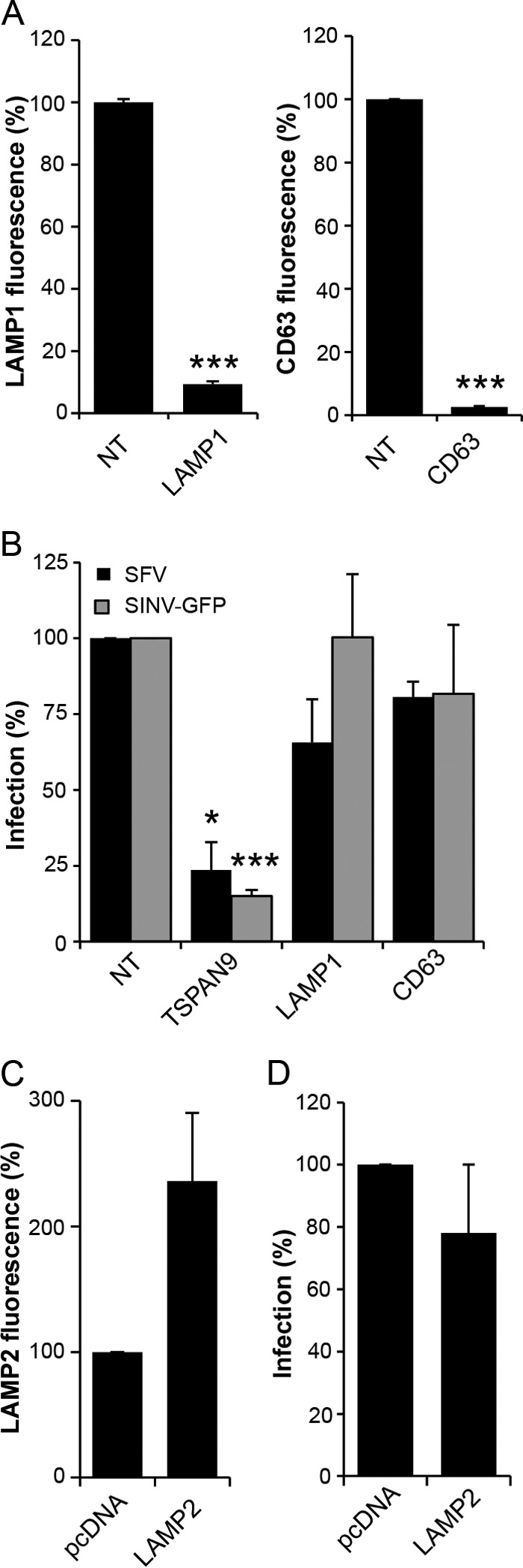
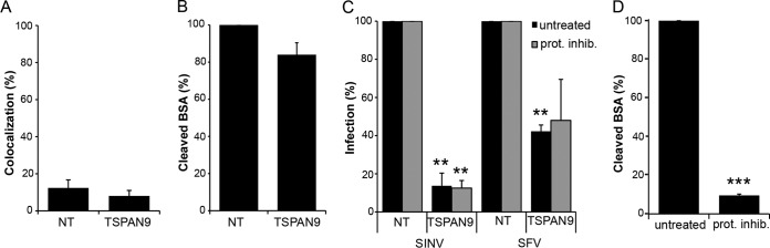
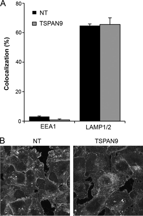
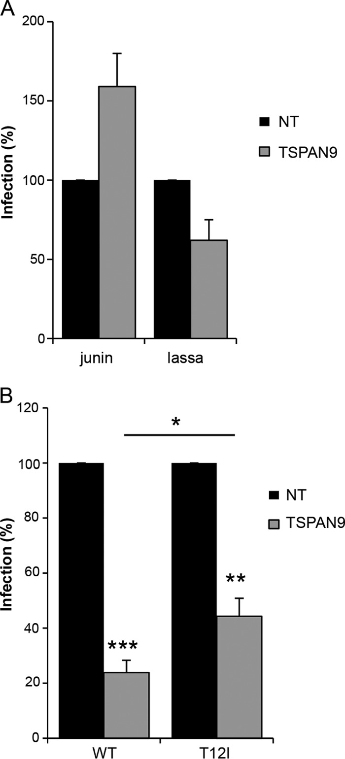
Alphaviruses are small enveloped RNA viruses that infect cells via clathrin-mediated endocytosis and low-pH-triggered fusion in the early endosome. Using a small interfering RNA (siRNA) screen in human cells, we previously identified TSPAN9 as a host factor that promotes infection by the alphaviruses Sindbis virus (SINV), Semliki Forest virus (SFV), and chikungunya virus (CHIKV). Depletion of TSPAN9 specifically decreases SFV membrane fusion in endosomes. TSPAN9 is a member of the tetraspanin family of multipass membrane proteins, but its cellular function is currently unknown. Here we used U-2 OS cells stably overexpressing TSPAN9 to show that TSPAN9 is localized at the plasma membrane and in early and late endosomes. Internalized SFV particles colocalized with TSPAN9 in vesicles early during infection. Depletion of TSPAN9 led to reductions in the amounts of the late endosomal proteins LAMP1 and CD63 and an increase in the amount of LAMP2. However, TSPAN9 depletion did not alter the delivery of SFV to early endosomes or change their pH or protease activity. Comparative studies showed that TSPAN9 depletion strongly inhibited infection by several viruses that fuse in early endosomes (SFV, SINV, CHIKV, and vesicular stomatitis virus [VSV]), while viruses that fuse in the late endosome (recombinant VSV-Lassa and VSV-Junin), including an SFV point mutant with a lower pH threshold for fusion (SFV E2 T12I), were relatively resistant. Our data suggest that TSPAN9 modulates the early endosome compartment to make it more permissive for membrane fusion of early-penetrating viruses.

Importance: Alphaviruses are spread by mosquitoes and can cause serious human diseases such as arthritis and encephalitis. Recent outbreaks of CHIKV infection are responsible for millions of cases of acute illness and long-term complications. There are no vaccines or antiviral treatments for these important human pathogens. Alphaviruses infect host cells by utilizing the endocytic machinery of the cell and fusing their membrane with that of the endosome. Although the mechanism of virus-membrane fusion is well studied, we still know relatively little about the host cell proteins that are involved in alphavirus entry. Here we characterized the role of the host membrane protein TSPAN9 in alphavirus infection. TSPAN9 was localized to early endosomes containing internalized alphavirus, and depletion of TSPAN9 inhibited virus fusion with the early endosome membrane. In contrast, infection of viruses that enter through the late endosome was relatively resistant to TSPAN9 depletion, suggesting an important role for TSPAN9 in the early endosome.


Figures

FIG 1
FIG 1
Intracellular localization of FR-TSPAN9. (A) U-2 OS cells stably expressing FR-TSPAN9 were stained with MAbs to the indicated proteins of the endosomal pathway, followed by anti-mouse Alexa 488 secondary antibody (green). FR-TSPAN9 is shown in red. A single confocal slice from a representative experiment of three is shown. (B) U-2 OS cells stably expressing FR-TSPAN9 were incubated with or without 0.2 μg SFV/well for 20 min at 37°C and then fixed and stained for the indicated proteins as described above for panel A. Colocalization between FR-TSPAN9 and endosome proteins was quantitated by using Cell Profiler in the presence and absence of SFV internalization. Data represent the means ± standard errors of the means of data from three independent experiments. No significant differences were observed between the results with and without SFV.
FIG 2
FIG 2
Colocalization of FR-TSPAN9 with SFV. U-2 OS cells stably expressing FR-TSPAN9 were incubated with 0.2 μg SFV/well for 20 min at 37°C and then fixed and stained with acid conformation-specific E1 MAb E1a-1 (green). Approximately 70% of the acid-exposed SFV colocalizes with punctate FR-TSPAN9 (red). A single confocal slice from a representative experiment of three is shown.
FIG 3
FIG 3
Effect of TSPAN9 depletion on the intracellular localization of SFV. U-2 OS cells were transfected with NT or TSPAN9 siRNA and cultured for 48 h. SFV was bound to cells for 1 h on ice, followed by warming to 37°C for 20 min before fixation. SFV was stained with anti-E1/E2 PAb before and after permeabilization, under conditions that differentiate virus remaining on the cell surface from internalized virus (9), and endosomal proteins were stained with the indicated MAbs. Colocalization of internalized virus and cellular proteins was quantitated by using Cell Profiler and expressed as a percentage of the total internalized SFV puncta. Data represent the means ± standard errors of the means of data from three independent experiments. No significant differences between the NT and TSPAN9 siRNA samples were observed.
FIG 4
FIG 4
Quantitation of endosomal proteins after TSPAN9 depletion. U-2 OS cells were transfected with NT or TSPAN9 siRNA for 48 h before fixation. Endosomal proteins were stained with the indicated MAbs. (A) Representative confocal extended-focus images from three experiments. The total integrated density of fluorescence per image was quantitated by using Image J and normalized to the number of cells per image. Fifty cells were counted in each of three independent experiments (bar graph). (B) U-2 OS cells transfected as described above for panel A were lysed and subjected to SDS-PAGE and Western blotting with the indicated antibodies. Protein amounts were quantitated and normalized to tubulin levels as the loading control. The bar graphs in panels A and B represent the means ± standard errors of the means for three independent experiments (*, P < 0.05; ***, P < 0.001).
FIG 5
FIG 5
Effect of depletion of LAMP1 and CD63 or overexpression of LAMP2. (A) U-2 OS cells were transfected with NT, LAMP1, or CD63 siRNA; cultured for 48 h; and stained with anti-LAMP1 antibody (left) or anti-CD63 antibody (right). The total integrated density of fluorescence per image was quantitated by using Image J and normalized to the number of cells per image. Fifty cells were quantitated under each condition in each of three independent experiments. (B) U-2 OS cells transfected as described above for panel A were cultured for 48 h and then infected with SFV (MOI = 0.1) or SINV-GFP (MOI = 10) for 1 h before the addition of 20 mM NH4Cl and incubation for 14 h (primary infection). Cells were then fixed, and SFV-infected cells were stained with PAb against E1/E2. Infected cells and nuclei were counted by using Cell Profiler to determine the percentage of infected cells, and values were normalized to those for the NT control. (C) U-2 OS cells were transfected with pcDNA or pcDNA-hLAMP2 for 24 h and stained with anti-LAMP2 MAb. The total integrated density of fluorescence per image was quantitated by using Image J and normalized to the number of cells per image. Fifty cells were quantitated under each condition in each of three independent experiments. (D) U-2 OS cells were transfected with pcDNA or pcDNA-hLAMP2 for 24 h before infection with SFV and staining as described above for panel B. Infected cells and nuclei were counted by using Cell Profiler, and values were normalized to values for the pcDNA control. Data in all bar graphs represent the means ± standard errors of the means for three independent experiments (*, P < 0.05; ***, P < 0.001).
FIG 6
FIG 6
Effect of TSPAN9 depletion on endosomal proteases. (A) U-2 OS cells were transfected with NT or TSPAN9 siRNA and cultured for 48 h, and 0.2 μg SFV/well was bound to cells for 1 h on ice, followed by warming to 37°C for 20 min. Cells were then fixed, permeabilized, and stained with SFV MAb E1a-1 and anti-cathepsin B PAb. Colocalization of SFV and cathepsin B staining was quantitated by using Cell Profiler. (B) U-2 OS cells were transfected as described above for panel A and incubated with 200 μg/ml DQ-BSA for 5 h at 37°C. The total integrated density of fluorescence per image was quantitated by using Image J and normalized to the number of cells per image for a total of 50 cells per experiment. (C) U-2 OS cells transfected as described above for panel A were pretreated with 5 μM E64d and 100 μM leupeptin for 5 h before the addition of SFV (MOI = 0.01) or SINV-GFP (MOI = 1). Infected cells were incubated for 12 h postinfection (SFV) or 24 h postinfection (SINV-GFP), and infection was quantitated by GFP expression or staining with SFV anti-E1/E2 PAb. (D) U-2 OS cells were treated with protease inhibitors as described above for panel C, incubated with 200 μg/ml DQ-BSA, and quantitated as described above for panel B. Bar graphs represent the means ± standard errors of the means for three independent experiments (**, P < 0.01; ***, P < 0.001).
FIG 7
FIG 7
Effect of TSPAN9 depletion on endosomal pH and cholesterol levels. (A) U-2 OS cells were transfected with NT or TSPAN9 siRNA, cultured for 48 h, and then incubated with LysoSensor Green DND-189 (pKa of ∼5.2) for 1 h at 37°C before fixation. Cells were then permeabilized and stained with either a MAb against EEA1 or MAbs against LAMP1 and LAMP2. Colocalization of LysoSensor and the indicated endosomal proteins was quantitated by using Cell Profiler. Bar graphs represent the means ± standard errors of the means of data from three independent experiments. No significant differences were observed between the NT and TSPAN9 siRNA samples. (B) Control or TSPAN9-depleted U-2 OS cells prepared as described above for panel A were fixed and stained with filipin (50 μg/ml). A single confocal slice from a representative experiment of three is shown.
FIG 8
FIG 8
Effect of TSPAN9 depletion on viruses entering from late endosomes. U-2 OS cells were transfected with NT or TSPAN9 siRNA, cultured for 48 h, and infected as described in the text. (A) Cells were infected with rVSV-Lassa (MOI = 0.5) or rVSV-Junin (MOI = 2) for 2 h before the addition of NH4Cl and incubation for 14 h (primary infection). GFP-positive cells and nuclei were counted by immunofluorescence. Bar graphs represent the means ± standard errors of the means of data from three independent experiments. No significant differences were observed between the NT and TSPAN9 siRNA samples. (B) Cells were infected with SFV or the SFV T12I mutant (MOI = 0.2) for 1 h before the addition of NH4Cl and incubation for 14 h (primary infection). Infected cells were stained with anti-E1/E2 PAb and counted by immunofluorescence analysis. Bar graphs represent the means ± standard errors of the means of data from three independent experiments (*, P < 0.05; **, P < 0.01; ***, P < 0.001).

Similar articles

Cited by

References

    1. Kuhn RJ. 2013. Togaviridae, p 629–650. In Knipe DM, Howley PM, Cohen JI, Griffin DE, Lamb RA, Martin MA, Racaniello VR, Roizman B (ed), Fields virology, 6th ed, vol 1 Lippincott Williams & Wilkins, Philadelphia, PA.
    1. Schwartz O, Albert ML. 2010. Biology and pathogenesis of chikungunya virus. Nat Rev Microbiol 8:491–500. doi:10.1038/nrmicro2368. - DOI - PubMed
    1. Enserink M. 2007. Infectious diseases. Chikungunya: no longer a third world disease. Science 318:1860–1861. doi:10.1126/science.318.5858.1860. - DOI - PubMed
    1. Morrison TE. 2014. Reemergence of chikungunya virus. J Virol 88:11644–11647. doi:10.1128/JVI.01432-14. - DOI - PMC - PubMed
    1. Johansson MA. 2015. Chikungunya on the move. Trends Parasitol 31:43–45. doi:10.1016/j.pt.2014.12.008. - DOI - PMC - PubMed

Publication types

MeSH terms

LinkOut - more resources