. 2016 Mar 28;90(8):4174-85.
doi: 10.1128/JVI.02891-15. Print 2016 Apr.

Hepatitis C Virus-Induced Degradation of Cell Death-Inducing DFFA-Like Effector B Leads to Hepatic Lipid Dysregulation

Affiliations

Hepatitis C Virus-Induced Degradation of Cell Death-Inducing DFFA-Like Effector B Leads to Hepatic Lipid Dysregulation

Emily M Lee et al. J Virol. .

Abstract

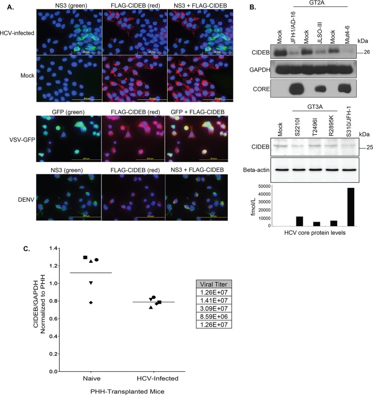
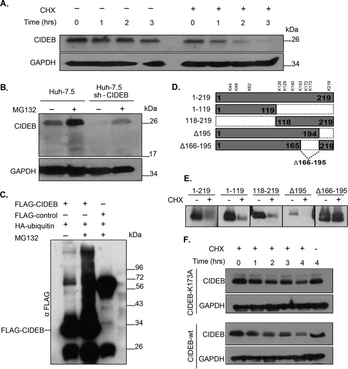
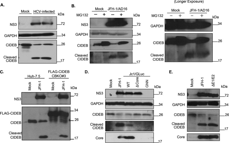
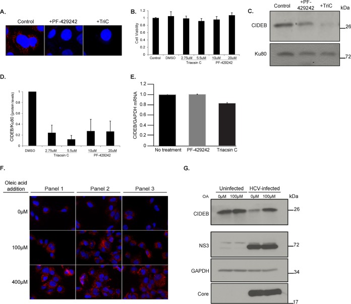
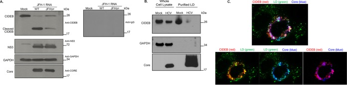
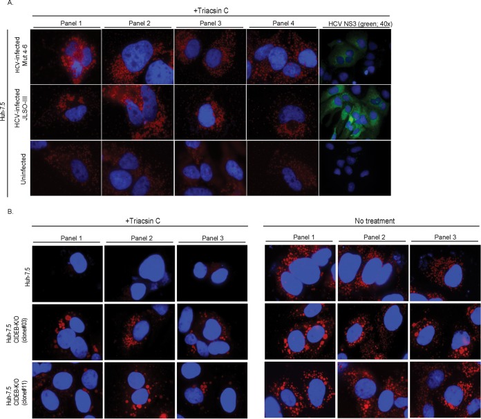
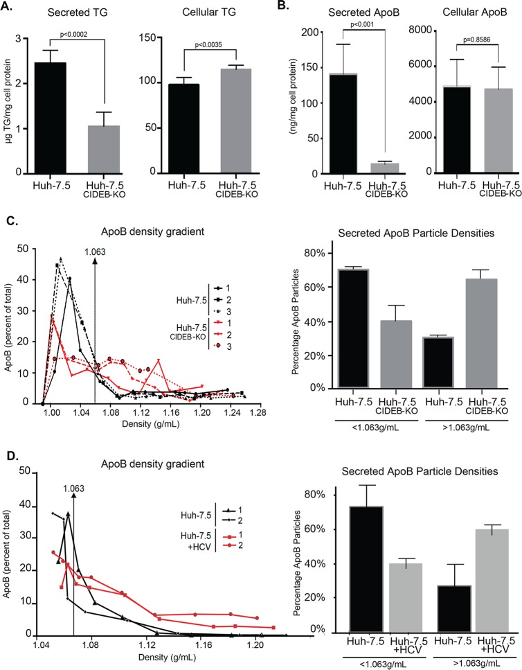
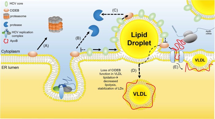
Individuals chronically infected with hepatitis C virus (HCV) commonly exhibit hepatic intracellular lipid accumulation, termed steatosis. HCV infection perturbs host lipid metabolism through both cellular and virus-induced mechanisms, with the viral core protein playing an important role in steatosis development. We have recently identified a liver protein, the cell death-inducing DFFA-like effector B (CIDEB), as an HCV entry host dependence factor that is downregulated by HCV infection in a cell culture model. In this study, we investigated the biological significance and molecular mechanism of this downregulation. HCV infection in a mouse model downregulated CIDEB in the liver tissue, and knockout of the CIDEB gene in a hepatoma cell line results in multiple aspects of lipid dysregulation that can contribute to hepatic steatosis, including reduced triglyceride secretion, lower lipidation of very-low-density lipoproteins, and increased lipid droplet (LD) stability. The potential link between CIDEB downregulation and steatosis is further supported by the requirement of the HCV core and its LD localization for CIDEB downregulation, which utilize a proteolytic cleavage event that is independent of the cellular proteasomal degradation of CIDEB.

Importance: Our data demonstrate that HCV infection of human hepatocytesin vitroandin vivoresults in CIDEB downregulation via a proteolytic cleavage event. Reduction of CIDEB protein levels by HCV or gene editing, in turn, leads to multiple aspects of lipid dysregulation, including LD stabilization. Consequently, CIDEB downregulation may contribute to HCV-induced hepatic steatosis.


Figures

FIG 1
FIG 1
HCV infection downregulates CIDEB in vitro and in vivo. (A) Immunofluorescence staining of viral proteins and CIDEB. Huh-7.5-based CIDEB-KO cells (clone 3) stably expressing FLAG-CIDEB were infected with a high-titer JFH-1 (HCV) variant, JFH-1/AD16 (genotype 2), for 3 days before costaining for FLAG and HCV NS3. FLAG-CIDEB CBKO#3 cells were infected with VSV-GFP for 16 h, stained for FLAG-CIDEB, and analyzed for coexpression of FLAG-CIDEB and GFP. FLAG-CIDEB CBKO#3 cells were infected with DENV for 48 h and costained for FLAG-CIDEB and DENV NS3. (B) Immunoblot of CIDEB in genotype 2-infected (top) or genotype 3-infected (bottom) cells. Cells were infected for 3 days with various genotype 2a viruses before analysis by Western blotting or for 8 days with various genotype 3a-based (S310) clones or the S310/JFH-1 chimera. On day 8 postinfection, core protein was measured from the supernatants by HCV core-specific ELISA, and cell lysates were analyzed by Western blotting for CIDEB. Beta-actin was included as a loading control. (C) CIDEB protein levels in the HCV-infected uPA/SCID humanized mouse model. Liver tissue lysates from primary human hepatocytes (PHH)-transplanted uPA/SCID mice, either uninfected (n = 5) or HCV infected (n = 5), were subjected to Western blotting to measure CIDEB protein levels. Data from individual mice are plotted as CIDEB protein normalized to GAPDH protein (control). Quantification of band intensity was performed using ImageJ software (National Institutes of Health). HCV-infected versus uninfected humanized mice showed statistically significant levels of CIDEB protein (P = 0.01). Measured HCV titers (in RNA copies per milliliter) are listed in the adjacent table.
FIG 2
FIG 2
Endogenous CIDEB is a short-lived protein that is posttranslationally regulated via the ubiquitin-proteasome pathway. (A) Cells were treated with the protein synthesis inhibitor CHX for 0, 1, 2, or 3 h, then lysed, and analyzed by Western blotting for CIDEB protein levels. GAPDH was included as a loading control. (B) Treatment with the proteasome inhibitor MG132 blocked CIDEB degradation in both wild-type cells and cells harboring a lentivirus encoding shRNA targeting CIDEB mRNA. (C) Analysis of CIDEB ubiquitination. Cells were cotransfected with FLAG-CIDEB or a FLAG-control protein (Prp31c) and HA-ubiquitin constructs, followed by MG132 treatment, coimmunoprecipitation with anti-FLAG beads, and Western blotting. (D) Schematic of FLAG-CIDEB deletion constructs used for stability analysis. (E) Stability of CIDEB deletion mutants. Cells were transfected with various FLAG-CIDEB-encoding constructs (represented in panel D) and subsequently treated with CHX, 24 h posttransfection, for 4 h, before immunoblot analysis using FLAG-specific antibody. (F) The point mutation K173A enhanced CIDEB stability. Cells transfected with wild-type or K173A CIDEB mutant cDNA were treated with CHX, collected at the indicated times, and analyzed similarly to the cells for panel A.
FIG 3
FIG 3
HCV-infected cells exhibit lower levels of CIDEB protein than do uninfected cells, likely through a proteolytic cleavage event. (A) Effect of HCV infection on endogenous CIDEB protein. Western blot analysis of Huh-7.5 cells infected with JFH-1/AD16 for 3 days revealed a faster-migrating band recognized by the CIDEB antibody, in addition to the full-length product. The asterisk indicates a degradation product occasionally seen from uninfected (mock) Huh-7.5 cells. (B) Effect of proteasome inhibition on CIDEB protein levels in HCV-infected cells. Huh-7.5 cells were infected with JFH-1/AD16 for 48 h and then treated with MG132 for 18 h before analysis by Western blotting. Results obtained with a longer exposure of GAPDH, CIDEB, and the CIDEB cleavage product are shown to the right. (C) Effect of HCV infection on N-terminally FLAG-tagged CIDEB. Shown are the results of Western blot analysis of CIDEB-KO cells (clone number 3) stably expressing FLAG-CIDEB and electroporated with JFH-1 for 3 days. FLAG-CIDEB was detected by anti-CIDEB antibody. (D) Western blot analysis of endogenous CIDEB and NS3 levels in Huh-7.5 cells with various HCV RNAs. Cells were electroporated with 10 μg of RNA generated from either wild-type or mutant genomes and then analyzed 48 h later for the ability to downregulate CIDEB. The Δcore mutant is a Jc1/GLuc variant lacking the intact HCV capsid protein; the GNN mutant is a Jc1/GLuc variant harboring a replication-deficient replicase. (E) Similar analysis of endogenous CIDEB and NS3 levels in Huh-7.5 cells 48 h after cells were electroporated with 10 μg of RNA generated from either wild-type JFH-1 or the ΔE1/E2-JFH-1 mutant, a viral construct lacking the two HCV envelope proteins.
FIG 4
FIG 4
Intracellular lipid abundance affects CIDEB protein stability. (A) Effect of two structurally distinct lipid droplet inhibitors, PF-429242, a reversible inhibitor of cholesterol synthesis, and Triacsin C, an inhibitor of long-chain-fatty-acid acyl-CoA synthetase, on LDs in Huh-7.5 cells. Cells were treated with 40 μM PF-429242 or 5.5 μM TriC for 24 h before staining with ORO. (B) Cell viability was measured after 24 h of treatment with the indicated compounds. (C and D) Western blot analysis of CIDEB protein levels in Huh-7.5 cells treated with PF-429242 or TriC for 24 h. Shown is quantification of CIDEB protein levels on a Western blot by ImageJ analysis (analyzed from biological triplicates). Ku80 was used as a loading control. (E) A 24-h treatment of Huh-7.5 cells with PF-429242 or TriC does not significantly alter CIDEB mRNA levels. (F and G) Effect of exogenous lipid loading on CIDEB protein in infected cells. Uninfected or (24-h) JFH-1/AD16-infected Huh-7.5 cells were treated with 100 μM OA for 20 h before being fixed for ORO staining or lysed for Western blot analysis.
FIG 5
FIG 5
(A) Effect of a JFH-1 core mutant deficient in LD-targeting on CIDEB cleavage. Western blot analysis of Huh-7.5 cells was performed 48 h after electroporation of cells with 10 μg of wild-type JFH-1 (WT) or JFHDP RNA. (B) Localization of endogenous CIDEB and HCV core after HCV infection assayed by LD fractionation. Fifteen micrograms of whole-cell lysate and 7.5 μg of purified LD-associated proteins were analyzed by Western blotting. (C) Colocalization of exogenously overexpressed FLAG-tagged CIDEB and HCV core protein on lipid droplets.
FIG 6
FIG 6
CIDEB modulates LD stability. (A) LD stability in naive or JFH-1/AD16-infected Huh-7.5 cells. Cells were incubated in lipid-rich medium for 14 h, washed with PBS, and cultured in DMEM with 5.5 μM TriC for 24 h before staining with ORO (red, 100×) and 4′,6-diamidino-2-phenylindole (DAPI) (blue) or with anti-NS3 (green, 40×) and DAPI. (B) Formation of LDs in Huh-7.5 or Huh-7.5 CIDEB-KO cells after lipid loading. Cells were incubated in lipid-rich medium for 20 h, washed with PBS, and stained for LDs using ORO (red, 100×). (C) LD stability in wild-type and CIDEB KO cells. After lipid loading as described above, cells were cultured in DMEM with or without 5.5 μM TriC for 24 h and then stained for LDs using ORO (red, 100×). Panels 1, 2, and 3 are separate frames. DAPI (blue) was used as a counterstain.
FIG 7
FIG 7
Knockout of CIDEB in human hepatoma cells results in altered VLDL secretion and TG storage. (A) Secreted and intracellular TG in Huh-7.5 or Huh-7.5 CIDEB-KO (clone 11) cells. Cells were cultured in a lipid-rich medium (supplemented with 375 μM OA) for 20 h, washed with PBS, and then either collected for lysate or further incubated in fresh DMEM for 2 h for supernatant collection. Both the lysate and supernatant were analyzed for TG contents. (B) Analysis of secreted and intracellular ApoB levels. Huh-7.5 cells or or Huh-7.5 CIDEB-KO (clone 11) cells were cultured in a lipid-rich medium (supplemented with 375 μM OA) for 14 h, washed with PBS, and then further incubated in fresh DMEM for 8 h for supernatant collection. (C) Lipid density profile of secreted ApoB-associated VLDL particles. Fractions of the supernatant collected as described above were analyzed for the amount of ApoB protein in each fraction. Distribution of ApoB-containing particles of different densities are quantified and plotted. Data from three replicate experiments are shown. (D) Lipid density profile of secreted ApoB-associated VLDL particles in HCV-infected cells. Naive Huh-7.5 cells and and Huh-7.5 cells 60 h post-JFH-1/AD16 infection were cultured in lipid-rich medium (supplemented with 375 μM OA) for 14 h, washed with PBS, and then further incubated in fresh DMEM for 8 h for supernatant collection. Fractions of the supernatant collected as described above were analyzed for the amount of ApoB protein in each fraction. Distributions of ApoB-containing particles of different densities are quantified and plotted. Data from two biological replicate experiments are shown.
FIG 8
FIG 8
Potential mechanism for HCV-mediated CIDEB downregulation. CIDEB protein levels are normally regulated by the ubiquitin-proteasome pathway (A). In the infected cells, HCV core is trafficked onto LDs (B), which may result in competitive displacement of CIDEB off the LD surface (C), exposing CIDEB to a protease. A reduced level of CIDEB results in an altered VLDL lipid profile (D), through a mechanism involving the reported CIDEB-ApoB interaction (E) (34). ER, endoplasmic reticulum.

Similar articles

Cited by

References

    1. Pawlotsky JM. 2013. Treatment of chronic hepatitis C: current and future. Curr Top Microbiol Immunol 369:321–342. - PubMed
    1. Schaefer EA, Chung RT. 2013. HCV and host lipids: an intimate connection. Semin Liver Dis 33:358–368. doi:10.1055/s-0033-1358524. - DOI - PubMed
    1. Miyanari Y, Atsuzawa K, Usuda N, Watashi K, Hishiki T, Zayas M, Bartenschlager R, Wakita T, Hijikata M, Shimotohno K. 2007. The lipid droplet is an important organelle for hepatitis C virus production. Nat Cell Biol 9:1089–1097. doi:10.1038/ncb1631. - DOI - PubMed
    1. Lindenbach BD, Rice CM. 2013. The ins and outs of hepatitis C virus entry and assembly. Nat Rev Microbiol 11:688–700. doi:10.1038/nrmicro3098. - DOI - PMC - PubMed
    1. Monazahian M, Bohme I, Bonk S, Koch A, Scholz C, Grethe S, Thomssen R. 1999. Low density lipoprotein receptor as a candidate receptor for hepatitis C virus. J Med Virol 57:223–229. doi:10.1002/(SICI)1096-9071(199903)57:3<223::AID-JMV2>3.0.CO;2-4. - DOI - PubMed

Publication types