. 2016 Mar 28;90(8):4067-4077.
doi: 10.1128/JVI.02449-15. Print 2016 Apr.

Porcine Sapelovirus Uses α2,3-Linked Sialic Acid on GD1a Ganglioside as a Receptor

Affiliations

Porcine Sapelovirus Uses α2,3-Linked Sialic Acid on GD1a Ganglioside as a Receptor

Deok-Song Kim et al. J Virol. .

Abstract

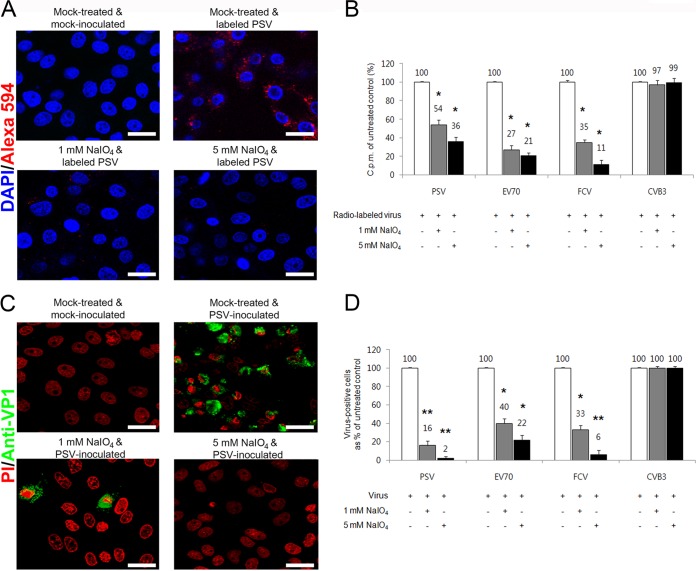
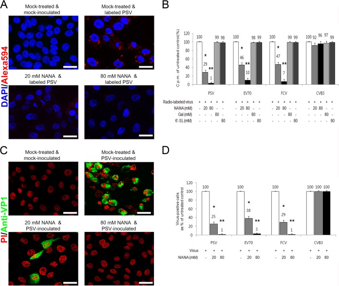
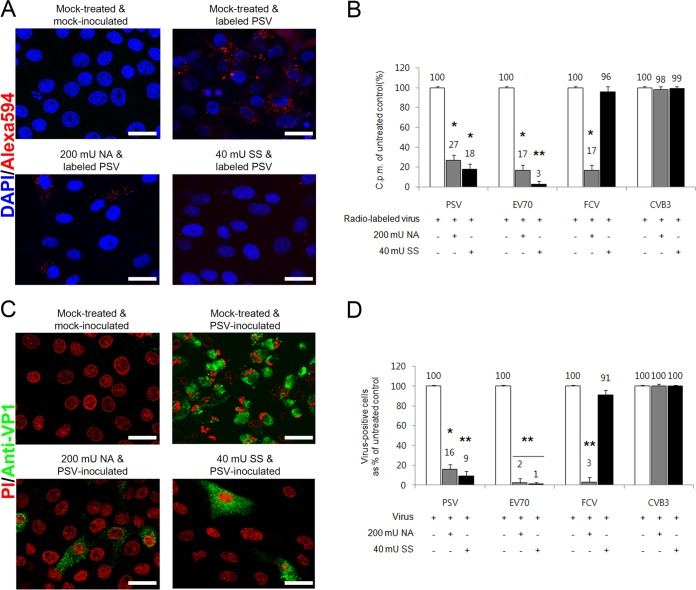
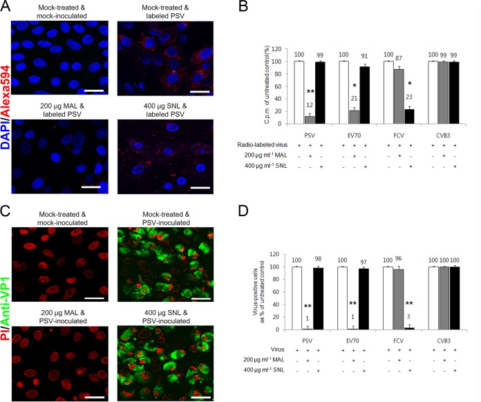
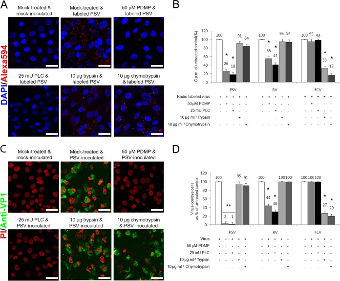
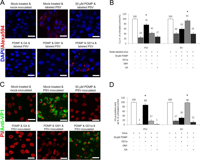
The receptor(s) for porcine sapelovirus (PSV), which causes diarrhea, pneumonia, polioencephalomyelitis, and reproductive disorders in pigs, remains largely unknown. Given the precedent for other picornaviruses which use terminal sialic acids (SAs) as receptors, we examined the role of SAs in PSV binding and infection. Using a variety of approaches, including treating cells with a carbohydrate-destroying chemical (NaIO4), mono- or oligosaccharides (N-acetylneuraminic acid, galactose, and 6'-sialyllactose), linkage-specific sialidases (neuraminidase and sialidase S), lectins (Maakia amurensislectin andSambucus nigralectin), proteases (trypsin and chymotrypsin), and glucosylceramide synthase inhibitors (dl-threo-1-phenyl-2-decanoylamino-3-morpholino-1-propanol and phospholipase C), we demonstrated that PSV could recognize α2,3-linked SA on glycolipids as a receptor. On the other hand, PSVs had no binding affinity for synthetic histo-blood group antigens (HBGAs), suggesting that PSVs could not use HBGAs as receptors. Depletion of cell surface glycolipids followed by reconstitution studies indicated that GD1a ganglioside, but not other gangliosides, could restore PSV binding and infection, further confirming α2,3-linked SA on GD1a as a PSV receptor. Our results could provide significant information on the understanding of the life cycle of sapelovirus and other picornaviruses. For the broader community in the area of pathogens and pathogenesis, these findings and insights could contribute to the development of affordable, useful, and efficient drugs for anti-sapelovirus therapy.

Importance: The porcine sapelovirus (PSV) is known to cause enteritis, pneumonia, polioencephalomyelitis, and reproductive disorders in pigs. However, the receptor(s) that the PSV utilizes to enter host cells remains largely unknown. Using a variety of approaches, we showed that α2,3-linked terminal sialic acid (SA) on the cell surface GD1a ganglioside could be used for PSV binding and infection as a receptor. On the other hand, histo-blood group antigens also present in the cell surface carbohydrates could not be utilized as PSV receptors for binding and infection. These findings should contribute to the understanding of the sapelovirus life cycle and to the development of affordable, useful and efficient drugs for anti-sapelovirus therapy.


Figures

FIG 1
FIG 1
PSV binding and infection require carbohydrate moieties. (A) The binding of AF-594-labeled PSV (MOI, 1,000 PFU/cell) was examined by confocal microscopy after pretreating LLC-PK cells with NaIO4. (B) Binding of [35S]methionine-cysteine-labeled PSV, EV70, FCV, or CVB3 (50,000 cpm) was measured by liquid scintillation counting after treating cell or not with NaIO4. The levels of bound virus were expressed as a percentage of the value for the mock-treated, virus-infected control. (C) The effect of NaIO4 pretreatment of permissive cells on the infectivity of PSV (MOI, 1 PFU/cell) was assessed by immunofluorescence using monoclonal antibody specific for PSV capsid protein at 15 h postinfection. (D) The levels of PSV, EV70, FCV, or CVB3 antigen-positive cells expressed as percentages of mock-treated, virus-infected cells were quantified from three independent fields of view. All experiments were performed three independent times. Panels A and C show representative sets of results. The scale bars corresponded to 20 μm. Error bars indicated standard deviations (SD) from triplicate experiments. *, P < 0.05; **, P < 0.005.
FIG 2
FIG 2
Binding of PSV to synthetic HBGAs. The binding ability of PSV strains (KS05151, KS05152, KS04105, or KS055217), the P domain of the human NV VA207 (GII.9) and VA387 (GII.4) strains, VP8* of RV strain Dhaka6, and influenza virus A/PR/8/34 (H1N1) were determined using HRP-conjugated streptavidin. Binding of HBGA was visualized using TMB and measured at 450 nm in three independent experiments. Error bars represent SD. The samples with absorbance greater than 0.62 of the cutoff value, indicated by red dash line (3 standard deviations above the mean OD of the wells with negative control H1N1 influenza virus), were considered positive. The mean ODs in the no-HBGA control and no-antigen control wells as a background are 0.04 and 0.06, respectively.
FIG 3
FIG 3
PSV binding and infection are blocked by N-acetylneuraminic acid (NANA). (A) The effect of a soluble form of NANA on the binding of AF-594-labeled PSV (MOI, 1,000 PFU/cell) to LLC-PK cells was examined by confocal microscopy. (B) The effect of soluble NANA, Gal, or 6′-SL on the binding of [35S]methionine-cysteine-labeled PSV, EV70, FCV, or CVB3 (50,000 cpm) to each corresponding cell was measured by liquid scintillation and expressed as a percentage of the value for the mock-treated, virus-infected control. (C) The effect of NANA of permissive cells on the infectivity of PSV (MOI, 1 PFU/cell) was assessed by immunofluorescence using monoclonal antibody specific for PSV capsid protein at 15 h postinfection. (D) The levels of PSV, EV70, FCV, or CVB3 antigen-positive cells expressed as percentages of the mock-treated, virus-infected control were quantified from three independent fields of view. All experiments were performed three independent times. Panels A and C show representative sets of results. Scale bars corresponded to 20 μm. Error bars indicate SD from triplicate experiments. *, P < 0.05; **, P < 0.005.
FIG 4
FIG 4
PSV binding and infection require terminal SA. (A) Binding of AF-594-labeled PSV (MOI, 1,000 PFU/cell) was examined by confocal microscopy after pretreating LLC-PK cells with NA or SS. (B) The effect of pretreament of NA or SS to each corresponding cell on the binding of [35S]methionine-cysteine-labeled PSV, EV70, FCV, or CVB3 (50,000 cpm) was examined by liquid scintillation counting and expressed as a percentage of the value for the mock-treated, virus-infected control. (C) The effect of NA or SS pretreatment of permissive cells on the infectivity of PSV (MOI, 1 PFU/cell) was assessed by immunofluorescence using monoclonal antibody specific for PSV capsid protein at 15 h postinfection. (D) The levels of PSV, EV70, FCV, or CVB3 antigen-positive cells expressed as percentages of mock-treated, virus-infected cells were quantified from three independent fields of view. All experiments were performed three independent times. Panels A and C show representative sets of results. The scale bars correspond to 20 μm. Error bars indicate SD from triplicate experiments. *, P < 0.05; **, P < 0.005.
FIG 5
FIG 5
PSV binding and infection require terminal α2,3-linked SA. (A) Binding of AF-594-labeled PSV (MOI, 1,000 PFU/cell) was examined by confocal microscopy after pretreating LLC-PK cells with MAL or SNL. (B) The effect of pretreament of MAL or SNL to each corresponding cell on the binding of [35S]methionine-cysteine-labeled PSV, EV70, FCV, and CVB3 strains (50,000 cpm) was examined by liquid scintillation counting and expressed as a percentage of the value for the mock-treated, virus-infected control. (C) The effect of MAL or SNL pretreatment of permissive cells on the infectivity of PSV (MOI, 1 PFU/cell) was assessed by immunofluorescence using monoclonal antibody specific for PSV capsid protein at 15 h postinfection. (D) The levels of PSV, EV70, FCV, or CVB3 antigen-positive cells expressed as percentages of the mock-treated, virus-infected cells were quantified from three independent fields of view. All experiments were performed three independent times. Panels A and C show representative sets of results. The scale bars correspond to 20 μm. Error bars indicate SD from triplicate experiments. *, P < 0.05; **, P < 0.005.
FIG 6
FIG 6
PSV interacts with SA on glycolipid. (A) The binding of AF-594-labeled PSV (MOI, 1,000 PFU/cell) was examined by confocal microscopy after pretreating LLC-PK cells with lipid metabolic inhibitors (PMDP and PLC) or proteases (trypsin and chymotrypsin). (B) Binding of [35S]methionine-cysteine-labeled PSV, RV, or FCV to each permissive cell was measured by liquid scintillation and expressed as a percentage of the value for the mock-treated, virus-infected cell control. (C) The effect of lipid metabolic inhibitor or protease pretreatment of each permissive cell on the infectivity of PSV (MOI, 1 PFU/cell) was assessed by immunofluorescence using a monoclonal antibody specific for PSV capsid protein at 15 h postinfection. (D) The levels of PSV, RV, or FCV antigen-positive cells expressed as percentages of the mock-treated, virus-infected control were quantified from three independent fields of view. All experiments were performed three independent times. Panels A and C show representative sets results. The scale bars correspond to 20 μm. Error bars indicate SD from triplicate experiments. *, P < 0.05; **, P < 0.005.
FIG 7
FIG 7
PSV binding and infection are rescued by addition of GD1a. (A) The binding of AF-594-labeled PSV (MOI, 1,000 PFU/cell) was examined by confocal microscopy after pretreating LLC-PK cells with PDMP for ganglioside depletion followed by the addition of various free gangliosides. (B) Binding of [35S]methionine-cysteine-labeled PSV or RV (50,000 cpm) was measured by liquid scintillation counting after pretreating cells or not with PDMP for ganglioside depletion, followed by the addition of various free gangliosides. The levels of bound virus were expressed as a percentage of the value for the mock-treated, virus-inoculated control. (C) The effect of PDMP for ganglioside depletion followed by the addition of various free gangliosides on the infection of cells was assessed by immunofluorescence using monoclonal antibody specific for PSV capsid protein at 15 h postinfection. (D) The levels of PSV or RV antigen-positive cells expressed as a percentage of the values for the mock-treated, virus-inoculated control were quantified from three independent fields of view. All experiments were performed three independent times. Panels A and C show representative sets of results. The scale bars correspond to 20 μm. Error bars indicate SD from triplicate experiments. The asterisks in panels B and D indicate the P value (<0.05) obtained when comparing the PDMP treatment groups with PDMP treatment following ganglioside replenishment.

Similar articles

Cited by

References

    1. Neu U, Bauer J, Stehle T. 2011. Viruses and sialic acids: rules of engagement. Curr Opin Struct Biol 21:610–618. doi:10.1016/j.sbi.2011.08.009. - DOI - PMC - PubMed
    1. Chen X, Varki A. 2010. Advances in the biology and chemistry of sialic acids. ACS Chem Biolol 5:163–176. doi:10.1021/cb900266r. - DOI - PMC - PubMed
    1. Nokhbeh MR, Hazra S, Alexander DA, Khan A, McAllister M, Suuronen EJ, Griffith M, Dimock K. 2005. Enterovirus 70 binds to different glycoconjugates containing α2,3-linked sialic acid on different cell lines. J Virol 79:7087–7094. doi:10.1128/JVI.79.11.7087-7094.2005. - DOI - PMC - PubMed
    1. Kim DS, Hosmillo M, Alfajaro MM, Kim JY, Park JG, Son KY, Ryu EH, Sorgeloos F, Kwon HJ, Park SJ, Lee WS, Cho D, Kwon J, Choi JS, Kang MI, Goodfellow I, Cho KO. 2014. Both α2,3- and 2,6-linked sialic acids on O-linked glycoproteins act as functional receptors for porcine sapovirus. PLoS Pathog 10:e1004172. doi:10.1371/journal.ppat.1004172. - DOI - PMC - PubMed
    1. Raman R, Tharakaraman K, Shriver Z, Jayaraman A, Sasisekharan V, Sasisekharan R. 2014. Glycan receptor specificity as a useful tool for characterization and surveillance of influenza A virus. Trends Microbiol 22:632–641. doi:10.1016/j.tim.2014.07.002. - DOI - PMC - PubMed

Publication types

LinkOut - more resources