. 2016 Feb 11;7(2):e143.
doi: 10.1038/ctg.2015.54.

The Effects of Bowel Preparation on Microbiota-Related Metrics Differ in Health and in Inflammatory Bowel Disease and for the Mucosal and Luminal Microbiota Compartments

Affiliations

The Effects of Bowel Preparation on Microbiota-Related Metrics Differ in Health and in Inflammatory Bowel Disease and for the Mucosal and Luminal Microbiota Compartments

Rima M Shobar et al. Clin Transl Gastroenterol. .

Abstract

Objectives: Bowel preparations (BPs) taken before colonoscopy may introduce a confounding effect on the results of gastrointestinal microbiota studies. This study aimed to determine the effect of bowel preparation on the mucosa-associated and luminal colonic microbiota in healthy subjects (HC) and inflammatory bowel disease (IBD) patients.

Methods: Biopsy samples (n=36) and fecal samples (n=30) were collected from 10 HC and 8 IBD subjects pre- and post-BP. 16S rRNA gene was pyrosequenced using 454 Titanium protocols. We compared the differences between the pre- and post-BP samples (i.e., comparisons-across-bowel-prep); we examined the effect of BP on the expected separation of the mucosal vs. the luminal compartments (i.e., comparisons-across-compartments). Last, we compared the baseline differences between the HC vs. IBD groups (a secondary analysis), and examined whether the differences between the HC vs. IBD changed after BP.

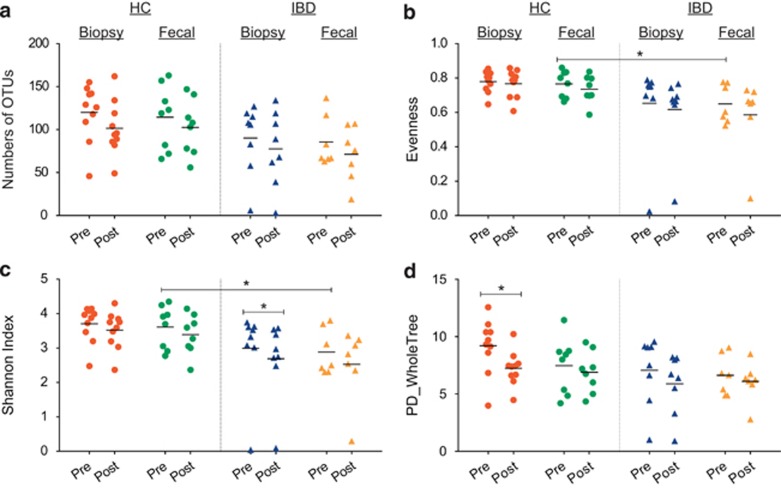
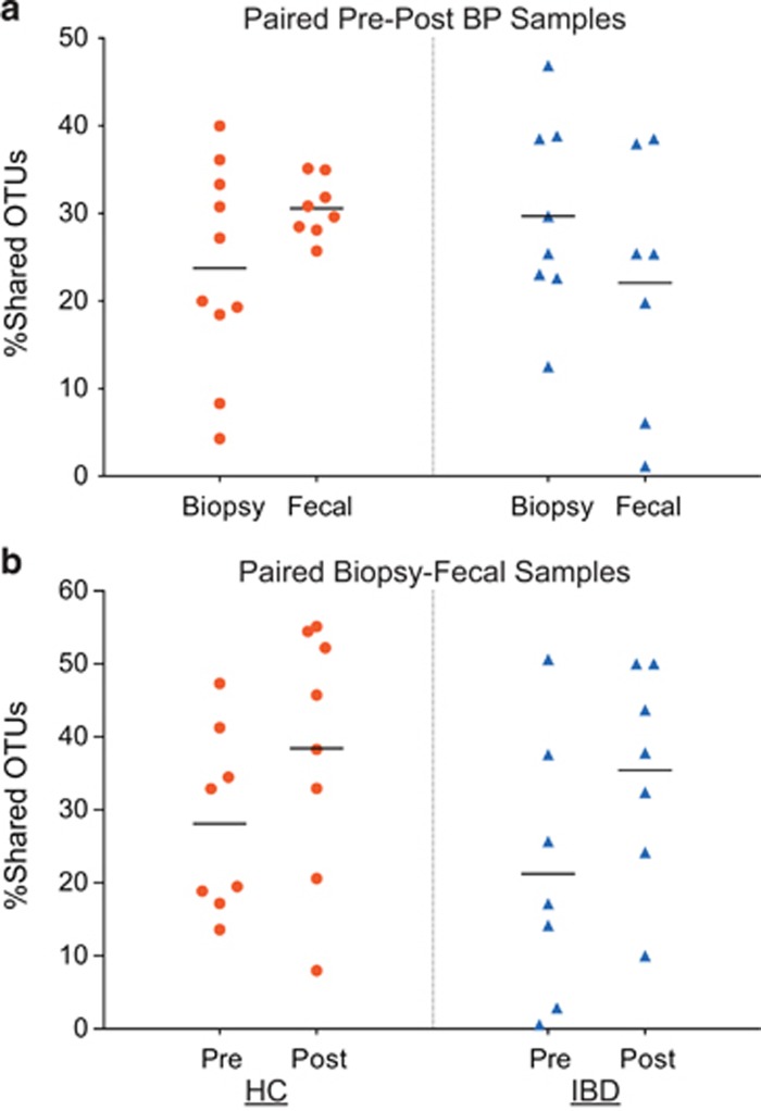
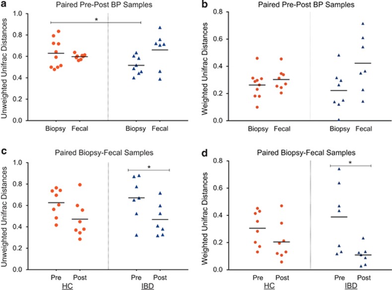
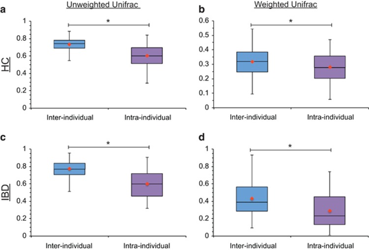
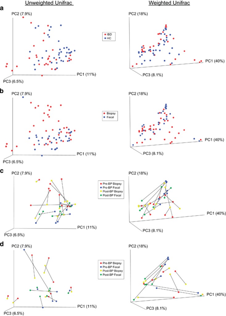
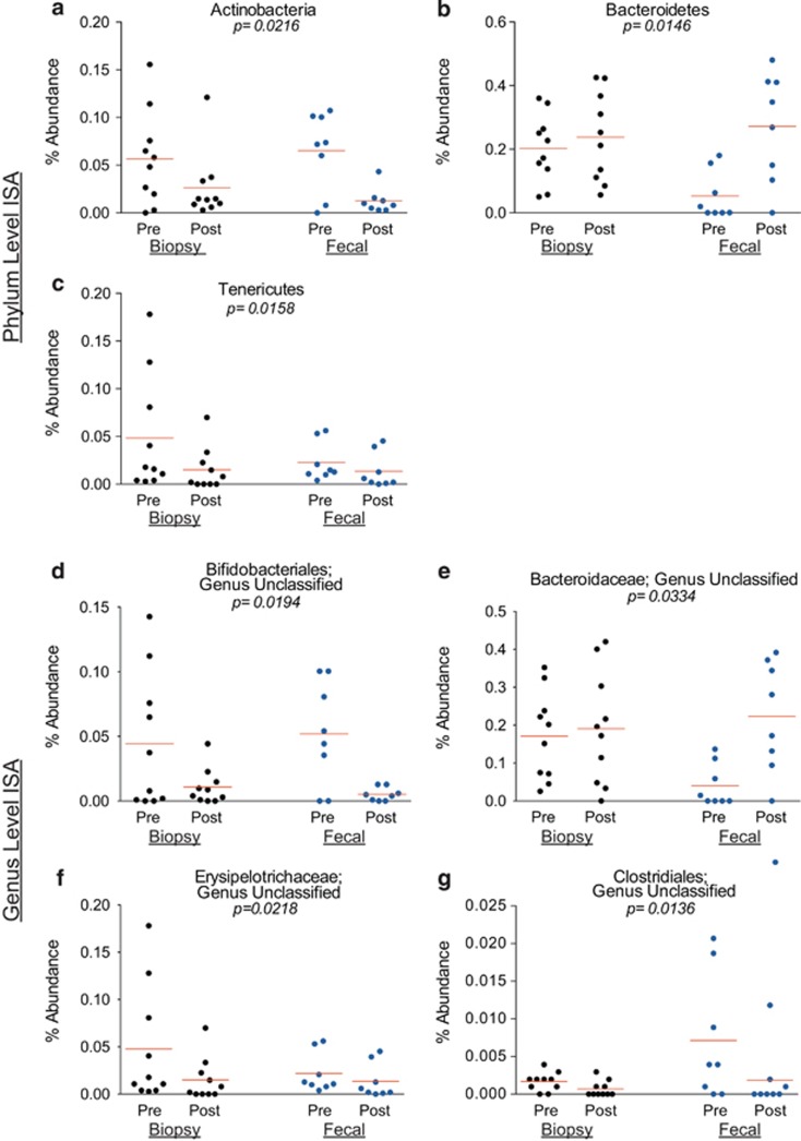
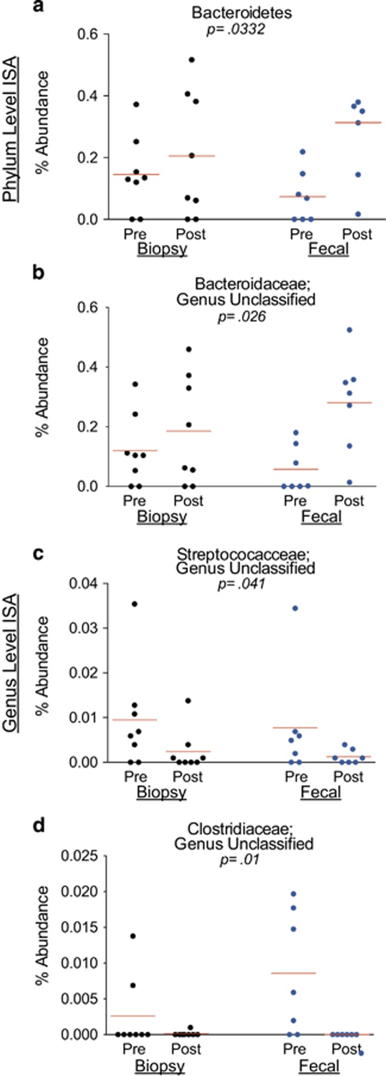
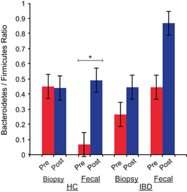
Results: In comparisons-across-bowel-prep, the Shannon's index (SI) decreased only in the biopsy samples of IBD subjects post-BP (P=0.025) and phylogenetic diversity-whole tree (PD-WT) metric decreased in biopsy samples of HC subjects post-BP (P=0.021). In secondary comparisons, the subtle differences between the fecal samples of the HC vs. IBD groups, in terms of evenness and the SI, were not apparent post-BP. In terms of β-diversity, in comparisons-across-bowel-prep, the proportion of shared operational taxonomic units (OTUs) in pre- and post-BP samples was low (~30%) and unweighted Unifrac distances between pre- and post-BP specimens ranged from 0.52 to 0.66. HC biopsies were affected more than IBD biopsies with BP (P=0.004). In comparisons-across-compartments, the proportion of shared OTUs between biopsy and fecal samples increased and Unifrac distances decreased post-BP in IBD subjects, reducing the differences between the mucosal and luminal compartments of the gut microbiota. Interindividual differences in Unifrac distances were preserved even with BP effects, although the effects were greater on weighted Unifrac distances. Bacteroidetes and its subtypes increased post-BP in both the luminal and mucosal compartments.

Conclusions: Bowel preparations affect the composition and diversity of the fecal and luminal microbiota in the short term, introducing potential bias into experiments examining the gut microbiota. The magnitude of the effect of BP is not greater than that of interindividual variation. Both the luminal and mucosal compartments of the gut microbiota get affected, and samples from controls and IBD subjects may get affected differently. Studies of the colonic microbiota should take into account the direction and the magnitude of the change introduced by BP during the design stage of the experiments, and consider sample sizes so that potential bias is minimized.


Figures

Figure 1
Figure 1
Operational taxonomic unit (OTU)-based diversity indices of pre- and post-bowel preparation (BP) samples. (a) Richness of OTUs pre- and post-BP. Each marker represents a single sample. The lines represent the mean value for each sample group. The color red represents biopsy samples of healthy controls (HCs). The color green represents fecal samples of HCs. The color blue represents biopsy samples of inflammatory bowel disease (IBD) subjects. The color yellow represents the fecal samples of IBD. In comparisons-across-bowel-prep, a trend towards reduced richness in samples collected post-BP is seen in both HC and IBD groups, but the differences are not statistically significant (P>0.05). (b) The evenness pre- and post-BP. There was a difference in the evenness between pre-BP fecal samples of HC vs. IBD subjects (P=0.033), and this difference was not apparent after BP. (c) The Shannon's diversity index pre- and post-BP. The baseline (pre-BP) Shannon's index for the fecal samples of IBD was significantly lower compared with those for HCs (P=0.04). This difference was not apparent after BP. Although there was a trend towards reduced Shannon's indices in the post-BP samples (compared with pre-BP samples) in general, this reduction was only significant in the biopsy samples of the IBD subjects (P=0.025, paired t-test). (d) The phylogenetic diversity-whole tree (PD-WT) index pre- and post-BP. The effect of BP was more pronounced in the biopsy samples, especially in the HC group (P=0.021, related-samples sign test). The apparent lesser effect on fecal samples may be explained by the greater inconsistency in the direction of response to BP among the fecal samples.
Figure 2
Figure 2
The proportion of shared operational taxonomic units (OTUs) between samples. (a) Shared OTUs between the pre- and post-bowel prep (BP) samples in the healthy control (HC) and inflammatory bowel disease (IBD) groups. Each marker plotted represents the proportion of shared OTUs between paired samples (before and after BP) of the same subject. Red markers represent shared OTUs in HCs. Blue markers represent shared OTUs in the IBD subjects. The lines represent the mean values for the proportion of shared OTUs. The proportion of shared OTUs between pre- and post-bowel prep samples in all sample sets was low (~20–30%). (b) Shared OTUs between the mucosal and luminal compartments (i.e., biopsy and fecal samples) collected at the same visit in the HC and IBD groups. Each marker plotted represents the proportion of shared OTUs between paired samples (fecal and biopsy samples in the same visit) of the same subject. Red markers represent shared OTUs in HCs. Blue markers represent shared OTUs in the IBD subjects. There was an increase in the proportion of shared OTUs between the biopsy and fecal samples after BP (P=0.016).
Figure 3
Figure 3
Unifrac distances between paired samples. (a) The unweighted Unifrac distances between the pre- and post-bowel prep (BP) samples in the healthy control (HC) and inflammatory bowel disease (IBD) groups. Each marker plotted represents the Unifrac distance between paired pre- and post-BP samples of the same type for the same subject. Red markers represent Unifrac distances in the HCs. Blue markers represent Unifrac distances in the IBD subjects. The lines represent the mean Unifrac distances. The unweighted Unifrac distances between the pre- and post-BP biopsy samples was significantly greater in the HC group, when compared with the IBD group (P=0.04). (b) The weighted Unifrac distances between the pre- and post-BP samples in the HC and IBD groups. (c) The unweighted Unifrac distances between the mucosal and luminal compartments (i.e., biopsy and fecal samples) collected at the same visit in the HC and IBD groups. Overall, the mean-unweighted Unifrac distance between the biopsy and fecal samples decreased after bowel prep (P=0.003). This reduction in unweighted Unifrac distances between sample types because of bowel prep was significant in the IBD group (P=0.03), but not in the HC group (P=0.068). (d) The weighted Unifrac distances between the mucosal and luminal compartments (i.e., biopsy and fecal samples) collected at the same visit in the HC and IBD groups. Although there was a trend towards reduced weighted Unifrac distances between the mucosal and luminal compartments after BP, this reduction was only significant in the IBD group (P=0.036).
Figure 4
Figure 4
Unifrac distances between samples from the same subject (i.e., intra-individual differences) vs. those from other subjects (i.e., inter-individual differences). (a) The unweighted Unifrac distances between samples of the same subject (intra-individual distances) vs. the unweighted Unifrac distances between samples of different subjects (inter-individual distances) in the healthy control (HC) group (P⩽0.0001). (b) The weighted Unifrac distances between samples of the same subject (intra-individual distances) vs. the weighted Unifrac distances between samples of different subjects (inter-individual distances) in the HC group (P=0.0047). (c) The unweighted Unifrac distances between samples of the same subject (intra-individual distances) vs. the unweighted Unifrac distances between samples of different subjects (inter-individual distances) in the inflammatory bowel disease (IBD) group (P≤0.0001). (d) The weighted Unifrac distances between samples of the same subject (intra-individual distances) vs. the weighted Unifrac distances between samples of different subjects (inter-individual distances) in the IBD group (P≤0.0001). The inter-individual distances were significantly greater than the intra-individual distances for all comparisons. The line in each box represents the median and the orange marker represents the mean.
Figure 5
Figure 5
Principal coordinate analysis (PCoA) plots of samples by Unifrac distances. (a) Plots of samples collected from both groups (healthy control (HC) and inflammatory bowel disease (IBD)) colored by disease state. Each marker denotes a single sample. PCoA plots for the unweighted Unifrac distances are shown on the left, whereas the PCoA plots for the weighted Unifrac distances are shown on the right. (b) Plots of all the samples colored by sample type (biopsy vs. fecal). (c) Plots of paired pre- and post-bowel prep samples of the HC group only. (d) Plots of paired pre- and post-bowel prep samples of IBD group only. The lines in (c and d) connect paired pre- and post-bowel prep samples of the same type for the same individual subject.
Figure 6
Figure 6
Relative abundances of significant indicator phyla and genera in healthy subjects. Each marker represents the relative (%) abundance of the denoted phylum or genus in one sample. Each panel shows the relative (%) abundance in the pre- and post-bowel prep samples collected from healthy subjects. The lines represent the mean relative abundance. Black circles represent the biopsy samples and blue circles represent the fecal samples. (a) The relative abundance of the phylum Actinobacteria (an indicator of the pre-bowel prep (BP) state). (b) The relative abundance of the phylum Bacteroidetes (an indicator of the post-BP state). (c) The relative abundance of the phylum Tenericutes (an indicator of the pre-BP state). (d) The relative abundance of an unclassified genus within the Bifidobacteriales order (an indicator of the pre-BP state). (e) The relative abundance of an unclassified genus within the Bacteroidaceae family (an indicator of the post-BP state). (f) The relative abundance of an unclassified genus within the Erysipelotrichaceae family (an indicator of the pre-BP state). (g) The relative abundance of an unclassified genus within the Clostridiales order (an indicator of the pre-BP state). ISA, indicator species analysis.
Figure 7
Figure 7
Relative abundances of significant indicator phyla and genera in inflammatory bowel disease (IBD) subjects. Each marker represents the relative (%) abundance of the denoted phylum or genus in one sample. Each panel shows the relative (%) abundance in the pre- and post-bowel prep samples collected from IBD subjects. The lines represent the mean relative abundance. Black circles represent the biopsy samples and blue circles represent the fecal samples. (a) The relative abundance of the phylum Bacteroidetes (the only significant indicator phylum in IBD subjects). (b) The relative abundance of an unclassified genus within the Bacteroidaceae family (an indicator of the post-bowel prep (BP) state). (c) The relative abundance of an unclassified genus within Streptococcaceae family (an indicator of the pre-BP state). (d) The relative abundance of an unclassified genus within the Clostridiaceae family (an indicator of the pre-BP state). ISA, indicator species analysis.
Figure 8
Figure 8
Bacteroidetes/Firmicutes ratio in samples. The bar chart shows the ratio of Bacteroidetes to Firmicutes in each sample group and compares the pre-bowel prep samples to the post-bowel prep samples. The average Bacteroidetes/Firmicutes ratio increased after bowel prep in the fecal samples of healthy control (HC) subjects (P=0.023, related-samples t-test). IBD, inflammatory bowel disease.

References

    1. Sears CL. A dynamic partnership: celebrating our gut flora. Anaerobe 2005; 11: 247–251. - PubMed
    1. Eckburg PB, Bik EM, Bernstein CN et al. Diversity of the human intestinal microbial flora. Science (New York, NY) 2005; 308: 1635–1638. - PMC - PubMed
    1. Backhed F, Ley RE, Sonnenburg JL et al. Host–bacterial mutualism in the human intestine. Science (New York, NY) 2005; 307: 1915–1920. - PubMed
    1. Rajilic-Stojanovic M, Smidt H, de Vos WM. Diversity of the human gastrointestinal tract microbiota revisited. Environ Microbiol 2007; 9: 2125–2136. - PubMed
    1. Rappe MS, Giovannoni SJ. The uncultured microbial majority. Annu Rev Microbiol 2003; 57: 369–394. - PubMed

LinkOut - more resources