. 2016 Aug 31;8(34):21923-32.
doi: 10.1021/acsami.5b11671. Epub 2016 Feb 11.

Nanotopography-Induced Structural Anisotropy and Sarcomere Development in Human Cardiomyocytes Derived from Induced Pluripotent Stem Cells

Affiliations

Nanotopography-Induced Structural Anisotropy and Sarcomere Development in Human Cardiomyocytes Derived from Induced Pluripotent Stem Cells

Daniel Carson et al. ACS Appl Mater Interfaces. .

Abstract

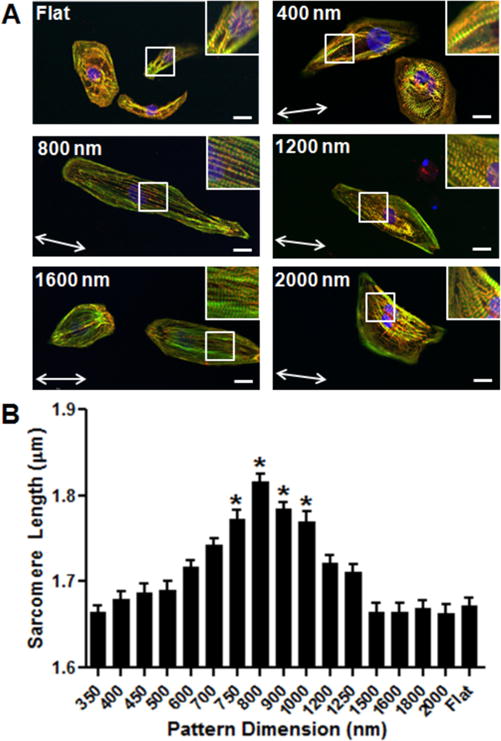
Understanding the phenotypic development of human induced pluripotent stem cell-derived cardiomyocytes (hiPSC-CMs) is a prerequisite to advancing regenerative cardiac therapy, disease modeling, and drug screening applications. Lack of consistent hiPSC-CM in vitro data can be largely attributed to the inability of conventional culture methods to mimic the structural, biochemical, and mechanical aspects of the myocardial niche accurately. Here, we present a nanogrid culture array comprised of nanogrooved topographies, with groove widths ranging from 350 to 2000 nm, to study the effect of different nanoscale structures on the structural development of hiPSC-CMs in vitro. Nanotopographies were designed to have a biomimetic interface, based on observations of the oriented myocardial extracellular matrix (ECM) fibers found in vivo. Nanotopographic substrates were integrated with a self-assembling chimeric peptide containing the Arg-Gly-Asp (RGD) cell adhesion motif. Using this platform, cell adhesion to peptide-coated substrates was found to be comparable to that of conventional fibronectin-coated surfaces. Cardiomyocyte organization and structural development were found to be dependent on the nanotopographical feature size in a biphasic manner, with improved development achieved on grooves in the 700-1000 nm range. These findings highlight the capability of surface-functionalized, bioinspired substrates to influence cardiomyocyte development, and the capacity for such platforms to serve as a versatile assay for investigating the role of topographical guidance cues on cell behavior. Such substrates could potentially create more physiologically relevant in vitro cardiac tissues for future drug screening and disease modeling studies.

Keywords: biomimetic surface; cardiomyocytes; chimeric self-assembling peptide; human induced pluripotent stem cells; nanotopography.


Conflict of interest statement

Notes

The authors declare the following competing financial interest(s): D.H. Kim is a co-founder and scientific board member of a start-up company, NanoSurface Biomedical, that aims to commercialize nanopatterned polymeric cultureware.

Figures

Figure 1
Figure 1
Fabrication and arrangement of nanopatterned substrates on the nanogrid cell culture array. (A) Schematic illustration of UV-assisted capillary force lithography (CFL) process used to generate nanotopographically defined PUA-based cell culture substrates. (B) Diagram of nanogrid array designed for high-throughput structural maturation analyses of cultured cells. SEM images illustrate the surface dimensions of small (400 μm), medium (800 and 1200 μm), and large (2000 μm) topographic features analyzed during this study.
Figure 2
Figure 2
Characterization of chimeric adhesion peptide affinity to PUA substrates. (A) Schematic illustration of assay to determine degree of peptide affinity for PUA substrates, in which PUABP-biotin is incubated with SA-Alexa and the resulting fluorescence emission is measured. (B) PUA binding assay for rational peptide library, each peptide was conjugated to biotin prior to use in assay. No fluorescence was detected for peptides #6–15. n = 4. (C, D) PUABP surface coverage characterization at 100 μM. Fluorescence microscopy was used to determine surface binding density compared to a blank surface (negative control; NC) and surfaces treated with SA-Alexa without PUABP-biotin. Average fluorescence intensity was measured at multiple locations on each examined substrate (n ≥ 5). PUABP2-biotin displayed similar surface coverage compared to PUABP1-biotin, but a higher average fluorescent intensity. Scale bar: 10 μm. (E) Binding curve of PUABP1 and 2 with saturation occurring for both compounds at 100 μM.
Figure 3
Figure 3
Directed cardiomyocyte differentiation from hiPSCs and comparative characterization of their adhesion to functionalized PUA substrates. (A) Representative schema of protocol for differentiating hiPSCs into cardiomyocytes. Prior to Day 0, undifferentiated IMR90 human iPSCs were cultured in mouse embryonic fibroblast-conditioned medium. At Day 0, undifferentiated cells were induced with activin-A, followed by BMP-4 between Days 1 and 3. Between Days 3 and 5, the cells were treated with XAV939 (XAV), a tankyrase inhibitor. During the first week of differentiation, cells were maintained in RPMI medium with B27 (without insulin). From Day 7 onward, insulin-containing B27 was used to supplement medium. Cells were maintained on Matrigel-coated surfaces until Day 21 before being harvested using trypsin and replated onto experimental surfaces. Medium was changed every other day throughout this time course. (B) Cell adhesion analysis of IMR90 cardiomyocytes 24 h post replating on the nanogrid array coated with 100 μM of PUABP1-RGD or PUABP2-RGD. Statistical analysis yielded no significant differences in cell adhesion among pattern dimensions and ECM coating. p > 0.05, n = 20 (distinct areas) per condition.
Figure 4
Figure 4
Quantitative analysis of cardiomyocyte morphology in response to different nanopatterned topographies. (A) Cell area analysis of IMR90 cardiomyocytes 3 weeks post replating pattern dimensions from 4 × 4-island platform coated with PUABP2-RGD (100 μM). *p < 0.05. (B) Cell perimeter analysis of IMR90 hiPSC-cardiomyocytes 3 weeks post plating on different nanotopographic surfaces from 4 × 4-island platform coated with PUABP2-RGD (100 μM). Statistically significant increases in cell perimeter were observed in cells maintained on 600–900 nm groove widths when compared to all other patterned conditions and flat controls. *p < 0.05. (C) Cell circularity analysis of IMR90 cardiomyocytes 3 weeks post replating pattern dimensions from 4 × 4-island platform coated with PUABP2-RGD (100 μM). Cells on flat and 2000 nm patterned surfaces had significantly higher circularity indices than all other patterned conditions, indicating less anisotropic development in these conditions. *p < 0.05. (D) Cell alignment analysis of IMR90 cardiomyocytes 3 weeks post replating on pattern dimensions from 4 × 4-island platform coated with PUABP2-RGD (100 μM). Alignment was determined by the percentage of cells whose major axis was within 5 degrees of the pattern direction. Cells on 800 nm nanogroove width exhibited the highest degree of cell alignment compared to all other surface conditions. For all the analyses, n = 60–95 cells per condition.
Figure 5
Figure 5
Analysis of sarcomere development in hiPSC-cardiomyocytes cultured on nanotopographic substrates of different dimensions. (A) Confocal images of IMR90 cardiomyocytes with inset image fixed 3 weeks post replating on PUABP2-RGD (100 μM) coated nanogrid array. Red, α-actinin; green, f-actin; blue, nuclei. Scale bar: 20 μm. (B) Sarcomere length analysis of IMR90 cardiomyocytes 3 weeks post replating on 4 × 4-island platform coated with 100 μM PUABP2-RGD. Statistically significant increases in sarcomere length were observed between cells on intermediate (750–1000 nm) groove widths. *p < 0.05.

References

    1. Liu WH, Chang YL, Lo WL, Li HY, Hsiao CW, Peng CH, Chiou SH, Ma HI, Chen SJ. Human Induced Pluripotent Stem Cell and Nanotechnology-Based Therapeutics. Cell Transplant. 2015;24:2185–2195. - PubMed
    1. Wikswo JP. The Relevance and Potential Roles of Microphysiological Systems in Biology and Medicine. Exp Biol Med. 2014;239:1061–1072. - PMC - PubMed
    1. Lancaster MA, Knoblich JA. Organogenesis in a Dish: Modeling Development and Disease Using Organoid Technologies. Science. 2014;345:1247125. - PubMed
    1. Acimovic I, Vilotic A, Pesl M, Lacampagne A, Dvorak P, Rotrekl V, Meli AC. Human Pluripotent Stem Cell-Derived Cardiomyocytes as Research and Therapeutic Tools. BioMed Res Int. 2014;2014:1. - PMC - PubMed
    1. Gherghiceanu M, Barad L, Novak A, Reiter I, Itskovitz-Eldor J, Binah O, Popescu LM. Cardiomyocytes Derived from Human Embryonic and Induced Pluripotent Stem Cells: Comparative Ultrastructure. J Cell Mol Med. 2011;15:2539–2551. - PMC - PubMed

Publication types