. 2016 Aug;68(8):1887-98.
doi: 10.1002/art.39618.

Aging Promotes Sirtuin 3-Dependent Cartilage Superoxide Dismutase 2 Acetylation and Osteoarthritis

Affiliations

Aging Promotes Sirtuin 3-Dependent Cartilage Superoxide Dismutase 2 Acetylation and Osteoarthritis

Yao Fu et al. Arthritis Rheumatol. 2016 Aug.

Abstract

Objective: To quantify functional age-related changes in the cartilage antioxidant network in order to discover novel mediators of cartilage oxidative stress and osteoarthritis (OA) pathophysiology.

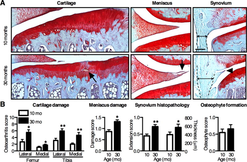
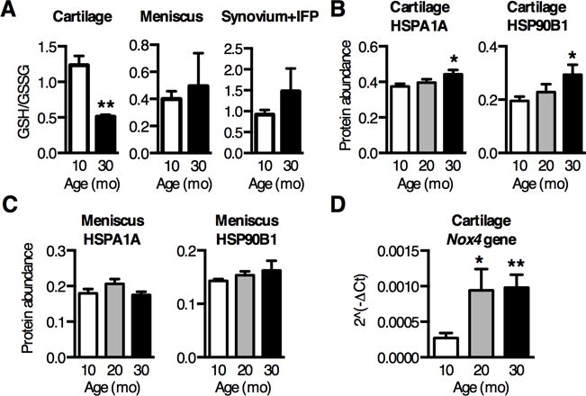
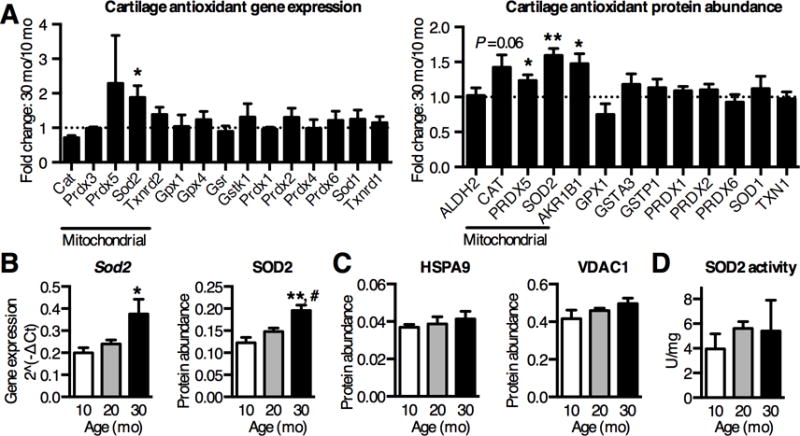
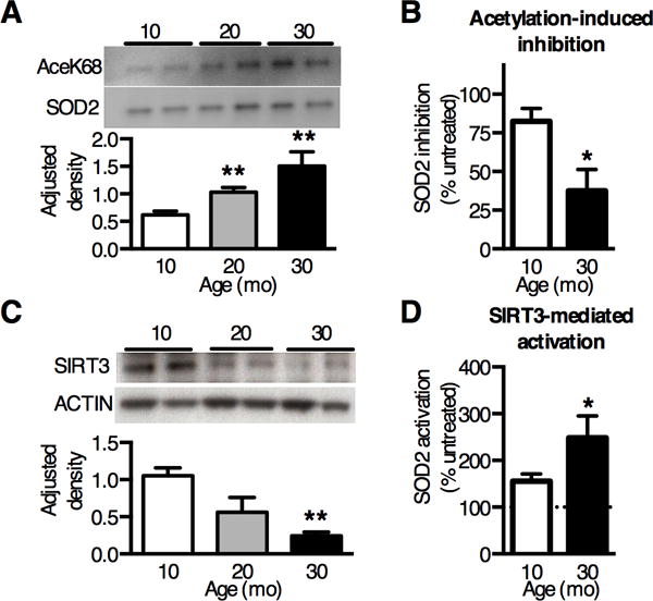
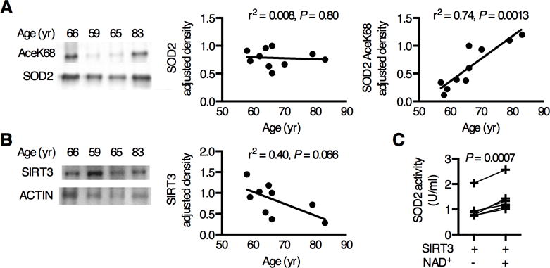
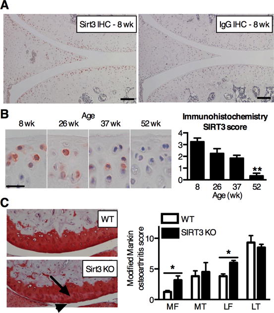
Methods: We evaluated histopathologic changes of knee OA in 10-, 20-, and 30-month-old male F344BN rats and analyzed cartilage oxidation according to the ratio of reduced to oxidized glutathione. Antioxidant gene expression and protein abundance were analyzed by quantitative reverse transcription-polymerase chain reaction and selected reaction-monitoring mass spectrometry, respectively. Superoxide dismutase 2 (SOD2) activity and acetylation were analyzed by colorimetric enzyme assays and Western blotting, respectively. We examined human OA cartilage to evaluate the clinical relevance of SOD2 acetylation, and we tested age-related changes in the mitochondrial deacetylase sirtuin 3 (SIRT-3) in rats and mice.

Results: Cartilage oxidation and OA severity in F344BN rats increased with age and were associated with an increase in SOD2 expression and protein abundance. However, SOD2-specific activity decreased with age due to elevated posttranslational lysine acetylation. Consistent with these findings, SIRT-3 levels decreased substantially with age, and treatment with SIRT-3 increased SOD2 activity in an age-dependent manner. SOD2 was also acetylated in human OA cartilage, and activity was increased with SIRT-3 treatment. Moreover, in C57BL/6J mice, cartilage SIRT-3 expression decreased with age, and whole-body deletion of SIRT-3 accelerated the development of knee OA.

Conclusion: Our results show that SIRT-3 mediates age-related changes in cartilage redox regulation and protects against early-stage OA. These findings suggest that mitochondrial acetylation promotes OA and that restoration of SIRT-3 in aging cartilage may improve cartilage resistance to oxidative stress by rescuing acetylation-dependent inhibition of SOD2 activity.


Figures

Figure 1
Figure 1. Age-dependent OA pathology in F344BN rats
(A) Representative histological images of coronal sections of the knee in aging F344BN rats. Aging increased cartilage and meniscus structural damage (arrows) and reduced Safranin-O staining, indicating proteoglycan loss. Aging also increased synovial thickness (double arrow between dotted lines) and growth (arrow head) into the joint space. Scale bars: 200μm. (B) Semi-quantitative histological grading for OA pathology by tissue type. OA pathology scores increased with age in all tissues except for osteophyte development (n=8). Values are mean ± SEM. *P < 0.05, **P < 0.01 versus 10-mo samples.
Figure 2
Figure 2. Aging increases oxidative stress specifically in cartilage joint tissues
(A) Ratio of reduced to oxidized glutathione (GSH/GSSG) decreased nearly 60% in cartilage isolated from 10 and 30-mo old rat knees, indicating an increase in cartilage glutathione oxidation with age (n=3). Glutathione oxidation did not increase with age in either meniscus or synovium and infrapatellar fat pad (IFP) tissues (n=3). (BC) Cartilage and meniscus stress response proteins were measured by selected reaction monitoring mass spectrometry and normalized to cellular reference proteins. Heat shock protein 70kDa 1A (HSPA1A), an oxidative stress marker, was upregulated 20% with age in cartilage (B) but not in meniscus (C). HSP 90kDa B1 (HSP90B1), an ER stress marker, was also upregulated over 50% with age in cartilage (B) but not meniscus (C) (n=6). (D) Nox4 gene expression was measured by qPCR array and showed the greatest upregulation with age out of 84 oxidative stress related genes (Table S2). Nox4 is a source of superoxide associated with the mitochondria, suggesting an age-dependent increase in superoxide production (n=4). Values are mean ± SEM. *P < 0.05, **P < 0.01 versus 10-mo samples.
Figure 3
Figure 3. Aging increases articular cartilage SOD2 expression but not activity
(A) Fold-change in antioxidant mRNA and protein levels from knee cartilage of 30-mo old F344BN rats, normalized to 10-mo old samples, measured by qRT-PCR array or SRM-MS. Sod2 gene expression (n=4) and protein abundance (n=6) were significantly upregulated with age. (B) Quantification of cartilage SOD2 gene and protein showed an age-dependent step-wise increase (n=4 & 6, respectively). (C) Increased SOD2 protein content was not associated with an age-dependent increase in mitochondrial reference protein levels (heat shock protein HSPA9 and mitochondrial membrane channel VDAC1), suggesting that overall mitochondrial abundance did not increase with age (n=6). (D) SOD2 activity normalized to total cartilage protein, as determined by inhibition of xanthine oxidase-mediated oxidation of Tetrazolium salt in the presence of KCN to inhibit SOD1 and SOD3. Despite increased protein abundance, SOD2 activity was not altered with age (n=6). Bar values are mean ± SEM. *P < 0.05, **P < 0.01 versus 10-mo samples, # P < 0.05 versus 20-mo samples.
Figure 4
Figure 4. Loss of SIRT3 protein with aging impairs cartilage SOD2 activity
(A) Representative Western blot images and densitometry analysis of SOD2 acetylated Lysine68 (AceK68) normalized to pair-matched SOD2 protein in cartilage. AceK68 expression showed a dose-response increase with age (n=4). (B) Inhibition of cartilage SOD2 activity following in vitro acetylation with 25μM acetic anhydride. Inhibition decreased with increasing age, consistent with elevated basal SOD2 acetylation with age (n=3). (C) Representative Western blot image and densitometry analysis of SIRT3 expression in knee cartilage of F344BN rats, normalized to β-ACTIN. SIRT3 decreased 77% with aging (P = 0.006, one-way ANOVA) (n=4). (D) SIRT3-mediated deacetylation increased SOD2 activity more in aged cartilage. Cartilage SOD2 activity was measured after 5 minutes in the presence of 1μg recombinant human SIRT3 protein with or without 1mM NAD+ at room temperature. Data presented as percentage of NAD+ dependent change in SOD2 activity (n=3). Bar values are mean ± SEM. *P < 0.05 versus 10-mo. **P < 0.01 versus 10-mo.
Figure 5
Figure 5. SOD2 acetylation is inversely correlated with SIRT3 protein content in an age-dependent manner in human OA cartilage
Acetylated SOD2 protein was detected in human OA cartilage by Western blot and compared to SIRT3 protein content as a function of age. (A) Representative SOD2 AceK68 and SOD2 Western blot images. SOD2 relative density was normalized to sample-matched β-ACTIN (r2=0.008, P = 0.80, n=10). SOD2 AceK68 normalized to SOD2 increased with age (r2=0.74, P = 0.0013, n=10). (B) Representative SIRT3 and β-ACTIN Western blot images. SIRT3 normalized to β-ACTIN showed a decreasing trend with age (r2=0.40, P = 0.066, n=9). (C) SIRT3-mediated deacetylation increased SOD2 activity in human OA cartilage (P = 0.0007, n=6).
Figure 6
Figure 6. SIRT3 is reduced in the cartilage of aging mice and protects against the onset of knee OA in mice
(A) SIRT3 and IgG control immunostaining in the knee joint of 8 wk old male C57BL/6J mice. SIRT3 staining is enriched in articular chondrocytes and surface-zone meniscal cells. Scale bar: 100μm. (B) Representative SIRT3 immunostaining and semi-quantitative scoring of tibial articular cartilage from mice ranging in age from 8 to 52 wks. Immunostaining scoring based on number and intensity of positively stained chondrocytes within the medial and lateral femoral condyles and tibial plateau (maximum = 4; see Table S1 for scoring details). Scoring shows a significant loss of SIRT3 with age (P = 0.0037, Kruskal-Wallis test) (n = 5). **P < 0.01 versus 8 wk. (C) Representative histological images of the lateral femoral condyles from 14-mo old WT and SIRT3 KO mice show characteristic differences, including a reduction in Safranin-O staining intensity (arrow) and surface fibrillation (arrowhead). Modified Mankin OA scores indicate a significant increase in femoral OA in SIRT3 KO mice. Values represent the per-animal averages of scores from left and right knees and two blinded graders (n=4, WT; n=3, KO). *P < 0.05 site-matched, Mann-Whitney test.

References

    1. Blaney Davidson EN, Scharstuhl A, Vitters EL, van der Kraan PM, van den Berg WB. Reduced transforming growth factor-beta signaling in cartilage of old mice: role in impaired repair capacity. Arthritis Res Ther. 2005;7:R1338–47. - PMC - PubMed
    1. Loeser RF, Carlson CS, Carlo MD, Cole A. Detection of nitrotyrosine in aging and osteoarthritic cartilage: Correlation of oxidative damage with the presence of interleukin-1β and with chondrocyte resistance to insulin-like growth factor 1. Arthritis Rheum. 2002;46:2349–2357. - PubMed
    1. Forsyth CB, Cole A, Murphy G, Bienias JL, Im H-J, Loeser RF. Increased matrix metalloproteinase-13 production with aging by human articular chondrocytes in response to catabolic stimuli. J Gerontol A Biol Sci Med Sci. 2005;60:1118–1124. - PMC - PubMed
    1. Loeser RF, Olex AL, McNulty MA, Carlson CS, Callahan MF, Ferguson CM, et al. Microarray analysis reveals age-related differences in gene expression during the development of osteoarthritis in mice. Arthritis Rheum. 2012;64:705–717. - PMC - PubMed
    1. Martin JA, Buckwalter JA. Aging, articular cartilage chondrocyte senescence and osteoarthritis. Biogerontology. 2002;3:257–264. - PubMed

Publication types