. 2016 Jun;34(6):1576-87.
doi: 10.1002/stem.2334. Epub 2016 Mar 11.

Wnt/β-Catenin Signaling Determines the Vasculogenic Fate of Postnatal Mesenchymal Stem Cells

Affiliations

Wnt/β-Catenin Signaling Determines the Vasculogenic Fate of Postnatal Mesenchymal Stem Cells

Zhaocheng Zhang et al. Stem Cells. 2016 Jun.

Abstract

Vasculogenesis is the process of de novo blood vessel formation observed primarily during embryonic development. Emerging evidence suggest that postnatal mesenchymal stem cells are capable of recapitulating vasculogenesis when these cells are engaged in tissue regeneration. However, the mechanisms underlining the vasculogenic differentiation of mesenchymal stem cells remain unclear. Here, we used stem cells from human permanent teeth (dental pulp stem cells [DPSC]) or deciduous teeth (stem cells from human exfoliated deciduous teeth [SHED]) as models of postnatal primary human mesenchymal stem cells to understand mechanisms regulating their vasculogenic fate. GFP-tagged mesenchymal stem cells seeded in human tooth slice/scaffolds and transplanted into immunodeficient mice differentiate into human blood vessels that anastomize with the mouse vasculature. In vitro, vascular endothelial growth factor (VEGF) induced the vasculogenic differentiation of DPSC and SHED via potent activation of Wnt/β-catenin signaling. Further, activation of Wnt signaling is sufficient to induce the vasculogenic differentiation of postnatal mesenchymal stem cells, while Wnt inhibition blocked this process. Notably, β-catenin-silenced DPSC no longer differentiate into endothelial cells in vitro, and showed impaired vasculogenesis in vivo. Collectively, these data demonstrate that VEGF signaling through the canonical Wnt/β-catenin pathway defines the vasculogenic fate of postnatal mesenchymal stem cells. Stem Cells 2016;34:1576-1587.

Keywords: Angiogenesis; Dental pulp stem cells; Multipotency; Self-renewal; Tissue engineering; Vasculogenesis.


Conflict of interest statement

Conflict of interest

The authors declare no conflict of interest.

Figures
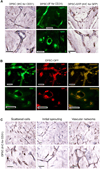
Figure 1
Figure 1
Dental pulp stem cells (DPSC) differentiate into blood vessels in vivo. 1×10⁶ DPSC or DPSC-GFP (stably transduced with GFP) cells were seeded in tooth slice/scaffolds and transplanted in the subcutaneous space of the dorsum of SCID mice (n=8). Five weeks later, tooth slice/scaffolds were retrieved and processed for imaging by immunohistochemistry (IHC) or immunofluorescence (IF). (A): Blood vessels originated from the human DPSC were detected by IHC for GFP (brown color) in tooth slice/scaffolds seeded with DPSC-GFP. Alternatively, we performed IHC or IF for CD31 in tooth slice/scaffolds seeded with DPSC cells. (B): DPSC-derived human blood vessels were detected by double staining with anti-human CD31 (green) and GFP (red) in tooth slice/scaffolds seeded with DPSC-GFP cells. (C): Stages of DPSC-mediated vasculogenesis as shown by IHC for anti-human CD31. Left panel: Scattered CD31-positive cells. Middle panel: Elongated CD31-positive cells connecting with each other. Right panel: Connected branches forming complex vascular networks. Scale bars: 25 µm.
Figure 2
Figure 2
Process of DPSC-derived vessel maturation, as determined by progressive investment with smooth muscle actin (SMA)-positive smooth muscle cells/pericytes. 1×10⁶ DPSC cells were seeded in tooth slice/scaffolds and transplanted in the subcutaneous space of the dorsum of SCID mice. Tooth slice/scaffolds were collected after 1–4 weeks (n=4), retrieved and processed for immunofluorescence. DPSC-derived human blood vessels were detected in the pulp chamber with anti-human CD31 (green) and anti-SMA antibody was used to detect smooth muscle cells/pericytes (red) lining the walls of these newly formed blood vessels. Green arrow points to a human CD31-positive cell and red arrow points to a SMA-positive smooth muscle cell. Scale bars: 25 ***m.
Figure 3
Figure 3
VEGF activates the canonical Wnt/β-catenin signaling in dental pulp stem cells. (A): Western blots for EGFR, E-cadherin, VEGFR-2, Tie-2, VE-cadherin and CD31 from whole cell lysates of DPSC, SHED, UM-SCC-1 and HDMEC. (B): RT-PCR for odontogenic/osteoblastic markers (DSPP, DMP-1), and for endothelial cell markers (VEGFR2, Tie-2, VE-cadherin, CD-31). Total RNA was prepared from odontoblasts retrieved from freshly extracted sound human 3rd molars, whole human pulp tissue, or from DPSC, SHED, HDMEC cells. (C): Western blot for Wnt pathway receptors LRP-5, LRP-6, Fzd-3, Fzd-4, Fzd-5 and Fzd-6 in DPSC and SHED, using as controls UM-SCC-1 and HDMEC. (D): Western blot for Wnt1, P-GSK-3β, GSK-3β, active-β-catenin and β-catenin. (E): Western blots for LRP-6, Fzd-6, active-β-catenin, β-catenin, Wnt1 and Bmi-1 in DPSC, SHED or HDMEC starved overnight and treated with 50 ng/ml rhVEGF165 or 50 ng/ml rhWnt1 for 24 hours. (F): Western blot for LRP-6, Fzd-6, active-β-catenin, β-catenin, Wnt1 and Bmi-1 in DPSC starved overnight and treated with 0–150 ng/ml rhVEGF165 for 24 hours. Numbers depict the band density normalized against untreated controls and GAPDH. (G): Western blot for Bmi-1 in DPSC starved overnight and treated with 50 ng/ml rhVEGF165 and/or 50 ng/ml rhWnt1 for 24 hours.
Figure 4
Figure 4
VEGF and Wnt mediate endothelial differentiation of dental pulp stem cells. (A): Western blots for VEGFR1, VEGFR2 and VE-cadherin from DPSC cells treated with 50 ng/ml rhVEGF165 or 50 ng/ml rhWnt1 for 5–15 days. (B, C): Western blots for VEGFR2 and Tie-2 from DPSC treated with 50 ng/ml rhWnt1 for 7–21 days (B) or with 50 ng/ml VEGF and/or 50 ng/ml rhWnt1 for 14 days (C). (D): Western blots for VEGFR2 and CD31 from SHED treated with 50 ng/ml VEGF and/or 50 ng/ml rhWnt1 for 14 days. Numbers depict the band density normalized against untreated controls and GAPDH. (E, F): Western blots for the self renewal marker Bmi-1 from DPSC (E) or SHED (F) treated with 50 ng/ml VEGF or 50 ng/ml rhWnt1 for the indicated time points.
Figure 5
Figure 5
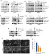
GSK-3β inhibition is sufficient to induce vasculogenic differentiation of dental pulp stem cells. (A): Western blots for Fzd-6, P-GSK-3β, and GSK-3β from DPSC treated with 10 µM CHIR99021 for up to 24 hours. Alternatively, we performed a dose-dependent experiment with DPSC cells treated with 0–10 µM CHIR99021 for 24 hours. Numbers depict the band density normalized against untreated controls and GAPDH. (B): Western blots for LRP-6, Fzd-6, active-β-catenin, β-catenin and Wnt1 from DPSC or SHED starved overnight, and treated with 0–10 µM CHIR99021 for 24 hours. (C): Western blots for endothelial differentiation markers (VEGFR2, VE-Cadherin, Tie-2 and CD31) from DPSC or SHED cells treated with 0–2.5 µM CHIR99021 for 14 days. (D): Western blots for Axin-2, P-GSK-3β, and GSK-3β from DPSC or SHED treated with 0–10 µM JW67 for 24 hours. (E): Western blots for vasculogenic differentiation markers (VEGFR2, Tie-2) from DPSC or SHED cells treated with 50 ng/ml rhWnt1 and/or 0–5 µM JW67 for 14 days. (F): Photomicrographs of DPSC cells seeded in 12-well plates (5×10⁴ cells/well) coated with growth factor-reduced Matrigel and incubated with EGM2-MV supplemented with 50 ng/ml rhVEGF165 with or without 0–5 µM JW 67 for up to 11 days. Scale bars: 100 µm. (G): Graph depicting the number of sprouts formed by DPSC treated with 0, 2.5 or 5 µM JW67 in EGM2-MV medium supplemented with 50 ng/ml rhVEGF165. Three independent experiments using triplicate wells per experimental condition were performed to verify reproducibility of the data. * p<0.01, ** p<0.001.
Figure 6
Figure 6
Knockdown of β-catenin inhibits angiogenic differentiation of DPSC in vitro(A–D): DPSC stably transduced with shRNA-scrambled or two different sequences (1 or 2) of shRNA-β-catenin were treated with 50 ng/ml rhWnt1 (A, B) or 50 ng/ml VEGF (C, D) for 14 days. Western blots were performed for active-β-catenin, β-catenin (A, C) or VEGFR2, Tie-2 (B, D). HDMEC cells were used as positive control for VEGFR2 and Tie-2. Numbers depict the band density normalized against untreated controls and GAPDH. (E): Photomicrographs of DPSC-shRNA-β-catenin (sequence 1 or 2) or control DPSC-shRNA-Scrambled cells seeded in 12-well plates (5×10⁴ cells/well) coated with growth factor-reduced Matrigel and incubated with EGM2-MV supplemented with 50 ng/ml rhVEGF165 for up to 16 days. Scale bars: 100 µm. (F) Graph depicting the number of sprouts formed by DPSC-shRNA-β-catenin (sequence 1 or 2) or control DPSC-shRNA-Scrambled cells (E). Three independent experiments using triplicate wells per experimental condition were performed to verify reproducibility of the data. Asterisk indicates p<0.01.
Figure 7
Figure 7
Knockdown of β-catenin inhibits angiogenic differentiation of DPSC in vivo(A): DPSC stably transduced with shRNA-β-catenin(1) or control shRNA-scrambled were seeded in tooth slice/scaffolds (n=8), and transplanted into the subcutaneous space of immunodeficient mice. Five weeks after transplantation, the tooth slice/scaffolds were retrieved from the mice, fixed, decalcified and prepared for IHC. Blood vessels (brown color) were detected with anti-factor VIII antibody. Scale bars: 50 µm. (B): Graph depicting the blood vessel density observed in the tissues generated with DPSC cells in (A). Asterisk indicates p<0.01.

Similar articles

Cited by

References

    1. Gronthos S, Mankani M, Brahim J, et al. Postnatal human dental pulp stem cells (DPSCs) in vitro and in vivo . Proc Natl Acad Sci USA. 2000;97:13625–13630. - PMC - PubMed
    1. Miura M, Gronthos S, Zhao M, et al. SHED: stem cells from human exfoliated deciduous teeth. Proc Natl Acad Sci USA. 2003;100:5807–5812. - PMC - PubMed
    1. Sonoyama W, Liu Y, Fang D, et al. Mesenchymal stem cell-mediated functional tooth regeneration in Swine. PLoS One. 2006;1:e79. - PMC - PubMed
    1. Sonayama W, Liu Y, Yamaza T, et al. Characterization of the apical and its residing stem cells from human immature permanent teeth: a pilot study. J Endod. 2008;34:166–171. - PMC - PubMed
    1. Huang GT, Gronthos S, Shi S. Mesenchymal stem cells derived from dental tissues vs. those from other sources: their biology and role in regenerative medicine. J Dent Res. 2009;88:792–806. - PMC - PubMed

MeSH terms