. 2016 Feb 11;11(2):e0148996.
doi: 10.1371/journal.pone.0148996. eCollection 2016.

Microtubule and Actin Interplay Drive Intracellular c-Src Trafficking

Affiliations

Microtubule and Actin Interplay Drive Intracellular c-Src Trafficking

Christopher Arnette et al. PLoS One. .

Abstract

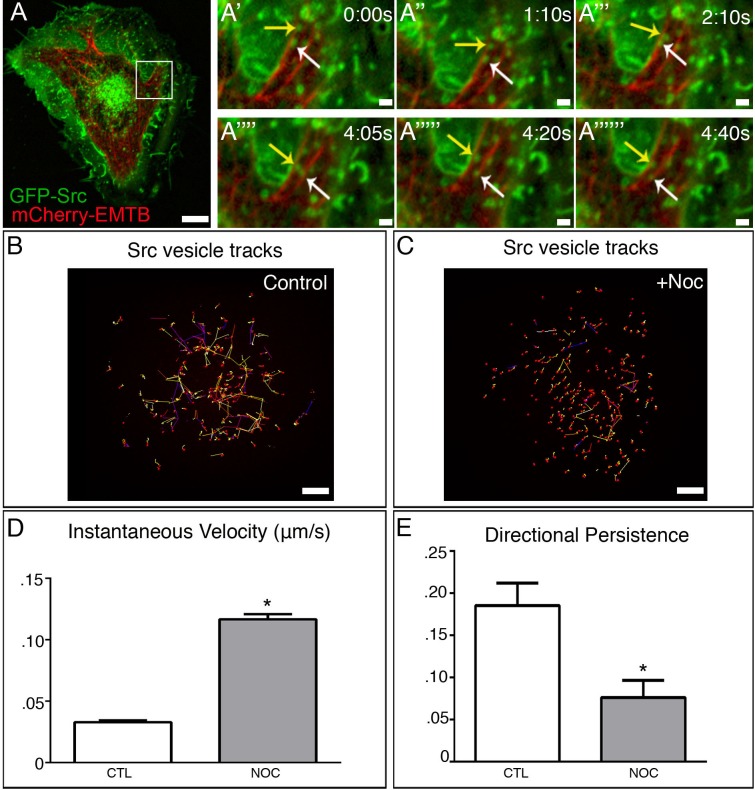
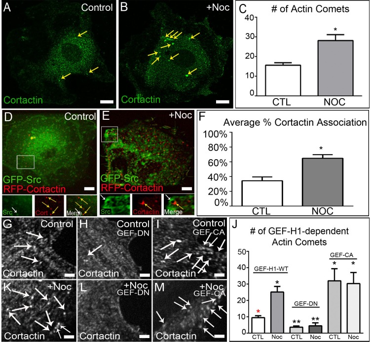
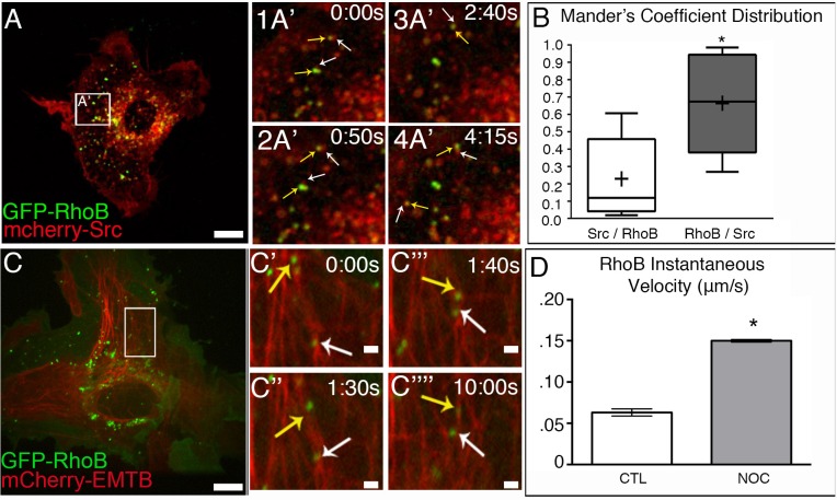
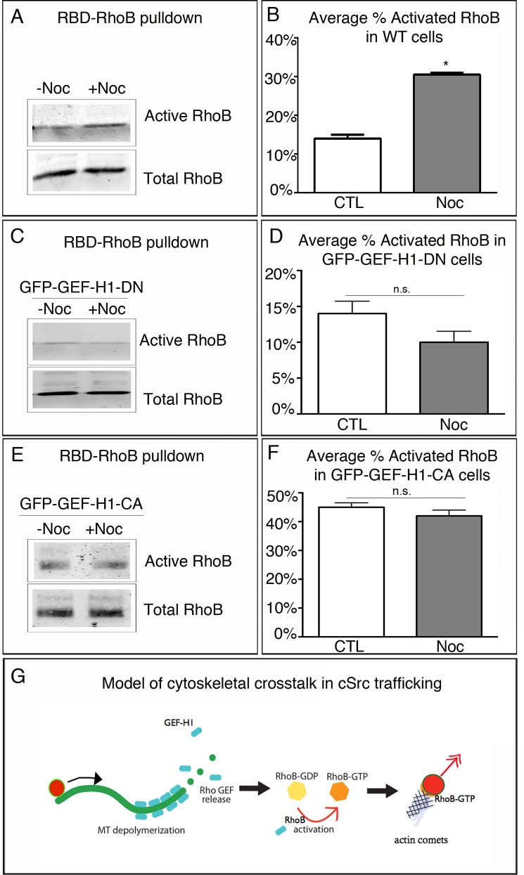
The proto-oncogene c-Src is involved in a variety of signaling processes. Therefore, c-Src spatiotemporal localization is critical for interaction with downstream targets. However, the mechanisms regulating this localization have remained elusive. Previous studies have shown that c-Src trafficking is a microtubule-dependent process that facilitates c-Src turnover in neuronal growth cones. As such, microtubule depolymerization lead to the inhibition of c-Src recycling. Alternatively, c-Src trafficking was also shown to be regulated by RhoB-dependent actin polymerization. Our results show that c-Src vesicles primarily exhibit microtubule-dependent trafficking; however, microtubule depolymerization does not inhibit vesicle movement. Instead, vesicular movement becomes both faster and less directional. This movement was associated with actin polymerization directly at c-Src vesicle membranes. Interestingly, it has been shown previously that c-Src delivery is an actin polymerization-dependent process that relies on small GTPase RhoB at c-Src vesicles. In agreement with this finding, microtubule depolymerization induced significant activation of RhoB, together with actin comet tail formation. These effects occurred downstream of GTP-exchange factor, GEF-H1, which was released from depolymerizing MTs. Accordingly, GEF-H1 activity was necessary for actin comet tail formation at the Src vesicles. Our results indicate that regulation of c-Src trafficking requires both microtubules and actin polymerization, and that GEF-H1 coordinates c-Src trafficking, acting as a molecular switch between these two mechanisms.


Conflict of interest statement

Competing Interests: The authors have declared that no competing interests exist.

Figures

Fig 1
Fig 1. c-Src exhibits bimodal trafficking.
(A) Detection of GFP-Src (green) and 3x-mCherry-EMTB (red) in time-lapse confocal movie of an A7r5 cell. Area in box is enlarged to the right. Bar, 5μm. (A’-A”””) Imaging sequence illustrates that GFP-Src is trafficked bidirectionally along MTs in A. Bar, 2μm (B) GFP-Src vesicles exhibit long, directional movement in time-lapse confocal movie of an A7r5 control (5s/frames). Representative tracks. Bar, 5μm. (C) GFP-Src vesicles exhibit short, randomized movement in time-lapse confocal movie of an A7r5 control (5s/frames). Representative tracks. Bar 5μm. (D) GFP-Src vesicles exhibit increased velocity in response to MT depolymerization. (E) GFP-Src vesicles show reduced directional persistence following nocodazole treatment. N = 5 cells/condition, 50 vesicles/cell. See also S1 Movie. Error bars present SEM. Asterisks indicate p-value < .05.
Fig 2
Fig 2. GEF-H1 regulates formation of actin comet tails at c-Src vesicle membranes.
(A-B) Control and nocodazole-treated A7r5 cells fixed and stained for cortactin (green). Immunostaining. Bar, 5μm. (C) Quantification of actin comet tail formation in control and nocodazole-treated cells reveals a two-fold increase upon MT depolymerization. Representative examples out of 30 cells/condition. Error bars indicate s.e.m. (D-E) Live-cell imaging of control and nocodazole-treated A7r5 cells expressing GFP-Src and RFP-Cortactin. Area in box is enlarged below. Bar 5μm. See also S3 Movie. (F) Quantification reveals that MT depolymerization induces increased actin comet formation at c-Src vesicle membranes. (G and K) Z-section of A7r5 cells expressing wild-type GEF-H1 pre- and post-nocodazole fixed and immunostained for cortactin. Images illustrate an increase in cortactin puncta (white arrows) following MT depolymerization Bar, 5μm. (H and L) Z-section of A7r5 cells expressing dominant-negative GEF-H1 pre- and post-nocodazole demonstrates the role of GEF-H1 in forming actin comets. Bar, 5μm. (I and M) Z-section of A7r5 cells expressing constitutively active GEF-H1 pre- and post-nocodazole show high number of actin comets in the presence or absence of MTs. Bar, 5μm. (J) Quantification of actin comet tail formation after expression of various GEF-H1 constructs. Representative examples out of 30 cells/condition. Error bars indicate SEM. Asterisks indicate p-value < .05.
Fig 3
Fig 3. RhoB associates with c-Src vesicles is trafficked along MTs.
(A) A7r5 cell expressing GFP-RhoB (green) and mCherry-Src (red). Area in box is enlarged to the right. Spinning disk confocal. Bar, 5μm. (A’1 and A’2) Time-lapse confocal microscopy shows that GFP-RhoB (yellow arrows) can associate with a subset of mcherry-Src vesicles (white arrows. See also S4 Movie. (B) Mander’s coefficient distribution reveals that the majority of GFP-RhoB vesicles are associated with mCherry-Src. N = 10 cells. (C) A7r5 cells expressing GFP-RhoB (green) and 3X-mCherry-EMTB (red). Area in box is enlarged to the right. Spinning disk confocal. Bar, 5μm. (C’–C””) Time-lapse confocal microscopy reveals vesicles GFP-RhoB vesicles move along MTs (yellow and white arrows). See also S5 Movie. (D) GFP-RhoB vesicles exhibit increased velocity in response to MT depolymerization. N = 5 cells/condition, 50 vesicles/cell. Bar, 2μm. Error bars represent SEM. Asterisks indicate p-value < .05.
Fig 4
Fig 4. RhoB is activated by MT depolymerization.
(A) Western blot analysis of RhoB activation in response to MT depolymerization in control and nocodazole-treated RPE1 cells. (B) Quantification of average percent of RhoB activation from A reveals that RhoB activation is significantly higher following MT depolymerization. (C) Western blot analysis of RhoB activation in GFP-GEF-H1-DN expressing cells pre- and post-nocodazole treatment. (D) Quantification of the average percent of activation under both conditions reveals that inhibition of GEF-H1 activity significantly impairs RhoB activation. (E) Western blot analysis of RhoB activation in GFP-GEF-H1 constitutively active expressing cells. (F) Quantification reveals that constitutive activation of GEF-H1 leads to high levels of RhoB even in the presence of MTs. Pulldowns repeated in triplicate. Error bars indicate SEM. Asterisks indicate p-value < .05. (G) Model of cytoskeletal crosstalk for c-Src delivery.

References

    1. Yeatman TJ. A renaissance for SRC. Nature reviews Cancer. 2004;4(6):470–80. 10.1038/nrc1366 . - DOI - PubMed
    1. Hanahan D, Weinberg RA. The hallmarks of cancer. Cell. 2000;100(1):57–70. . - PubMed
    1. Seddon B, Zamoyska R. TCR signals mediated by Src family kinases are essential for the survival of naive T cells. Journal of immunology. 2002;169(6):2997–3005. . - PubMed
    1. Giannoni E, Fiaschi T, Ramponi G, Chiarugi P. Redox regulation of anoikis resistance of metastatic prostate cancer cells: key role for Src and EGFR-mediated pro-survival signals. Oncogene. 2009;28(20):2074–86. 10.1038/onc.2009.77 . - DOI - PubMed
    1. Armaiz-Pena GN, Allen JK, Cruz A, Stone RL, Nick AM, Lin YG, et al. Src activation by beta-adrenoreceptors is a key switch for tumour metastasis. Nature communications. 2013;4:1403 10.1038/ncomms2413 - DOI - PMC - PubMed

Publication types

MeSH terms