. 2016 Oct;18(10):1374-89.
doi: 10.1111/cmi.12578. Epub 2016 Mar 18.

Heme oxygenase 1 controls early innate immune response of macrophages to Salmonella Typhimurium infection

Affiliations

Heme oxygenase 1 controls early innate immune response of macrophages to Salmonella Typhimurium infection

Anna-Maria Mitterstiller et al. Cell Microbiol. 2016 Oct.

Abstract

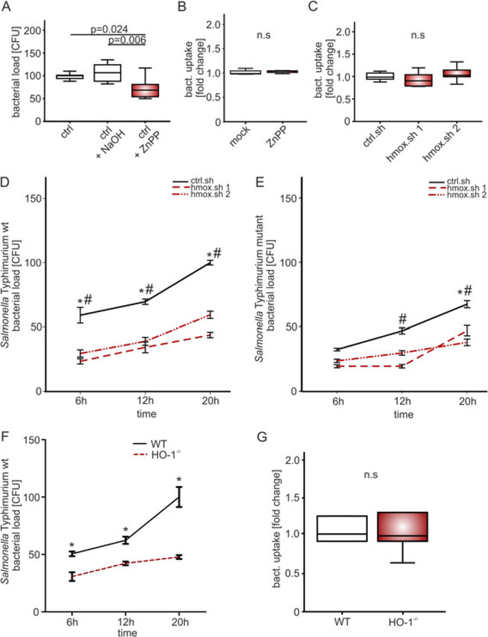
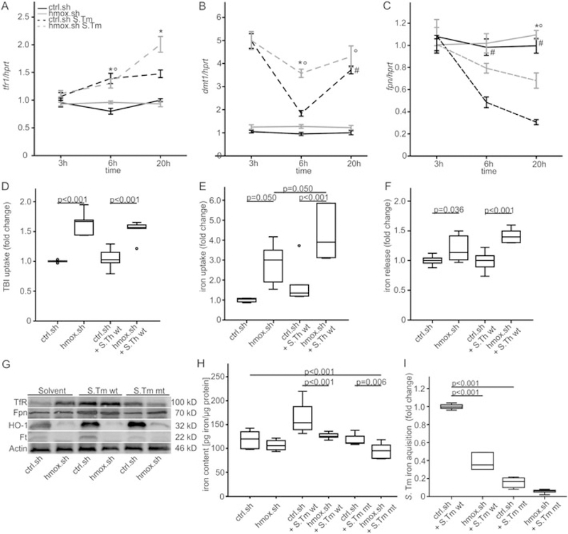
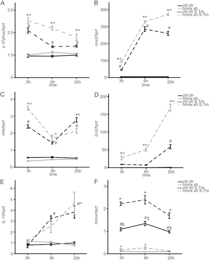
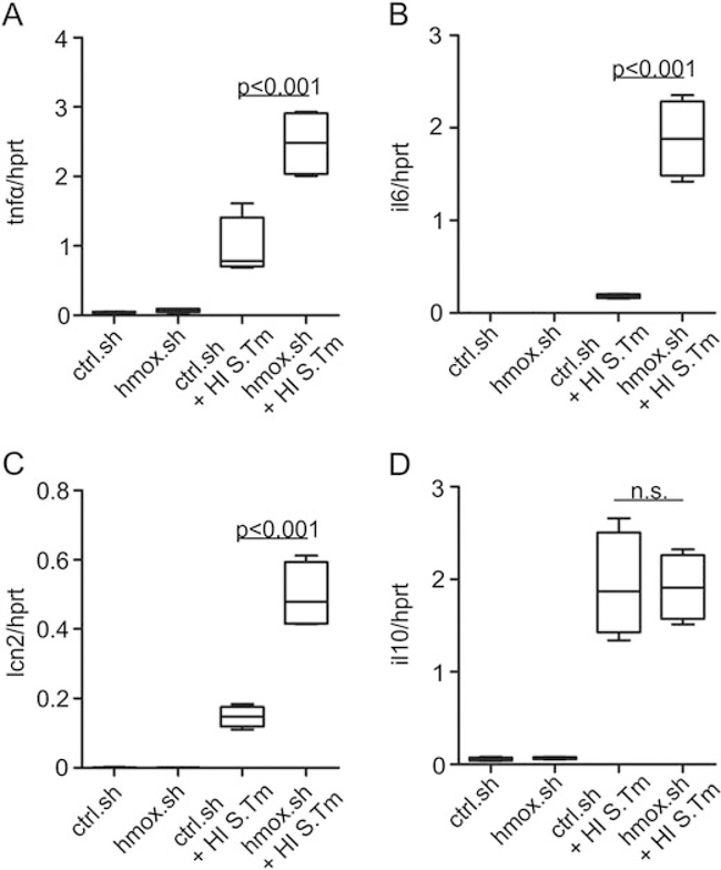
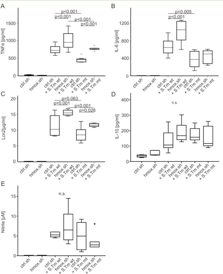
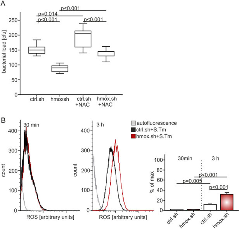
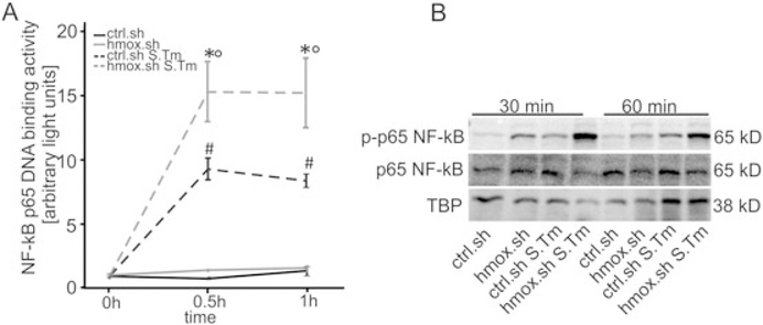
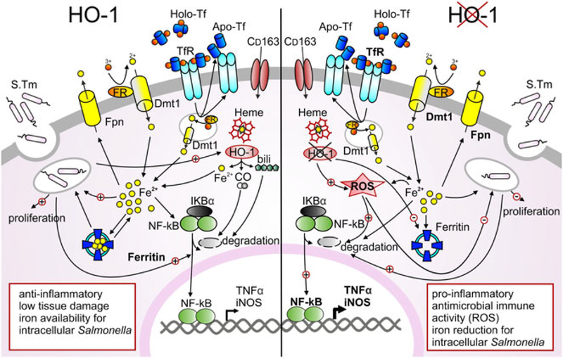
Macrophages are central for the immune control of intracellular microbes. Heme oxygenase 1 (HO-1, hmox) is the first and rate limiting enzyme in the breakdown of heme originating from degraded senescent erythrocytes and heme-proteins, yielding equal amounts of iron, carbon monoxide and biliverdin. HO-1 is strongly up-regulated in macrophages in response to inflammatory signals, including bacterial endotoxin. In view of the essential role of iron for the growth and proliferation of intracellular bacteria along with known effects of the metal on innate immune function, we examined whether HO-1 plays a role in the control of infection with the intracellular bacterium Salmonella Typhimurium. We studied the course of infection in stably-transfected murine macrophages (RAW264.7) bearing a tetracycline-inducible plasmid producing hmox shRNA and in primary HO-1 knockout macrophages. While uptake of bacteria into macrophages was not affected, a significantly reduced survival of intracellular Salmonella was observed upon hmox knockdown or pharmacological hmox inhibition, which was independent of Nramp1 functionality. This could be traced to limitation of iron availability for intramacrophage bacteria along with enhanced stimulation of innate immune effector pathways, including the formation of reactive oxygen and nitrogen species and increased TNF-α expression. Mechanistically, these latter effects result from intracellular iron limitation with subsequent activation of NF-κB and further inos, tnfa and p47phox transcription along with reduced formation of the anti-inflammatory and radical scavenging molecules, CO and biliverdin as a consequence of HO-1 silencing. Taken together our data provide novel evidence that the infection-driven induction of HO-1 exerts detrimental effects in the early control of Salmonella infection, whereas hmox inhibition can favourably modulate anti-bacterial immune effector pathways of macrophages and promote bacterial elimination.


Figures

Fig. 1.
Fig. 1.. Heme oxygenase gene knockdown reduces the survival of intracellular Salmonella Typhimurium.
(A) RAW 264.7, (B-E) RAW.sh macrophages (ctrl.sh, hmox.sh 1 and 2) and (F-G) BMDM from LysM-Cre+/+ HO-1fl/fl (HO-1−/−) and LysM-Cre−/− HO-1fl/fl (wt) mice were infected with (A-D, F-G) Salmonella enterica serovar Typhimurium (S. Tm) wild-type (wt) or (E) an isogenic Salmonella Typhimurium mutant (ΔentCΔsitΔfeo) strain at a MOI of 10 for the indicated period of time. (A) Bacterial burden (CFU) was determined 24 h post-infection following treatment with 10 μM of HO-1 Inhibitor ZnPP or solvent (NaOH, PBS). (B-C, G) Bacterial uptake relative to mock/control (fold change) was measured one hr post infection to determine phagocytosis. (A-C, G) Data from three independent experiments performed in triplicate are shown (n = 9). Boxes depict lower quartile, mean and upper quartile; whiskers show maximum and minimum range. P-values are reported as determined by statistical analysis using ANOVA and Bonferroni correction. (D-F) Bacterial burden (CFU) was determined as mean ± SEM of at least three independent experiments performed in triplicate (n = 9) and normalized (%) relative to control. Whiskers show maximum and minimum range. P-values are reported as determined by statistical analysis using ANOVA and Bonferroni’s correction. * P < 0.001 for crtl.sh versus hmox.sh 1 or control (wt) BMDM versus HO-1−/− cells; # P < 0.001 for ctrl.sh versus hmox.sh 2 macrophages.
Fig. 2.
Fig. 2.. Effects of hmox on iron acquisition and iron release in Salmonella infected macrophages.
(A-B, D-E) Iron import. Ctrl.sh cells (black line) and hmox knockdown macrophages (hmox.sh; grey line) were infected with Salmonella Typhimurium wild-type (S. Tm wt; dashed line) strain at a MOI of 10 for the indicated period of time. (A) tfr1 and (B) dmt1 levels were determined by qRT-PCR. Values were normalized to the housekeeping gene hprt, and fold-changes relative to the untreated control (20 h) are shown. (D, E) The uptake of (D) TBI (transferrin loaded with 59Fe) and (E) iron (59Fe) for 4 h following an 18 h infection period was determined as described in Materials and methods. (C, F) Iron export. Macrophages were treated exactly as described above. (C) fpn1 levels by qRT-PCR and (F) iron (59Fe) release 20 h post infection were determined. (A-C) Data from at least three independent experiments are shown (ctrl.sh n =10; hmox.sh n = 9) and expressed as mean ± SEM. P-values are reported as determined by post hoc with Bonferroni correction. *# ° P < 0.001 (* ctrl.sh vs. hmox.sh infected with S. Tm; # ctrl.sh vs. ctrl.sh S.Tm; ° hmox.sh vs. hmox.sh S.Tm). (D-F) Data were compared and are depicted of three independent experiments (D, E) n = 6, (F) n = 12 ctrl.sh, n = 8 hmox.sh. P-values are reported as determined by post hoc analysis with Bonferroni’s correction following ANOVA. (G) Western blot analysis of whole cell lysates using specific antibodies to TfR1, Fpn1, heme oxygenase (HO-1), Ferritin (Ft) and the control β-Actin. One of three representative western blot experiments is shown. (H) Cellular iron content in phagocytes was measured by atomic absorption spectrometry. Macrophages were infected with Salmonella wt or mt (ΔentCΔsitΔfeo) for 48 h or left untreated. Results were normalized to protein content. Data from three independent experiments are shown (n = 8 untreated and S. Tm mt infected, n = 10 infected with S. Tm wt). (I) Bacterial iron acquisition within macrophages was determined after loading of infected macrophages with 59Fe. Data were normalized for Salmonella numbers determined by CFU count. Three independent experiments were carried out in duplicate (n = 6). Data were compared by ANOVA using Bonferroni’s correction (P-values). Values (boxes) are depicted as lower quartile, median and upper quartile with maximum and minimum range.
Fig. 3.
Fig. 3.. Effects of hmox gene knockdown on p47phox, nos2, tnfa, II-10 and lcn2 transcript expression.
Ctrl.sh (black line) and hmox.sh (grey line) macrophages infected with Salmonella (black or grey dashed line) were subjected to qRT-PCR at indicated time points. Expression of genes of interest was normalized to hprt expression and fold-change relative to untreated control is shown. Data from three independent experiments (n = 10 ctrl.sh; n = 8 hmox. sh) are shown. Error bars represent means ± SEM, values are reported as determined by ANOVA using Bonferroni’s correction. * $ P < 0.001 for hmox.sh versus ctrl.sh, both infected with S. Tm (*) or both uninfected ($); ° P < 0.001 for hmox.sh uninfected versus infected with S. Tm; # P < 0.001 for comparison of ctrl.sh and ctrl.sh infected with S. Tm.
Fig. 4.
Fig. 4.. Early response to Salmonella is improved in hmox knockdown macrophages.
Ctrl.sh and hmox.sh macrophages supplemented with an identical number of heat-inactivated Salmonella (HI S. Tm; as a defined inflammatory stimulus not compromised by bacterial number) as described in Fig. 3 were subjected to qRT-PCR. Gene expression of (A) tnfa, (B) il6, (C) lcn2 and (D) il10 was determined 6 h post-infection and normalized to hprt expression levels. (A-D) Fold changes relative to untreated controls are shown. Data from three independent experiments are shown. Error bars represent means ± SEM; values are reported as determined by ANOVA using Bonferroni’s correction.
Fig. 5.
Fig. 5.. Productionofpro-inflammatory mediators, Lcn2, IL-10 and nitrate during Salmonella infection following hmox gene knockdown.
(A-E) Ctrl.sh and hmox.sh cells were infected with Salmonella wt (S. Tm wt) or mutant ΔentCΔsitΔfeo (S. Tm mt) for 20 h before culture supernatants were analyzed for formation of (A) TNFα, (B) IL-6, (C) Lcn2, (D) IL-10 and (E) nitrate. (A-E) Boxes depict lower quartile, mean and upper quartile; whiskers show minimum and maximum range. Data are shown as mean ± SEM from at least three independent experiments. P-values are reported as determined by ANOVA using Bonferroni’s correction.
Fig. 6.
Fig. 6.. Increased ROS production by hmox knockdown macrophages reduces their bacterial burden.
(A) Macrophages (hmox.sh versus ctrl.sh) were infected with Salmonella Typhimurium wt (S. Tm) and thereafter treated with the radical scavenger N-acetyl-L-cysteine (NAC) for 20 h. Boxes are depicted as lower quartile, median and upper quartile with minimum and maximum range. Data of at least three independent experiments are shown (n = 10). P-values are reported as determined by ANOVA using Bonferroni’s correction. (B) ROS levels were measured via FACS analysis in ctrl.sh and hmox.sh cells infected with Salmonella at 30 min and 3 h post infection. The histograms represent the average ROS levels measured by CellROX™ fluorescence. Data of three independent experiments performed in triplicate are shown. P-values are reported as determined by ANOVA using Bonferroni’s correction.
Fig. 7.
Fig. 7.. Knockdown of hmox in macrophages show increased NF-κB activation.
RAW.sh macrophage-like cells were infected with Salmonella for the indicated periods of time. NF-κB DNA binding activity in nuclear protein extracts was evaluated by means of a specific p-65 NF-κB chemo-luminescence transcription factor assay. Data from three independent experiments are shown and expressed as arbitrary light units. Error bars are depicted as means ± SEM and were compared by ANOVA with Bonferroni’s correction. Asterisks indicate statistically significant differences between Salmonella infected control and hmox knockdown macrophages: * p < 0.001 for comparison of ctrl.sh versus hmox.sh infected with S. Tm; # p < 0.001 ctrl.sh versus ctrl.sh S. Tm;°p < 0.001 between hmox.sh and hmox.sh S. Tm. (B) Protein extracts from parallel experiments described in (A) were used for evaluation of phosphorylated (p-) p65 NF-κB by means of Western blot analysis as described in Material and methods. Antibodies to p65 NF-κB and TATA-binding protein (as loading control) were used. One of three representative Western blot experiments is shown.
Fig. 8.
Fig. 8.. Effects of heme oxygenase in regulating iron homeostasis and innate immune response of macrophages during Salmonella infection.
Macrophages play an important role in infected tissue. Within these cells heme bound iron is degraded via HO-1 to equal amounts of iron, carbon monoxide (CO) and biliverdin (bili). Ferric iron is acquired via transferrin receptor 1 (TfR1) mediated endocytosis of holoTf, reduced within the vesicle by a ferric reductase (FR) and released to the cytoplasmic pool via divalent metal transporter (Dmt1), while Tf and TfR are recycled to the surface. During infection, bacterial pathogens may employ mechanisms to increase the abundance of potential iron sources. Transitory iron accumulation in the cytosol may promote storage into ferritin, ferroportin (Fpn1)-mediated export and reduction of TfR1 expression, thereby limiting iron for the pathogen or lowering intracellular iron levels. Both LPS and iron stimulate NF-κB expression, resulting in an inflammatory response that includes expression of iNOS, TNFα and other cytokines. Macrophages with absent HO-1 function show an increased iron transport activity, strong iron utilization via TfR1 and Dmt1 and increased iron export via Fpn1. Cells may simultaneously restrict iron levels during Salmonella infection and increase production of reactive oxygen species (ROS). Restriction of iron and increased ROS production promote NF-κB mediated activation of a pro-inflammatory immune response that results in improved Salmonella control and may also result in tissue injury.

Similar articles

Cited by

References

    1. Abdalla MY, Ahmad IM, Switzer B, and Britigan BE (2015) Induction of heme oxygenase-1 contributes to survival of Mycobacterium abscessus in human macrophages-like THP-1 cells. Redox Biol 4: 328–339. - PMC - PubMed
    1. Ables GP, Takamatsu D, Noma H, El-Shazly S, Jin HK, Taniguchi T, et al. (2001) The roles of Nramp1 and Tnfa genes in nitric oxide production and their effect on the growth of Salmonella typhimurium in macrophages from Nramp1 congenic and tumor necrosis factor-alpha−/− mice. J Interferon cytokine Res: Off J Int Soc Interferon Cytokine Res 21: 53–62. - PubMed
    1. Andrews-Polymenis HL, Baumler AJ, McCormick BA, and Fang FC (2010) Taming the elephant: Salmonella biology, pathogenesis, and prevention. Infect Immun 78: 2356–2369. - PMC - PubMed
    1. Arezes J, Jung G, Gabayan V, Valore E, Ruchala P, Gulig PA, et al. (2015) Hepcidin-induced hypoferremia is a critical host defense mechanism against the siderophilic bacterium Vibrio vulnificus. Cell Host Microbe 17: 47–57. - PMC - PubMed
    1. Armitage AE, Eddowes LA, Gileadi U, Cole S, Spottiswoode N, Selvakumar TA, et al. (2011) Hepcidin regulation by innate immune and infectious stimuli. Blood 118: 4129–4139. - PubMed

Publication types