. 2016 Jul;34(7):1922-33.
doi: 10.1002/stem.2339. Epub 2016 Mar 4.

Tumor Necrosis Factor α Regulates Endothelial Progenitor Cell Migration via CADM1 and NF-kB

Affiliations

Tumor Necrosis Factor α Regulates Endothelial Progenitor Cell Migration via CADM1 and NF-kB

Anthony R Prisco et al. Stem Cells. 2016 Jul.

Abstract

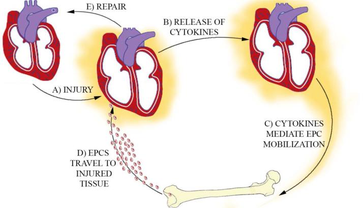
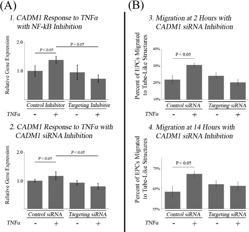
Shortly after the discovery of endothelial progenitor cells (EPCs) in 1997, many clinical trials were conducted using EPCs as a cellular based therapy with the goal of restoring damaged organ function by inducing growth of new blood vessels (angiogenesis). Results were disappointing, largely because the cellular and molecular mechanisms of EPC-induced angiogenesis were not clearly understood. Following injection, EPCs must migrate to the target tissue and engraft prior to induction of angiogenesis. In this study EPC migration was investigated in response to tumor necrosis factor α (TNFα), a pro-inflammatory cytokine, to test the hypothesis that organ damage observed in ischemic diseases induces an inflammatory signal that is important for EPC homing. In this study, EPC migration and incorporation were modeled in vitro using a coculture assay where TNFα treated EPCs were tracked while migrating toward vessel-like structures. It was found that TNFα treatment of EPCs increased migration and incorporation into vessel-like structures. Using a combination of genomic and proteomic approaches, NF-kB mediated upregulation of CADM1 was identified as a mechanism of TNFα induced migration. Inhibition of NF-kB or CADM1 significantly decreased migration of EPCs in vitro suggesting a role for TNFα signaling in EPC homing during tissue repair. Stem Cells 2016;34:1922-1933.

Keywords: CADM1; Cellular migration; Endothelial progenitor cells; TNFα.


Figures

Figure 1
Figure 1. Bone Marrow Stem Cell Repair Axis
EPCs are transported from the bone marrow to the myocardium. Injured myocardium releases inflammatory signals that mobilize EPCs from the bone marrow. Cells are released into the circulation and blood flow through damaged organs induces recruitment of cells to target tissue. These cells can then repair the damaged myocardium and suppress the inflammatory signal, completing the feedback loop.
Figure 2
Figure 2. TNFα Produced in an In Vivo Model of Angiogenesis Increases the Migratory Activity of EPCs In Vitro
(A) TNFα expression is increased in an in vivo model of angiogenesis. A hind limb muscle stimulator that has previously been shown to induce angiogenesis was implanted and run for 7 days. After 7 days, RNA expression of TNFα was significantly increased in the stimulated limb vs the non-stimulated control (n = 6 per group). TNFα protein expression in homogenized hindlimb muscle (pg/mg of homogenized protein) was also found to be significantly upregulated in the stimulated vs the non-stimulated control (n = 6 per group). (B) In vitro EPC migration assay. Because TNFα expression was found to be increased in the angiogenic limb, EPCs were treated with TNFα/control and their locations were tracked with respect to capillary-like tubes in vitro. (C) 3hr, 1ng/mL TNFα/vehicle pre-treatment of EPCs increased the fraction that migrated towards tubes (P < 0.05) at 2 hours and 14 hours (n = 16 per group).
Figure 3
Figure 3. TNFα Pathway Analysis Results by qPCR
To determine which TNF receptor was driving the migratory phenotype, expression of key pathway genes was measured in response to TNFα. Significantly upregulated genes are displayed in green and significantly down regulated genes are displayed in red. Key genes down regulated in the TNFR1 family are members of the caspase family. Key genes upregulated in the TNFR2 family are NF-kB and TNFR2 suggesting that the migratory phenotype is being signaled through the TNFR2 family via NF-kB (n = 6 per group).
Figure 4
Figure 4. Inhibition of NF-kB Reduces TNFα Induced Migration
Prior to TNFα administration, EPCs were pretreated for 1 hour with a 1μg/mL dose of a peptide inhibitor of NF-kB or a scrambled control. The TNFα induced migratory phenotype was still observed in the presence of the scrambled inhibitor at 2 hour and 14 hours (P < 0.05) but the phenotype was abolished at 2 hours and 14 hours when the NF-kB inhibitor was administered (n = 16 per group).
Figure 5
Figure 5. Validation of CADM1 as a Candidate Protein Pair
(A) Example LC-MS/MS Spectra. 16 scans of the peptide sequenced ‘VSLTNVSISDEGR’ were observed in the CSC TNFα treated EPC data set. The peptide has 12 potential peptide fragmentation sites and therefore 12 potential ‘B’ and ‘Y’ ions. Following peptide fragmentation, the following m/z spectra was obtained. 11 ‘B’ ions were observed (11 shown) and 9 ‘Y’ ions were observed (5 shown). (B) Western blot analysis was conducted to confirm quantitation via spectral counting of CADM1 in EPCs (1) and RCMVECs (2). Statistically significant regulation was observed in EPCs, but not RCMVECs confirming proteomics results. Quantitation was performed by normalizing CADM1 bands to total protein detected on the membrane via coomassie staining.
Figure 6
Figure 6. Analysis of CADM1 for a Function Role in EPC Migration in Response to TNFα
(A) PCR Analysis of CADM1 Expression in Response to Inhibitors. (1) CADM1 gene expression was measured in response to TNFα in the presence of a synthetic peptide inhibitor of NF-kB or the scrambled control. In presence of the scrambled control TNFα induced up regulation of CADM1 (P < 0.05) whereas in presence of the inhibitor this response was abolished. (2) In the presence of scrambled siRNA, TNFα was able to increase gene expression of CADM1 (P < 0.05) whereas in the presence of targeting siRNA this phenotype was abolished (n = 16 per group). (B) EPC Migration with CADM1 Knockdown. Prior to TNFα administration, EPCs were transfected with a CADM1 siRNA or a scrambled control. The TNFα induced migratory phenotype was still observed in the presence of the scrambled inhibitor at 2 hour and 14 hours (P < 0.05) but the phenotype was abolished at 2 hours and 14 hours when the targeted siRNA was transfected (n = 16 per group).

References

    1. Asahara T, Murohara T, Sullivan A, et al. Isolation of putative progenitor endothelial cells for angiogenesis. Science. 1997;275:964–967. - PubMed
    1. Zuk PA, Zhu M, Mizuno H, et al. Multilineage cells from human adipose tissue: implications for cell-based therapies. Tissue Eng. 2001;7:211–228. - PubMed
    1. Kawamoto A, Gwon HC, Iwaguro H, et al. Therapeutic potential of ex vivo expanded endothelial progenitor cells for myocardial ischemia. Circulation. 2001;103:634–637. - PubMed
    1. Wollert KC, Meyer GP, Lotz J, et al. Intracoronary autologous bone-marrow cell transfer after myocardial infarction: the BOOST randomised controlled clinical trial. Lancet. 2004;364:141–148. - PubMed
    1. Kang HJ, Kim HS, Zhang SY, et al. Effects of intracoronary infusion of peripheral blood stem-cells mobilised with granulocyte-colony stimulating factor on left ventricular systolic function and restenosis after coronary stenting in myocardial infarction: the MAGIC cell randomised clinical trial. Lancet. 2004;363:751–756. - PubMed

Publication types

MeSH terms