. 2016 Feb 11;11(2):e0149020.
doi: 10.1371/journal.pone.0149020. eCollection 2016.

Age-Dependent Changes in Geometry, Tissue Composition and Mechanical Properties of Fetal to Adult Cryopreserved Human Heart Valves

Affiliations

Age-Dependent Changes in Geometry, Tissue Composition and Mechanical Properties of Fetal to Adult Cryopreserved Human Heart Valves

Daphne van Geemen et al. PLoS One. .

Abstract

There is limited information about age-specific structural and functional properties of human heart valves, while this information is key to the development and evaluation of living valve replacements for pediatric and adolescent patients. Here, we present an extended data set of structure-function properties of cryopreserved human pulmonary and aortic heart valves, providing age-specific information for living valve replacements. Tissue composition, morphology, mechanical properties, and maturation of leaflets from 16 pairs of structurally unaffected aortic and pulmonary valves of human donors (fetal-53 years) were analyzed. Interestingly, no major differences were observed between the aortic and pulmonary valves. Valve annulus and leaflet dimensions increase throughout life. The typical three-layered leaflet structure is present before birth, but becomes more distinct with age. After birth, cell numbers decrease rapidly, while remaining cells obtain a quiescent phenotype and reside in the ventricularis and spongiosa. With age and maturation-but more pronounced in aortic valves-the matrix shows an increasing amount of collagen and collagen cross-links and a reduction in glycosaminoglycans. These matrix changes correlate with increasing leaflet stiffness with age. Our data provide a new and comprehensive overview of the changes of structure-function properties of fetal to adult human semilunar heart valves that can be used to evaluate and optimize future therapies, such as tissue engineering of heart valves. Changing hemodynamic conditions with age can explain initial changes in matrix composition and consequent mechanical properties, but cannot explain the ongoing changes in valve dimensions and matrix composition at older age.


Conflict of interest statement

Competing Interests: The authors have declared that no competing interests exist.

Figures

Fig 1
Fig 1. Schematic overview of the leaflet sectioning and assignment to the different experiments.
(A) Schematic overview of sectioning of one heart valve for analyses. Samples for biaxial tensile testing are indicated in green; parts for histology in red. The right coronary cusp (RCC) and right facing cusp (RFC) were used for indentation tests (purple). Afterwards, this leaflet was used for biaxial tensile testing. Leftover tissue (blue) was freeze-dried for biochemical assays. LCC: Left coronary cusp; LFC: Left facing cusp; NCC: non-coronary cusp; AC: anterior cusp. (B) The size of the leaflets was measured in circumferential and radial direction (arrows). (C) Indentation tests were performed in the commissural (c1 –c4) and belly (b1 –b7) region of the RCC/RFC. (D) Schematic cross-section of the postnatal heart valve used for histology, depicting the wall, leaflet and hinge regions.
Fig 2
Fig 2. Evolution of valve dimensions.
(A) Increase in annulus diameter in the aortic (grey) and pulmonary (black) valve. The diameter increases rapidly early in life and slowly, but continuously, thereafter. The pulmonary valve is slightly larger compared to the aortic valve. (B) The thickness of the leaflets is heterogenous and larger in the belly compared to the commissures, which is similar for all age groups. Aortic valves are slightly thicker than pulmonary valves, especially in the belly. (C+D) Changes in leaflet geometry (left side: all data points for correlation analysis with age; right side: grouped data). The leaflet size measured in circumferential (C) and radial (D) direction increases with age. Significant differences between groups (p<0.05) are indicated by paired symbols. AV: aortic valve, PV: pulmonary valve.
Fig 3
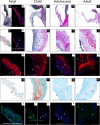
Fig 3. Representative histological and immunofluorescent stainings on aortic valves.
Figures A-E represent the whole aortic valve (leaflet, hinge region, and wall, while in E-W a representative part of the leaflet is shown. (A, C-F, H-J) Verhoeff-Van Gieson staining for collagen (red) and elastin (black). (B, G) Elastin was observed in the fetal valve using immunofluorescence (red). (K-N) Collagen type I (red) was predominant in the fibrosa. (O-R) Safranin-O staining showed proteoglycan presence (red/orange) mainly in the spongiosa and the hinge region. (S-W) αSMA (immunofluorescence; green) with cell nuclei (in blue). In the leaflets of the fetal (S) and 8-month old donor (T) αSMA-positive cells were observed, while in the older leaflets almost no αSMA-positive cells were observed (U-W). Scale bar: 500 μm. l: leaflet; w: wall; f: fibrosa; s: spongiosa; v: ventricularis.
Fig 4
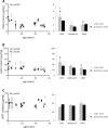
Fig 4. Changes in DNA-, sGAG-, and hydroxyproline (HYP) content.
Changes in DNA- (A), sGAG- (B), and hydroxyproline (HYP) content (C) in μg/mg dry weight (left side: all data points used for correlation analysis with age; right side: grouped data). DNA content is higher in children, especially in the first years of life (0–4 years), compared to the adolescents and adults. sGAG content decreases with age. Hydroxyproline content increases with age in the aortic valve. Differences between groups (p<0.05) are indicated by paired symbols. AV: aortic valve, PV: pulmonary valve.
Fig 5
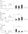
Fig 5. Changes in collagen HP, LP cross-links, and the HP-to-LP ratio.
Changes in collagen HP (A), LP cross-links (B), and the HP-to-LP ratio (C) in μg/mg dry weight (left side: all data points used for correlation analysis with age; right side: grouped data). HP cross-links density increases with age in the aortic valve. In the pulmonary valve, the HP cross-link density increases from adolescent to adult age. LP cross-link density increases with age in both valves. Only in the adult valves, LP cross-link density is different between the aortic and pulmonary valve. In both valves, the HP-to-LP ratio decreases with age, but particularly between childhood and adolescence. Differences between groups (p<0.05) are indicated by paired symbols. AV: aortic valve, PV: pulmonary valve.
Fig 6
Fig 6. Changes in stiffness at low strains as measured with indentation tests.
The stiffness was computed in the belly (A) and commisures (B) (left side: all data points used for correlation analysis with age; at the right side). In the aortic valve, the stiffness in the belly and commissures increase with age, whereas in the pulmonary valve only the commissural stiffness increases with age. In both valves, E-moduli show a steep increase from adolescence to adulthood. Significant differences between groups (p<0.05) are indicated by paired symbols. AV: aortic valve, PV: pulmonary valve.
Fig 7
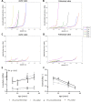
Fig 7. Averaged stress-strain curves, E-modulus at high strains, and extensibility obtained from biaxial tensile tests.
Averaged stress-strain curves (A, B), E-modulus at high strains (C), and extensibility (D) obtained from biaxial tensile tests. The E-moduli (E) in both circumferential and radial direction increase with age in the aortic valve (p<0.05). In the pulmonary valve the E-modulus does not change with age. The leaflets of the 11 and 20-year-old donors are more extensible than the adult leaflets (F). In addition, in these young donors, the pulmonary leaflets were slightly more extensible than the aortic leaflets. As the data of the 8-month old donor could be considered as an outlier, we also performed linear regression analysis of the data set excluding the 8-month data and added the corresponding linear regression lines to Figs E and F. This reveals that after the age of 11, leaflet extensibility significantly decreases with age in circumferential (AV: p<0.01, PV: p<0.05) and radial direction (PV: p<0.01). AV: aortic valve, PV: pulmonary valve.
Fig 8
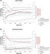
Fig 8. Valve hemodynamics during postnatal life in the aortic and pulmonary valve.
Trend lines for reported data on valve hemodynamics (black) and our measured data on annulus diameter (grey), matrix composition, and stiffness of the belly (red) during postnatal life in the aortic (A) and pulmonary valve (B). Left-sided pressures increase during childhood, while the right-sided pressures decrease to adult values rapidly after birth. Hemodynamic data are collected from several databases, including The National Heart Lung and Blood Institute, MedScape and literature. Trend lines for our experimental data were obtained using the mean values of the data sets depicted in Figs 2, 4 and 6.

References

    1. Ross D, Jackson M, Davies J. The pulmonary autograft—a permanent aortic valve. Eur J cardio-thoracic Surg. 1992;6: 113–116. - PubMed
    1. Schoen FJ. Heart valve tissue engineering: quo vadis? Curr Opin Biotechnol. 2011;22: 698–705. 10.1016/j.copbio.2011.01.004 - DOI - PubMed
    1. Moretti A, Whitehouse MW. Changes in the mucopolysaccharide composition of bovine heart valves with age. Biochem J. 1963;87: 396–402. - PMC - PubMed
    1. Stephens EH, Grande-Allen KJ. Age-related changes in collagen synthesis and turnover in porcine heart valves. J Heart Valve Dis. 2007;16: 672–682. - PubMed
    1. Stephens EH, Chu C-K, Grande-Allen KJ. Valve proteoglycan content and glycosaminoglycan fine structure are unique to microstructure, mechanical load and age: Relevance to an age-specific tissue-engineered heart valve. Acta Biomater. 2008;4: 1148–1160. 10.1016/j.actbio.2008.03.014 - DOI - PMC - PubMed

Publication types

MeSH terms

Substances